





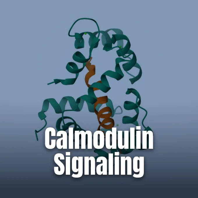
Asking what is calmodulin leads into the molecular machinery that governs calcium signal paths inside living cells. Calmodulin, or calcium-modulated protein, is a small intermediate messenger present throughout the cytosol in every eukaryotic lineage. When a cell receives an electrical or chemical stimulus, calcium ions enter the cytosol and attach to this protein, switching its physical shape into a receptive state. It doesn’t generate signals on its own, but it senses minute shifts in ion levels and binds hundreds of partner enzymes [10].
- What Is Calmodulin in Cellular Signaling?
- Molecular Architecture of the Flexible Central Linker
- How Do EF-Hand Motifs Bind Calcium Ions?
- Conformational Shift from Apo to Holo State
- Activation of Target Enzymes via Myosin Light Chain Kinase
- Modulation of Cardiac Channels in Synaptic Plasticity
- Diverse Roles of CaM in Plant Stress Responses
- Why Calmodulin Conservation Matters in Human Genetics
What Is Calmodulin in Cellular Signaling?
Calcium serves as a common second messenger in eukaryotic biology. Cells keep a steep ionic gradient across the plasma membrane. In resting cells, free calcium in the cytosol stays below 0.1 micromolar, whereas extracellular fluids carry roughly 1 millimolar [1]. Outside calcium levels remain about 10,000 times higher than internal levels [3].
Because most internal enzymes can’t sense calcium on their own, they rely on calmodulin to translate incoming ion surges into metabolic action. Calmodulin acts as a universal sensor that couples ion influx with enzymatic activation, allowing a brief calcium influx to trigger multiple coordinated cellular processes. Through this structural response, a single calcium pulse coordinates glycogen breakdown, cellular movement, and hormone release [10].
Calmodulin appears in every eukaryote studied to date [10]. Yeasts like Saccharomyces cerevisiae carry only one gene for this protein, while vertebrates maintain three non-allelic genes—CALM1, CALM2, and CALM3—that produce the exact same protein sequence across all tissues [5]. Genetic damage to calmodulin calcium-binding sites is lethal to the cell [1]. The sensor operates in the cytosol, inside the cell nucleus, and along organelle membranes [10]. In resting cells, calcium sits in the endoplasmic reticulum or exits through pumps, keeping cytosolic levels low [1]. By reading calcium signals from surface ion channels or internal stores, calmodulin keeps complex cells in homeostatic balance, regulating basic metabolic paths that have remained unchanged since the origin of vertebrate animals [10].
Molecular Architecture of the Flexible Central Linker
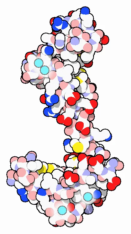
Calmodulin is a compact protein whose single polypeptide chain contains 148 amino acids with a molecular mass of 16.7 kilodaltons [10]. The molecule resembles a tiny dumbbell, joining an N-terminal domain and a C-terminal domain through a central helical linker that keeps each two-loop binding lobe in an extended spatial orientation. In crystal structures like PDB entry 3CLN, all four binding sites are filled with calcium ions, and the linker forms an extended alpha helix separating the two globular ends. Although X-ray crystallography depicts this central segment as a stiff rod, nuclear magnetic resonance data from solution studies show that the linker remains flexible when unattached to target proteins [3].
Flexible linker movement lets the protein grip partner peptides of varied dimensions [3]. When calcium enters the cytosol, the linker bends and rotates. The two globular lobes then clamp around a target peptide. Inside the cell, the C-terminal domain binds calcium with higher affinity than the N-terminal domain [10].
Single-molecule force spectroscopy reveals how individual calmodulin molecules fold under mechanical loads. In 2011, J. Stigler, F. Ziegler, and M. Rief published an optical tweezers study in Science tracking single protein transitions. Their hidden Markov models identified four distinct intermediates during the folding process. Two off-pathway intermediates display non-native interactions across domain boundaries. Competing directly with the productive folding route, these off-pathway intermediates show that domain interactions guide the folding network through cooperative and anticooperative transitions under applied mechanical loads [9].
How Do EF-Hand Motifs Bind Calcium Ions?
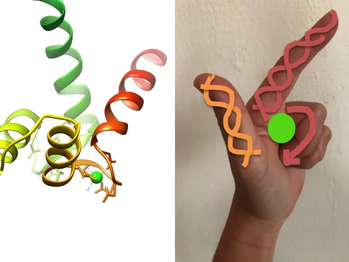
Calcium sensing in calmodulin relies on four evolutionarily conserved EF-hand motifs distributed evenly between its two globular lobes. The motif is named after its resemblance to an L-shaped human hand, where an index finger and thumb represent two alpha helices flanking a central twelve-amino-acid loop that cradles the metal ion [2]. The first such motif was identified in troponin C in the late 1960s before the discovery of calmodulin [3]. With two such motifs in the N-terminal lobe and two in the C-terminal lobe, a single calmodulin molecule captures four calcium ions during signaling events, switching its conformation into a fully saturated state [1].
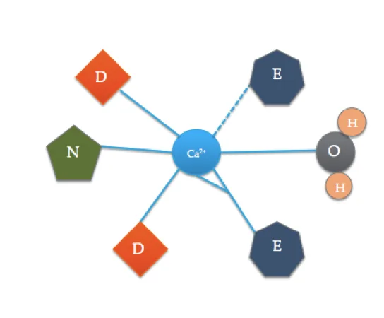
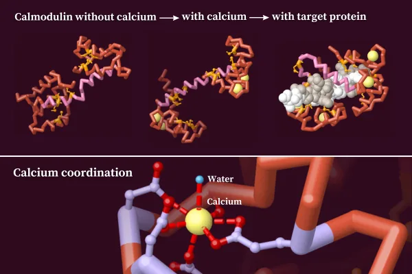
Six ligands surround each metal ion to form seven coordinate bonds. The resulting heptadentate coordination sphere includes oxygen-bearing side chains from three aspartates, one bidentate glutamate, one peptide carbonyl oxygen, and one water molecule from the surrounding aqueous solution. Because glutamate contributes two oxygens to the metal center, the complex forms a distorted octahedral geometry [2].
Thermodynamics and the chelate effect help calmodulin select calcium over magnesium in the cytosol [2]. Cytosolic magnesium is abundant, staying between 0.5 and 1.0 millimolar in resting cells [10]. Because calcium ions are larger than magnesium ions, they reduce steric clash among the six coordinating oxygen ligands inside the binding loop. As a result, the binding association constant for calcium far exceeds that of magnesium, allowing calcium surges to displace bound magnesium ions rapidly during cellular activation [2].
Conformational Shift from Apo to Holo State
In the calcium-free apo state, helical segments across all four binding motifs stay collapsed in a compact shape [10]. Packing these helices tightly keeps nonpolar contact surfaces buried safely inside the protein core [3]. When calcium saturates the loops, electrostatic forces rotate the helices into an open holo state where neighboring helices stand perpendicular to each other, uncovering two hydrophobic binding clefts [10].
The exposed binding pockets are packed with methionine residues. Methionine carries a flexible, unbranched thioether side chain with a polarizable sulfur atom. These sulfur-bearing residues form malleable nonpolar grooves that fit around varied target peptides without requiring strict chemical matches [3]. Target proteins typically present an amphipathic alpha helix with bulky hydrophobic residues separated by polar amino acids, which calmodulin grips securely using these methionine-lined contact surfaces. This flexible hydrophobic surface lets calmodulin interact with more than 300 target proteins in eukaryotic cells [10].
Mapping domain orientations in solution demanded fresh biophysical methods. In 2012, A. Grishaev, N. J. Anthis, and G. M. Clore reported a contrast-matched X-ray scattering study on a lead-substituted complex in the Journal of the American Chemical Society. By swapping four calcium ions for lead atoms in a 65% sucrose buffer, the team made the protein invisible to scattering rays. This heavy-atom labeling strategy yielded pairwise distance data that positioned both protein domains with one-angstrom precision. Their structural model, deposited as PDB entry 2LV6, proved that combining contrast-matched scattering with NMR residual dipolar couplings resolves domain geometry that ordinary solution measurements miss [4].

Activation of Target Enzymes via Myosin Light Chain Kinase
In smooth muscle, calmodulin directs excitation-contraction coupling. Unlike skeletal muscle fibers that rely on troponin C to move tropomyosin off actin filaments, smooth muscle cells depend on myosin light chain kinase for contraction [1]. When cytosolic calcium levels rise, calcium-saturated calmodulin binds an autoinhibitory segment on MLCK, relieving its inhibition [10]. The activated kinase transfers a phosphate group from ATP onto the regulatory light chain of myosin. This phosphorylation initiates cross-bridge cycling between myosin heads and actin filaments, generating the mechanical force that drives smooth muscle shortening in blood vessels and digestive organs [1]. Through this mechanism, calmodulin controls blood vessel tone, arterial blood pressure, and gastrointestinal motility [10].
Beyond muscle action, calmodulin regulates cellular energy metabolism as an integral subunit of phosphorylase kinase. In this complex, it serves as the delta subunit that drives glycogen breakdown into glucose [10]. Calcium-bound calmodulin also stimulates plasma membrane calcium ATPase pumps, clearing cytosolic calcium into the endoplasmic reticulum [1].
Because bacteria lack calmodulin, some pathogens exploit host calmodulin to cause disease. A prime example is the edema factor toxin from Bacillus anthracis, the bacterium responsible for anthrax. When the anthrax toxin enters a host cell, it binds host calmodulin with high affinity, as seen in PDB entry 1K93. Calmodulin binding alters the toxin shape and turns on its adenylyl cyclase activity. The activated toxin converts cellular ATP into cyclic AMP at pathological rates, stripping host tissues of metabolic fuel and crippling immune responses during severe anthrax infection [3].
Modulation of Cardiac Channels in Synaptic Plasticity
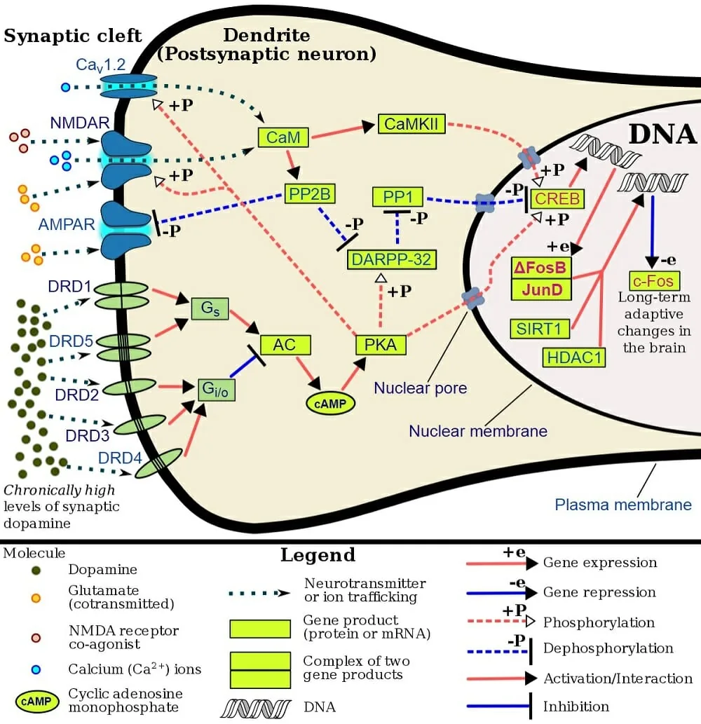
In brain tissue, calmodulin controls synaptic plasticity through calcium/calmodulin-dependent protein kinase II. During high-frequency neural firing, calcium influx through NMDA receptors activates CaMKII, which subsequently phosphorylates AMPA receptors to boost glutamate transmission between synapses during memory storage. Blocking CaMKII halts long-term potentiation, which impairs memory storage in the hippocampus [10].
Some brain proteins bind calmodulin without calcium. Presynaptic GAP-43 and postsynaptic neurogranin carry specialized IQ motifs that attach to apo-calmodulin. GAP-43 sequesters calmodulin near axonal growth cones during rest, acting as a local buffer. When protein kinase C phosphorylates GAP-43 or neurogranin, the modified proteins release calmodulin to engage downstream kinases during axonal growth and synaptic remodeling in developing circuits [12].
In heart muscle, calmodulin modulates cardiac ion channels to keep electrical rhythms regular. The protein binds directly to the pore-forming subunit of voltage-gated CaV1.2 calcium channels to drive calcium-dependent inactivation. It also binds to ryanodine receptor 2 channels, designated as RyR2, on the sarcoplasmic reticulum membrane, helping to suppress aberrant calcium leakage during the resting phase between successive heartbeats in cardiac muscle. Disrupting these channel interactions destabilizes cardiac repolarization and triggers irregular rhythms. Through these dual contacts on CaV1.2 and RyR2, calmodulin prevents calcium overload in cardiac myocytes and ensures that electrical repolarization finishes cleanly after every ventricular beat [6].
Diverse Roles of CaM in Plant Stress Responses
Plants carry both true calmodulins and an expanded family of calmodulin-like proteins [8]. True plant calmodulins match vertebrate sequences closely, but calmodulin-like proteins, known as CMLs, share only about 15% amino acid identity with the classical sensor protein. The model plant Arabidopsis thaliana carries about 50 CML genes in addition to true CaM genes [10]. These diverse CML isoforms display different calcium affinities and distinct locations within plant cells, helping crops handle environmental stress like cold, freezing temperatures, and severe drought. Target proteins regulated by plant calmodulins include ion transporters, transcription factors like AtSR1/CAMTA3, protein kinases, and metabolic enzymes [8].
Genomic comparisons across 41 plant species published by T. K. Mohanta and colleagues in BMC Plant Biology revealed key evolutionary distinctions between these two related sensor families. Phylogenetic analysis indicated that CMLs evolved earlier in plant history than true calmodulins. Most plant CML genes lack introns, whereas true calmodulin genes possess several introns. Furthermore, true calmodulins conserve four D-x-D motifs across all four EF-hands, while CML proteins usually keep only a single D-x-D-x-D motif in their fourth loop [7].
Plant calmodulins also regulate developmental milestones like brassinosteroid hormone synthesis through contacts with the DWF1 enzyme in Arabidopsis. In legumes like Medicago and Lotus, calcium signals guide symbiotic nodule development with nitrogen-fixing Rhizobium bacteria, where the DMI3 gene acts downstream of root hair calcium spiking [10].
Why Calmodulin Conservation Matters in Human Genetics
Human genetics features an unusual setup: three distinct genes—CALM1, CALM2, and CALM3—code for the identical 148-amino-acid protein [5]. CALM1 sits on chromosome 14, CALM2 on chromosome 2, and CALM3 on chromosome 19. Their coding sequences diverge by 13% to 15% at the nucleotide level [11]. Yet natural selection has purged every synonymous mutation that would alter the resulting amino acid chain, leaving the protein sequence completely unchanged across all vertebrate species [5].
Human calmodulin genes generate distinct transcripts through alternative polyadenylation. In CALM1, cells produce a ubiquitous major transcript measuring 1.7 kilobases, while brain and skeletal muscle express a longer minor transcript measuring either 4.1 or 4.4 kilobases. The human genome also contains two non-functional pseudogenes. CALMP1 lies on chromosome 7, and CALMP2 sits on chromosome X. Both pseudogenes lack introns and contain premature stop mutations that eliminate protein production [11].
Because calmodulin is so conserved, scientists once believed CALM mutations were incompatible with human life. In 2012, researchers identified the first human CALM mutations in patients with severe cardiac arrhythmias [6]. CALM1 missense mutations cause long QT syndrome type 14, characterized by prolonged cardiac repolarization, T-wave alternans, and severe atrioventricular block in young infants under ten years old. Other variants trigger catecholaminergic polymorphic ventricular tachycardia, causing exertion-induced syncope or cardiac arrest [11]. Studies confirm that these mutations disrupt CaV1.2 and RyR2 channel regulation, demonstrating the life-or-death role of calcium sensing in the human heart. Biochemists have shown that these mutations alter calcium binding affinities, proving that even subtle disruptions in calmodulin function can destabilize electrical signaling throughout cardiac muscle [6].
- WEBSITE BD Editors. (2017). Calmodulin – Definition, Function and Structure. Biology Dictionary. [Article Link]
- WEBSITE Haas, K. (2016). Signaling: Calcium and Calmodulin. Chemistry LibreTexts. [Article Link]
- WEBSITE Goodsell, D. S. (2017). Calmodulin. RCSB PDB-101. [Article Link]
- ACADEMIC JOURNAL Grishaev, A., Anthis, N. J., & Clore, G. M. (2012). Contrast-Matched Small-Angle X-ray Scattering from a Heavy-Atom-Labeled Protein in Structure Determination: Application to a Lead-Substituted Calmodulin–Peptide Complex. Journal of the American Chemical Society, 134(36), 14686-14689. [Article Link]
- ACADEMIC JOURNAL Sørensen, A. B., Søndergaard, M. T., & Overgaard, M. T. (2013). Calmodulin in a Heartbeat. FEBS Journal, 280(21), 5511-5532. [Article Link]
- ACADEMIC JOURNAL Jensen, H. H., Brohus, M., Nyegaard, M., & Overgaard, M. T. (2018). Human Calmodulin Mutations. Frontiers in Molecular Neuroscience, 11, 396. [Article Link]
- ACADEMIC JOURNAL Mohanta, T. K., Kumar, P., & Bae, H. (2017). Genomics and evolutionary aspect of calcium signaling event in calmodulin and calmodulin-like proteins in plants. BMC Plant Biology, 17(1), 38. [Article Link]
- ACADEMIC JOURNAL Zeng, H., Xu, L., Singh, A., Wang, H., Du, L., & Poovaiah, B. W. (2015). Involvement of calmodulin and calmodulin-like proteins in plant responses to abiotic stresses. Frontiers in Plant Science, 6, 600. [Article Link]
- ACADEMIC JOURNAL Stigler, J., Ziegler, F., Gieseke, A., Gebhardt, J. C. M., & Rief, M. (2011). The Complex Folding Network of Single Calmodulin Molecules. Science, 334(6055), 512-516. [Article Link]
- WEBSITE Wikipedia contributors. Calmodulin. Wikipedia. [Article Link]
- WEBSITE Wikipedia contributors. Calmodulin 1. Wikipedia. [Article Link]
- WEBSITE Wikipedia contributors. Calmodulin-binding proteins. Wikipedia. [Article Link]


