





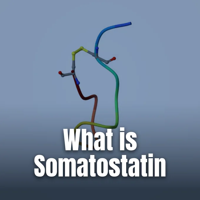
What is somatostatin, and how does it balance hormone output across human organs? Delta cells in the digestive tract and specialized neuroendocrine neurons in the brain synthesize this regulatory peptide to stop excessive glandular activity while preserving metabolic communication among distant organ systems. When released into tissue fluid or blood, it binds to surface G protein-coupled receptors to stop the release of secondary hormones. The peptide halts Growth Hormone output from the pituitary gland and curbs both insulin and glucagon in the Pancreatic Islets. Through these targeted blocking actions, the hormone prevents endocrine overproduction and stabilizes metabolic signaling across physiological networks.
What Is Somatostatin?
Scientists discovered somatostatin over four decades ago while purifying hypothalamic extracts that could halt Growth Hormone release from anterior pituitary cells. Because the peptide silenced somatotropin discharge, early medical literature introduced several descriptive synonyms, including Growth Hormone-Inhibiting Hormone (GHIH), Growth Hormone Release-Inhibiting Hormone (GHRIH), Somatotropin Release-Inhibiting Hormone (SRIH), and Somatotropin Release-Inhibiting Factor (SRIF). GHIH acts as a systemic hormonal brake. Its primary role stays inhibitory in every examined tissue [3].
In humans, somatostatin production begins from a single gene designated as SST. The cell translates this code into a larger precursor called pre-prosomatostatin, which undergoes enzymatic cleavage at specific sites to yield two biologically active peptides. The first active form is somatostatin-14 (SS-14), an amino acid chain of 14 residues that predominates in the central nervous system and mediates rapid synaptic responses. The second form, somatostatin-28 (SS-28), contains 28 amino acids and extends from the same carboxyl-terminal sequence, showing higher abundance in the mucosal lining of the intestine [3, 11]. Alternative processing of the same precursor protein also yields Neuronostatin, producing active peptides known as Neuronostatin-13 and Neuronostatin-19 that bind separate cellular targets [3].
Vertebrate lineages display considerable genetic divergence in how they produce these regulatory peptides. While humans and tetrapods rely on a single SST gene, teleost fish possess multiple distinct genes [1, 3]. Zebrafish carry all six genes: SS1, SS2, SS3, SS4, SS5, and SS6. These ancestral whole-genome duplications provide teleost species with flexible regulatory control over separate tissues, allowing specialized organ expression that differs from mammalian systems [1, 3]. In every vertebrate lineage, however, the peptide retains its core structure to govern glandular secretion and cellular turnover [1].
Peptide Structure and Cleavage
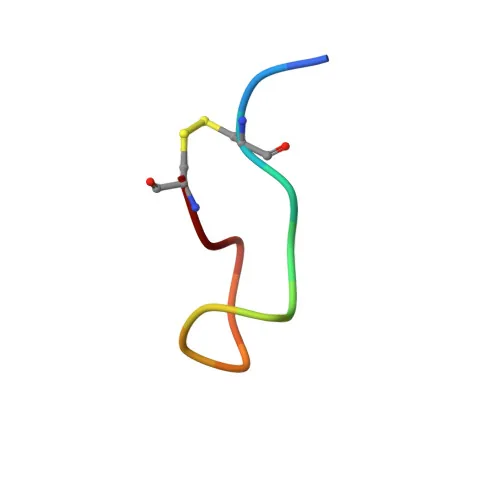
The biological activity of somatostatin-14 depends on a cyclic molecular conformation held together by an internal covalent link. Anoop, Ranganathan, Dhaked, Jha, Pratihar, and Maji at the Indian Institute of Technology Bombay resolved the solution structure of somatostatin-14 in five percent D-mannitol for the Worldwide Protein Data Bank (PDB 2MI1). Their work in the Journal of Biological Chemistry mapped its nuclear magnetic resonance coordinates. Shape directs hormone docking [2].
Within the cyclic sequence of somatostatin-14, an intramolecular disulfide bridge connects cysteine residues at positions 3 and 14, designated chemically as Cys(3)-Cys(14). In healthy endocrine tissues, peptide hormones face an apparent paradox where safe storage needs physical aggregation into granules, yet hormone release demands monomer supply on short notice. The Mumbai team showed that somatostatin forms reversible amyloid fibrils in vivo within endocrine secretory granules. Cleavage of the Cys(3)-Cys(14) disulfide bridge disrupts native folding, which increases peptide flexibility and drives rapid amyloid formation that resists monomer dissolution [2].
Evolutionary analyses published in the Annals of the New York Academy of Sciences by Gahete, Cordoba-Chacón, Duran-Prado, Malagón, Martinez-Fuentes, Gracia-Navarro, Luque, and Castaño traced the origin of these peptides to an ancestral somatostatin and urotensin II (SST/UII) gene. Local duplication first split that precursor into separate SST and UII genes before whole-genome duplication rounds expanded the family in early vertebrates. Tetrapods preserved two diverging lines from these events, retaining the classic hormone as well as cortistatin (CST), an allied peptide encoded by the SS2 gene [1, 3]. Gahete and co-authors observed that somatostatin and its receptors form “a molecular family with unique functional complexity and versatility” across vertebrates. These variations shape how different peptide forms dock into cell surface receptors in modern animals [1].
Hypothalamic Control of Growth Hormone
In the brain, neuroendocrine neurons in the Ventromedial Nucleus of the Hypothalamus produce somatostatin to govern pituitary hormone discharge. Axons from these neurosecretory cells project straight to the Median Eminence, releasing peptide packets into hypophyseal portal vessels. Portal blood flow delivers the hormone directly to somatotrope cells in the Anterior Pituitary gland, where it counters Growth Hormone-Releasing Hormone (GHRH) to halt systemic Growth Hormone discharge. Hypothalamic axons drive this release [3].
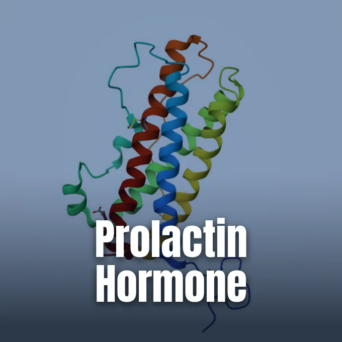
Beyond curbing Growth Hormone, hypothalamic somatostatin restricts the output of Thyroid-Stimulating Hormone (TSH) and Prolactin (PRL) from pituitary cells. The endocrine axis depends on negative feedback loops to prevent excessive body growth. When circulating levels of Growth Hormone and somatomedins rise in peripheral blood, neuroendocrine cells in the Periventricular Nucleus increase their secretory rate to suppress further pituitary somatotrope output. This heightened peptide release reaches somatotrope cells and tempers hormone synthesis. Negative feedback curbs Growth Hormone surges. Hypothalamic signaling thus enforces strict physiological control over physical development and energy expenditure [3].
Somatostatin also acts within central neural networks that operate away from neuroendocrine blood vessels. Distinct populations of somatostatin-positive neurons reside in the Cerebral Cortex, Hippocampus, Arcuate Nucleus, and the Brainstem Nucleus of the Solitary Tract [3, 6]. In the neocortex, somatostatin-containing interneurons (SOM-INs) form inhibitory circuits that synapse onto the dendrites of excitatory pyramidal cells. Liguz-Lecznar, Urban-Ciećko, and Kossut reported in Frontiers in Neural Circuits that somatostatin acts both pre-synaptically and post-synaptically on cortical circuits to fine-tune synaptic plasticity, memory formation, and sensory learning [6]. In Biomolecules, Liguz-Lecznar, Dobrzański, and Kossut showed that loss of somatostatin interneurons alters the excitation-inhibition balance, contributing to cognitive deficits across neurodegenerative conditions [10].
Delta Cells in Digestive Tissues
The digestive tract is the largest reservoir of somatostatin in the human body. Endocrine Delta cells reside throughout the mucosa of the gastric Pyloric Antrum, Duodenum, and Pancreatic Islets [3, 8]. From these mucosal positions, the peptide enters local tissue fluid to act on neighboring cells and enters venous vessels that drain into portal blood heading toward the liver. Delta cells release SS-14 into mucosal tissue [3].
In the stomach, luminal acidity stimulates Delta cells to release somatostatin, whereas Vagus Nerve stimulation suppresses its discharge. Once released into the gastric mucosa, the hormone binds surface receptors on acid-producing parietal cells to inhibit adenylate cyclase, effectively antagonizing histamine-driven hydrochloric acid secretion. It also suppresses the secretion of gastrin from G cells and histamine from enterochromaffin-like cells, which slows gastric acid output. Within Pancreatic Islets, Delta cells release somatostatin when triggered by the Beta-cell peptide Urocortin3 (Ucn3). This local secretion exerts paracrine suppression over adjacent Alpha and Beta cells, curbing the discharge of both insulin and glucagon to stabilize blood sugar levels [3, 8].
Somatostatin exerts broad inhibitory control over digestive transit. The peptide slows gastric emptying, dampens circular smooth muscle contractions in the bowel wall, and reduces splanchnic blood flow during the active stages of nutrient breakdown [3, 8]. It also suppresses the exocrine function of the pancreas, lowering the discharge of bicarbonate, water, and digestive enzymes like Amylase [3, 5]. In Frontiers in Endocrinology, Shamsi, Chatoo, Xu, and Chen highlighted that somatostatin signaling in mucosal tissues coordinates digestion, reduces inflammation, and regulates intestinal motility [8].

Somatostatin Receptor Family
All biological actions of somatostatin depend on five distinct G protein-coupled receptors labeled SSTR1 through SSTR5 (also called sst1 to sst5) [1, 5]. Each receptor spans the plasma membrane seven times and couples to inhibitory Gi alpha subunits that decrease intracellular cyclic adenosine monophosphate levels upon hormone binding to stop vesicle exocytosis. Receptor binding silences cellular discharge [5]. Five distinct genes encode SSTR1 through SSTR5 [1].
Tissue-specific receptor distribution allows somatostatin to exert selective control over different organ systems. Mouse knock-out studies demonstrate that SSTR2 and SSTR5 mediate Growth Hormone suppression in the pituitary gland, whereas SSTR1 and SSTR5 combine to block Prolactin secretion. Pituitary corticotrope cells use SSTR2 and SSTR5 to restrict Adrenocorticotropic Hormone (ACTH) release. In Pancreatic Islets, SSTR2 directs glucagon inhibition, while both SSTR2 and SSTR5 cooperate to shut down insulin release. In the stomach, SSTR2 suppresses parietal acid secretion, while SSTR5 curtails pancreatic Amylase and GLP-1 output. Thyroid C-cells rely on SSTR1 to halt Calcitonin secretion, and immune cells utilize SSTR2 to dampen Interferon Gamma production [5].
The promoter region of the human SST gene responds to specific transcriptional regulators that direct hormone synthesis. In the International Journal of Molecular Sciences, Ampofo, Nalbach, Menger, and Laschke reviewed the enhancers and silencers that modulate somatostatin expression [11]. In fact, biochemists first discovered the transcription factor CREB (cyclic AMP response element-binding protein) based on its direct binding to the regulatory promoter region of the somatostatin gene [5]. Extracellular signals activate CREB, initiating peptide synthesis in response to physiological cues [11].
Inhibitory Mechanisms in Secretory Cells
Why does somatostatin act as a universal inhibitor across human tissues? The answer lies in how its five receptor subtypes link to core intracellular signaling cascades in target cells [1, 5]. Rather than exciting target tissues, hormone binding to SSTR subtypes couples to inhibitory Gi alpha proteins that shut down intracellular second messengers, preventing vesicle exocytosis and stopping secretion. This receptor coupling silences hormone discharge across diverse endocrine organs [5].
At the molecular level, somatostatin receptor activation directly suppresses the enzyme Adenylate Cyclase, decreasing cyclic adenosine monophosphate (cAMP) production and shutting off Protein Kinase A activity [3, 5]. At the same time, receptor signaling opens inward-rectifying potassium channels to hyperpolarize the plasma membrane while closing voltage-dependent calcium channels. Because vesicle exocytosis requires calcium influx, this electrical silencing stops hormone release within seconds [2, 5]. Shamsi, Chatoo, Xu, and Chen documented how these signaling events suppress cellular secretion, dampen mucosal inflammation, and reduce visceral pain pathways throughout the digestive tract [8].
In addition to stopping hormone release, somatostatin exerts clear antiproliferative effects on normal and neoplastic tissues. In the International Journal of Molecular Sciences, Kumar detailed how somatostatin activates surface receptor subtypes to induce cell cycle arrest and trigger apoptosis in multiple neuroendocrine tumor lineages. Indirectly, the peptide curtails tumor growth by suppressing circulating growth factors and blocking tumor angiogenesis. Somatostatin thus functions as both a secretory brake and a tumor suppressor in human tissues [9].
Synthetic Somatostatin Analogues in Medicine
Natural somatostatin poses practical clinical limitations due to its swift breakdown in the human bloodstream. The endogenous hormone has a circulation half-life of only 2 to 3 minutes in plasma, requiring continuous intravenous infusion to produce sustained clinical effects in patients. To solve this pharmacokinetic challenge, pharmaceutical chemists developed synthetic analogues with greater metabolic stability. The first widely adopted synthetic analogue was Octreotide (Sandostatin, Novartis Pharmaceuticals), an octapeptide that mimics natural somatostatin while resisting enzymatic cleavage [3].
Octreotide displays an elimination half-life of approximately 90 minutes, allowing subcutaneous or intramuscular injection for outpatient treatment. A second major analogue, Lanreotide (marketed as Somatuline by Ipsen Pharmaceuticals), gained approval from the United States Food and Drug Administration on August 30, 2007. Lanreotide acts as a long-acting SSTR2 analogue, providing symptom relief for patients with Carcinoid Syndrome and Acromegaly in the United States, the United Kingdom, Canada, and Australia. A third analogue, Pasireotide (marketed as Signifor by Novartis), was engineered with 40-fold increased binding affinity for SSTR5. The US FDA and European Union approved Pasireotide as an orphan drug for Cushing’s Disease in patients who fail pituitary surgery [3, 5].
These synthetic analogues have transformed the clinical management of neuroendocrine neoplasms. Medical data show that survival rates for patients with Neuroendocrine Tumors (NETs) improved 3-fold following the introduction of somatostatin analogue therapy [5]. In the Cochrane Database of Systematic Reviews, Gurusamy, Koti, Fusai, and Davidson evaluated prophylactic somatostatin analogues in pancreatic surgery, assessing whether perioperative analogue treatment lowers post-resection complications such as pancreatic fistulas [7]. Synthetic somatostatin mimics remain a cornerstone of endocrine oncology [5].
Radionuclide Imaging and Antagonist Therapies
Neuroendocrine tumors strongly overexpress somatostatin receptors on their plasma membranes, particularly the SSTR2 subtype [4, 9]. Nuclear medicine teams exploit this high receptor density by coupling somatostatin peptide vectors to diagnostic radionuclides, enabling sensitive Positron Emission Tomography (PET) scans for detecting primary tumors and occult metastases. Macrocyclic chelators such as DOTA, NODAGA, and CB-TE2A anchor radioactive isotopes to targeting peptides. Non-radioactive antagonists such as cyclosomatostatin also serve as experimental tools in molecular pharmacology. SSTR2 guides these diagnostic radioactive probes [4].
While early radiopharmaceuticals relied on receptor agonists, clinical oncology has increasingly shifted toward somatostatin receptor antagonists. First-generation antagonists included sst2-ANT, BASS, and sst3-ODAN-8. Subsequent research yielded second-generation candidates with higher binding affinity, including LM3, JR10, and JR11. Among these compounds, JR11 emerged as the most effective antagonist for clinical imaging and therapy. Unlike traditional agonists that require receptor activation and internalization, antagonist molecules bind surface receptors regardless of their activation state, achieving superior tumor uptake in neoplastic lesions. Pairing JR11 with Gallium-68 yields Ga-NODAGA-JR11 for diagnostic PET imaging, whereas conjugating it with Lutetium-177 produces Lu-DOTA-JR11 for targeted tumor irradiation [4].
Peptide Receptor Radionuclide Therapy (PRRT) couples therapeutic beta-emitting radioisotopes to somatostatin vectors, delivering targeted ionizing radiation directly to malignant cells while sparing distant healthy organ structures. Clinicians use radionuclides including Lutetium-177, Yttrium-80, Copper-64, and Indium-111, adjusting dosage to patient characteristics to avoid renal damage. The low-energy electrons emitted by Lutetium-177 destroy malignant cells while minimizing radiation exposure to adjacent healthy tissues. By combining high-affinity receptor targeting with radionuclide payloads, somatostatin-directed therapies continue to advance precision oncology for patients with neuroendocrine tumors [4, 9].
- ACADEMIC JOURNAL Gahete, M. D., Cordoba‐Chacón, J., Duran‐Prado, M., Malagón, M. M., Martinez‐Fuentes, A. J., Gracia‐Navarro, F., Luque, R. M., & Castaño, J. P. (2010). Somatostatin and its receptors from fish to mammals. Annals of the New York Academy of Sciences, 1200(1), 43-52. [Article Link]
- ACADEMIC JOURNAL Anoop, A., Ranganathan, S., Dhaked, B. D., Jha, N. N., Pratihar, S., Ghosh, S., Sahay, S., Kumar, S., Das, S., Kombrabail, M., Agarwal, K., Jacob, R. S., Singru, P., Bhaumik, P., Padinhateeri, R., Kumar, A., & Maji, S. K. (2014). Elucidating the role of disulfide bond on amyloid formation and fibril reversibility of somatostatin-14. Journal of Biological Chemistry, 289(24), 16884-16903. [Article Link]
- WEBSITE Wikipedia contributors. (2026). Somatostatin. Wikipedia, The Free Encyclopedia. [Article Link]
- WEBSITE Wikipedia contributors. (2026). Somatostatin inhibitor. Wikipedia, The Free Encyclopedia. [Article Link]
- WEBSITE Wikipedia contributors. (2026). Somatostatin receptor. Wikipedia, The Free Encyclopedia. [Article Link]
- ACADEMIC JOURNAL Liguz‐Lecznar, M., Urban‐Ciećko, J., & Kossut, M. M. (2016). Somatostatin and somatostatin-containing neurons in shaping neuronal activity and plasticity. Frontiers in Neural Circuits, 10, 48. [Article Link]
- ACADEMIC JOURNAL Gurusamy, K. S., Koti, R. S., Fusai, G., & Davidson, B. R. (2013). Somatostatin analogues for pancreatic surgery. Cochrane Database of Systematic Reviews, 2013(4), CD008370. [Article Link]
- ACADEMIC JOURNAL Shamsi, B. H., Chatoo, M., Xu, X. K., Xu, X., & Chen, X. Q. (2021). Versatile functions of somatostatin and somatostatin receptors in the gastrointestinal system. Frontiers in Endocrinology, 12, 652363. [Article Link]
- ACADEMIC JOURNAL Kumar, U. (2023). Somatostatin and somatostatin receptors in tumour biology. International Journal of Molecular Sciences, 25(1), 436. [Article Link]
- ACADEMIC JOURNAL Liguz‐Lecznar, M., Dobrzański, G., & Kossut, M. M. (2022). Somatostatin and somatostatin-containing interneurons—From plasticity to pathology. Biomolecules, 12(2), 312. [Article Link]
- ACADEMIC JOURNAL Ampofo, E., Nalbach, L., Menger, M. D., & Laschke, M. W. (2020). Regulatory mechanisms of somatostatin expression. International Journal of Molecular Sciences, 21(11), 4170. [Article Link]


