





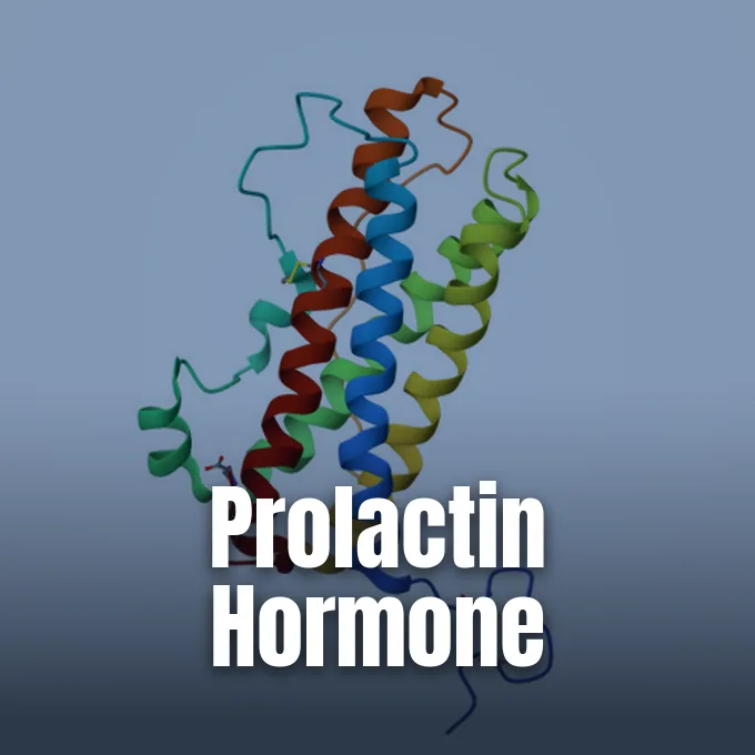
Learning what is prolactin begins with its source in the anterior pituitary gland, a small endocrine organ resting at the base of the human brain. Prolactin (also called lactotropin or PRL) is a peptide hormone that guides breast tissue growth and triggers milk flow after childbirth [3]. Beyond motherhood, it influences fluid balance, metabolic regulation, and immune defenses in hundreds of vertebrate species [6]. Clinicians at institutions such as Cleveland Clinic and MedlinePlus explain that blood tests check pituitary health, evaluate unexpected breast leakage, and diagnose fertility issues in both women and men [1].
What Is Prolactin?
Biologists first found prolactin in non-human animals around 1930 through lab studies by Oscar Riddle, who observed its capacity to stimulate crop milk in pigeons. Four decades later, Canadian medical researcher Henry Friesen identified the hormone in human blood in 1970. The molecule is a peptide hormone encoded by the single PRL gene located on human chromosome 6 [6]. Medical textbooks call it lactotropin or mammotropin because of its classic milk-making actions in mammalian biology [3]. Endocrine cells in the anterior pituitary gland release the protein into systemic blood channels, sending signals to distant target organs that carry matching surface receptor proteins [6].
How does one protein manage so many different tasks across the animal kingdom? The hormone influences over 300 separate body processes in vertebrates. In freshwater fish, it controls salt and water balance across cell membranes. In birds, it prompts brooding in nesting hens and helps flamingos and male emperor penguins produce crop milk to nourish their young. In male mammals, normal levels enhance luteinizing hormone receptors on testicular Leydig cells to support testosterone production and sperm maturation [6].
Prolactin acts through three distinct signaling routes rather than functioning solely through general blood flow. In addition to moving in the bloodstream, it works as an autocrine messenger on its source cell and as a paracrine signal (reaching nearby cells) in local tissue pockets. Receptors for the hormone belong to the class 1 cytokine receptor family, activating cell Janus kinase and signal transducer pathways to guide cellular growth [6].
Lactotroph Production of PRL
The anterior pituitary gland houses endocrine units known as prolactin cells, which medical texts designate as lactotrophs (or mammotrophs). Lactotrophs make up 20% to 55% of anterior pituitary cells, with shares shifting based on sex, age, and reproductive stage. Women show higher baseline cell activity than men. Endocrine cells synthesize the hormone on rough endoplasmic reticulum before transferring the newly formed polypeptide to the trans-Golgi zone for packaging into dense storage granules. Granules store the hormone during active phases until calcium signals prompt release into neighboring capillary networks, while lysosomal enzymes degrade surplus granules when release needs subside [8].
During pregnancy, lactotrophs undergo marked growth to prepare the body for future milk making. Placental estrogens trigger both hyperplasia, which multiplies cell numbers, and hypertrophy, which expands cell volume. Estrogen stimulation causes the maternal pituitary gland to enlarge clearly throughout the second and third trimesters of gestation [8].
Although the pituitary gland makes most blood prolactin, several peripheral tissues produce the peptide on their own. Peripheral synthesis occurs in the uterine decidua, myometrium, breast glandular tissue, prostate, and immune cells like T cells and macrophages. Gene transcription in these local tissues relies on an alternative superdistal promoter located 5.8 kilobases upstream of the primary pituitary transcription start site within the human genome. The distal promoter responds to cyclic adenosine monophosphate signals via C/EBP and Oct-1 factors rather than responding to dopamine [6].
Molecular Architecture of Lactotropin
Human prolactin consists of a single protein chain of 199 amino acids folded into a compact globular structure. Three internal disulfide bonds link six cysteine residues (Cys4–Cys11, Cys58–Cys174, and Cys191–Cys199) to keep the peptide stable [8]. The most common blood form is an unglycosylated monomer with a molecular weight of 23 kilodaltons, known in laboratory reports as little prolactin [6].
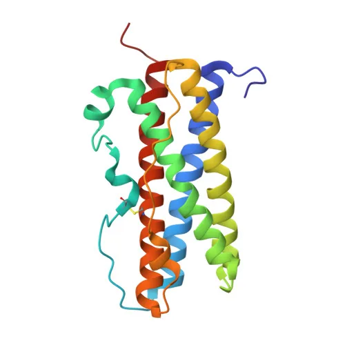
Researchers clarified the three-dimensional form of the hormone when K. Teilum, J. C. Hoch, V. Goffin, S. Kinet, J. A. Martial, and B. B. Kragelund solved its solution structure using NMR tests at the University of Copenhagen. Cataloged as entry 1RW5 in the RCSB PDB and indexed on PubMed, the atomic model revealed a classic four-helix bundle fold. Unbound prolactin keeps roughly the same spatial geometry as receptor-bound prolactin, meaning that no major shape shift takes place during initial docking. One hormone molecule links to two separate receptor units, assembling a stable ternary complex that initiates downstream intracellular signaling cascades. Gel filtration and electrophoresis clearly detected both binary and ternary complexes [4].
Scientists have also engineered altered versions of the molecule to test potential medical therapies. At the Necker research center in Paris, J. B. Jomain, E. Tallet, I. Broutin, S. Hoos, J. van Agthoven, A. Ducruix, P. A. Kelly, P. England, and V. Goffin studied prolactin receptor antagonists. The team solved the crystal structure of an antagonist named Del1-9-G129R-hPRL, cataloged in the RCSB PDB under code 2Q98 and listed in PubMed. By cutting nine amino acids from the N-terminus and placing an arginine at position 129, the authors blocked receptor dimerization and stopped downstream activation in target cells [5].
Hypothalamic Inhibition by Dopamine
Unlike other anterior pituitary hormones that rely on stimulating factors from the brain, prolactin stays under steady brake control. Neurosecretory tuberoinfundibular dopamine neurons (TIDA neurons) in the arcuate nucleus of the hypothalamus release dopamine into hypophyseal portal vessels. Dopamine locks onto D2 receptors on lactotroph membranes to dampen adenylate cyclase activity, cut calcium flow into the cytoplasm, and curb both hormone synthesis and granule release. When hypothalamic dopamine release decreases under physiological demand, lactotroph cells escape inhibition and rapidly elevate hormone output into the systemic bloodstream [6].
The body keeps blood hormone levels steady through a fast short-loop feedback path. When blood prolactin levels climb, molecules cross into the brain and bind receptors in hypothalamic centers. Elevated prolactin prompts arcuate neurons to release more dopamine, which travels down portal vessels to the pituitary and shuts down further prolactin output [6].
A few chemical signals stimulate hormone release when the body requires elevated levels. Thyrotropin-releasing hormone (TRH) from the hypothalamus binds lactotrophs and prompts rapid secretion, while vasoactive intestinal peptide (VIP) also shows stimulatory properties in laboratory models [7]. Estrogens boost gene activity through the Pit-1 factor in pituitary cells [6]. Scientists have spent decades searching for a dedicated prolactin-releasing hormone in humans. A candidate peptide emerged in 1998, but its physiological role remains unverified, leaving TRH as the best-known stimulator [7].


Mammary Stimulation and Milk Production
The primary role of prolactin in female mammals is encouraging mammary alveoli to make breast milk. During gestation, rising hormone levels work with estrogen and progesterone to build lobuloalveolar structures [3]. However, high circulating concentrations of progesterone and estrogen during pregnancy prevent early milk secretion by strongly blocking prolactin actions at the breast receptor site [6].
When the placenta delivers during childbirth, maternal estrogen and progesterone levels drop sharply within hours. The postpartum drop removes the biochemical block on breast cells, launching the phase of active lactogenesis [6]. Prolactin binds to alveolar receptors, turning on genes for casein protein, lactose sugar, and milk fats. Alveolar epithelial cells rapidly fill with milk for newborn nourishment [8]. If nursing stops, maternal prolactin levels slide back to baseline nonpregnant numbers within one to two weeks, and breast glandular tissue begins to reset through natural involution [6].
Keeping up ongoing milk yield depends on a sensory reflex started by an infant suckling at the breast [3]. Nipple nerve endings send signals up the spinal cord to the hypothalamus, pausing dopamine release and triggering a fresh wave of prolactin into the blood. At the same time, suckling prompts the posterior pituitary to release oxytocin, which contracts myoepithelial cells around alveolar pockets to eject milk down the ducts [6].

Metabolic and Immune System Signaling
Outside the reproductive tract, prolactin works as an immune cytokine that guides white blood cell responses [6]. T lymphocytes, B lymphocytes, and macrophages carry prolactin receptors and can release modest quantities of the hormone. Binding of the protein activates protein kinase C and tyrosine kinase signaling pathways, helping white blood cells survive, divide, and coordinate immune responses against harmful microbes [8].
Because prolactin spurs immune activity, altered hormone amounts can connect to autoimmune troubles. When levels rise too high, the hormone stops the normal removal of self-attacking B cells, letting autoreactive immune clones slip past safety stops. Impaired B-cell selection helps explain why hyperprolactinemia often links to symptom flare-ups in conditions like systemic lupus erythematosus, rheumatoid arthritis, and multiple sclerosis. Changes during pregnancy also reshape the maternal immune system, contributing to gender differences observed across autoimmune illnesses [6].
Prolactin also helps steer daily metabolism and systemic energy balance. Fat cells and pancreatic beta cells carry both prolactin receptors and type 2 dopamine receptors on their surfaces [6]. In pregnancy, higher hormone levels expand beta-cell numbers and raise insulin output, ensuring steady glucose flows to the unborn baby [8]. In adipose tissue, the hormone slows lipid storage in maternal fat while speeding fat production in the breast. Outside pregnancy, chronically high hormone levels can promote abdominal weight gain, impair glucose-insulin profiles, and alter blood lipid concentrations. Medical treatment with dopamine agonists like bromocriptine or cabergoline improves insulin response and restores metabolic balance [6].
Clinical Causes of Hyperprolactinemia
When blood prolactin levels stay above standard reference intervals, doctors diagnose hyperprolactinemia. The most common non-cancerous growth is a pituitary adenoma called a prolactinoma [3]. Pituitary prolactinomas release the hormone without feedback control, disrupting the hypothalamic-pituitary-gonadal axis. High prolactin suppresses gonadotropin-releasing hormone from the hypothalamus, which lowers downstream secretion of luteinizing hormone and follicle-stimulating hormone [6]. In women, this change causes missed menstrual periods (amenorrhea) and difficulty conceiving, while in men it causes lower testosterone, reduced sex drive, and erectile dysfunction. Both women and men may notice unprompted breast milk leakage, a clinical symptom designated as galactorrhea. Large pituitary adenomas can also press on the neighboring optic chiasm, causing visual field defects and steady headaches in addition to systemic reproductive disruptions [1].
Several health conditions outside the brain can push hormone levels up. In primary hypothyroidism, an underactive thyroid gland leads the hypothalamus to release extra thyrotropin-releasing hormone (TRH), which prompts the pituitary to pump out more prolactin. Kidney disease also raises numbers because damaged kidneys cannot filter the hormone out of circulation. Some patients test high because of macroprolactin, a harmless condition where circulating hormone molecules bind antibody proteins without provoking clinical symptoms or organ damage [1].

Prescription drugs cause many mild hormone increases. Antipsychotic drugs like risperidone, phenothiazines, and butyrophenones block D2 dopamine receptors in the pituitary gland, lifting the natural brake on lactotrophs. Antidepressants, metoclopramide, verapamil, opioid painkillers, and marijuana products can also elevate numbers. Physical stress, heavy exercise, and high-protein meals can cause brief spikes as well [1].
Diagnostic Blood Testing Standards
Doctors order a prolactin blood test to check pituitary health and investigate fertility problems or unexpected milk leakage. The test requires a fasting blood draw taken from an arm vein, usually scheduled in the early morning when hormone concentrations reach baseline diurnal stability. Patients do not need complicated prep, but clinical guidance from MedlinePlus suggests skipping strenuous workouts and breast stimulation before the appointment to prevent false spikes [1].
Laboratories measure hormone concentrations in nanograms per milliliter (ng/mL) or micrograms per liter (µg/L). In adult men, healthy numbers fall less than 20 ng/mL (20 µg/L). In nonpregnant women, standard values remain below 25 ng/mL (25 µg/L). During pregnancy, maternal hormone levels climb by ten to twenty times, reaching physiological peaks between 100 ng/mL and 500 ng/mL (500 µg/L) to support breast tissue preparation. As clinical references in Goldman-Cecil Medicine by Elsevier and reviews edited by Sandeep K. Dhaliwal, David C. Dugdale, and Brenda Conaway note, patients should evaluate numbers against the specific limits printed on their laboratory report [1].
To keep test results reliable around the world, the World Health Organization Expert Committee on Biological Standardization (ECBS) coordinates shared reference materials. In 2016, an international collaborative study across ten laboratories in seven countries tested candidate material coded 83/573. The ECBS adopted this sample as the 4th International Standard for Prolactin, human, setting an assigned strength of 67 mIU per ampoule. The preparation replaced the older 3rd International Standard, coded 84/500, after heat tests confirmed its chemical stability. Immunoassay calibration using this reference material ensures that clinical laboratories obtain consistent measurements across diverse patient blood samples and competing commercial diagnostic platforms [2].
- WEBSITE MedlinePlus. (2024, July 21). Prolactin blood test. U.S. National Library of Medicine. [Article Link]
- REPORT World Health Organization. (2016, October 25). WHO international collaborative study of the proposed 4th IS for prolactin, human (WHO/BS/2016.2292). Expert Committee on Biological Standardization. [Article Link]
- WEBSITE Cleveland Clinic. (2023, August 21). What does prolactin do? Cleveland Clinic. [Article Link]
- ACADEMIC JOURNAL Teilum, K., Hoch, J. C., Goffin, V., Kinet, S., Martial, J. A., & Kragelund, B. B. (2005). Solution structure of human prolactin. Journal of Molecular Biology, 351(4), 810–823. [Article Link]
- ACADEMIC JOURNAL Jomain, J. B., Tallet, E., Broutin, I., Hoos, S., van Agthoven, J., Ducruix, A., Kelly, P. A., Kragelund, B. B., England, P., & Goffin, V. (2007). Structural and thermodynamic bases for the design of pure prolactin receptor antagonists: X-ray structure of Del1-9-G129R-hPRL. Journal of Biological Chemistry, 282(45), 33118–33131. [Article Link]
- WEBSITE Wikipedia contributors. (2026). Prolactin. Wikipedia, The Free Encyclopedia. [Article Link]
- WEBSITE Wikipedia contributors. (2026). Prolactin-releasing hormone. Wikipedia, The Free Encyclopedia. [Article Link]
- WEBSITE Wikipedia contributors. (2026). Prolactin cell. Wikipedia, The Free Encyclopedia. [Article Link]


