






Readers learning about Wilson’s disease encounter a rare genetic disorder that impairs how human cells handle copper [2]. In a healthy body, liver transport proteins excrete surplus dietary copper through bile into the gut for safe disposal [12]. Gene variants in ATP7B disrupt this pathway, so copper ions build up inside liver cells before spilling into the blood to damage the brain, corneas, and kidneys [2]. Without lifelong medical care, this copper buildup causes liver failure and permanent neurological harm [4].
How Copper Overload Begins
Copper is a trace mineral. Human cells need small amounts of copper from food to make enzymes and keep nerves healthy [6]. However, excess free copper acts as a potent cellular poison when normal liver clearance stops, allowing unchecked metal stores to injure hepatocytes and leak into general circulation [12].
Older population studies estimated that Wilson’s disease affected about 1 in 30,000 people worldwide [2]. However, modern gene screening shows that disease-causing mutations occur more often than old clinic registries recorded. In the United Kingdom, a team led by Dr. A. J. Coffey sequenced the ATP7B coding region in 1,000 control subjects and found pathogenic variants in roughly 1 in 7,000 people [10]. Geneticists think this gap occurs because some people carrying two mutations have mild signs and never get tested. Symptoms most often appear between ages 5 and 40. Yet doctors have found symptoms in infants as young as 9 months and in adults older than 70 years [2].
Inheritance follows an autosomal recessive pattern. A child must inherit an abnormal copy of the ATP7B gene from each parent to develop the illness. Parents who carry one mutated copy are healthy carriers [3]. In the United States, carrier rates reach about 1 in 90 people, while global carrier rates sit near 1 in 100, though consanguinity raises rates in select populations. Japan records 1 in 30,000 cases, while Australia records 1 in 100,000 [6].
Normal Copper Metabolism
The adult human body holds between 50 and 100 milligrams of copper. Daily food intake supplies 2 to 5 milligrams through meats, legumes, mushrooms, and shellfish [6]. Gut cells absorb 50% to 75% of this dietary copper using the cell membrane transporter Ctr1, also known as SLC31A1 [12]. Once absorbed, copper acts as a helper for cellular enzymes, such as cytochrome c oxidase for energy production, superoxide dismutase for cell protection, and dopamine beta-hydroxylase for brain chemistry. Free copper ions spark harmful chemical reactions [6]. Because free metal harms cells, liver tissue uses chaperone proteins to move copper safely between organelles [12].
Inside liver cells, the chaperone protein ATOX1 binds copper ions and carries them to the trans-Golgi network, where ATP7B loads copper onto ceruloplasmin for distribution to peripheral organs [13]. Ceruloplasmin carries most circulating copper in healthy blood. When copper levels climb, ATP7B moves to vesicle membranes to push excess metal into bile, which accounts for nearly 95% of all copper leaving the human body. But when ATP7B fails, bile cannot clear the metal [6].
Protein structures explain this cellular action. In Italy, researcher Lucia Banci and colleagues at the Magnetic Resonance Center CERM at the University of Florence solved the NMR structure of domains 3 and 4 of human ATP7B. Their work showed that both domains bind copper delivered by the chaperone HAH1 [8].
Mutations in the ATP7B Gene
The gene that causes Wilson’s disease sits on human chromosome 13 at position 13q14.3 [12]. The ATP7B gene covers about 80 kilobases of DNA across 21 exons and produces an mRNA of 7.5 kilobases that encodes a transport protein of 1,465 amino acids. This protein weighs 165 kilodaltons and shares 57% sequence homology with ATP7A, the protein altered in Menkes disease [13]. Medical researchers have cataloged more than 800 distinct mutations in ATP7B [7]. These changes include missense errors, small deletions, and frame shifts [13]. Most patients carry two different mutant alleles [6].
Mutation types differ across world regions. In Western Europe and North America, the most common mutation is H1069Q in exon 14. Stapelbroek and colleagues showed that H1069Q accounts for 37% to 63% of European disease alleles and links to later onset with brain symptoms [6, 12]. In China, researchers find the R778L mutation in exon 8 far more often, while European populations show high frequencies of the H1069Q variant linked to later neurological symptoms [12]. Some patients also carry variations in the MURR1 gene, which causes copper toxicosis in Bedlington terriers [6].
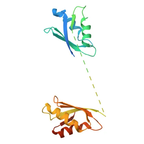
Protein folding decides whether the copper pump reaches its post. Oleg Dmitriev at the University of Saskatchewan showed that the H1069Q mutation destabilizes a helical hairpin at body heat, keeping ATP7B from reaching the trans-Golgi network [9]. Meanwhile, variations at codon 129 of PRNP modify onset timing [3].
Hepatic and Neurological Signs
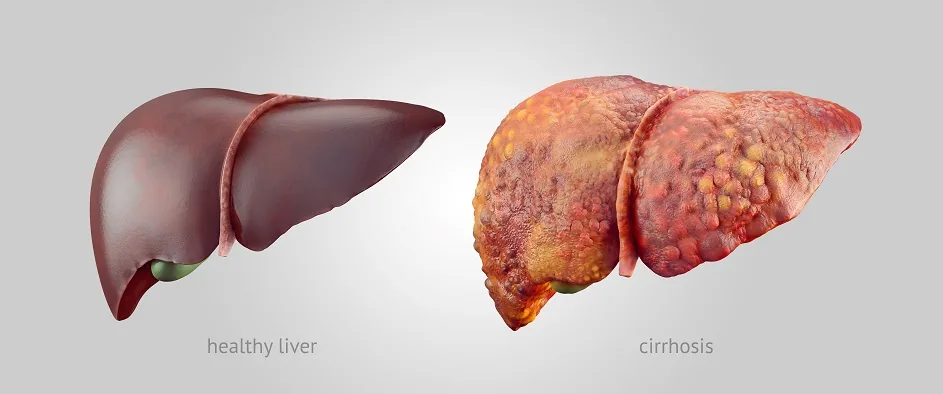
Liver illness is the most common first presentation in children and teens [3, 7]. When liver copper exceeds storage limits, free metal ions drive oxidative cell injury through Fenton chemistry, destroying lipids and proteins [6, 12]. Clinical signs range from tired feelings and jaundice to chronic active hepatitis, and roughly 35% to 45% of patients already have cirrhosis when doctors first diagnose them. In about 5% of cases, patients present with rapid acute liver failure and Coombs-negative hemolytic anemia [2, 6].
Neurological and psychiatric signs usually appear during the teens or twenties [3, 4]. Copper spills into the blood and crosses the blood-brain barrier to settle in the lenticular nucleus, mainly in the putamen and globus pallidus. These basal ganglia regions coordinate movement and mood. Patients often develop hand tremors, slurred speech, slow movements, clumsy hands, and a blank face, with some showing dystonia and a wing-beating tremor brought out by holding arms wide [7, 12]. Psychiatric issues occur in 10% to 20% of patients and include depression, anxiety, personality swings, and loss of impulse control, which clinicians sometimes mistake for primary psychiatric illness [4, 6].
Blood tests can track active nerve cell injury. Neurologist S. Shribman and colleagues at University College London reported in the journal Movement Disorders that plasma neurofilament light levels rise in active brain illness. This blood marker gives clinicians a practical tool to track nerve damage during chelation therapy [11].


Diagnostic Tests for Wilson’s Disease
Eye checks provide a famous visual clue. Copper deposits in the Descemet’s membrane of the cornea form a brown or green band called the Kayser-Fleischer ring, appearing in about 66% of all cases and in over 90% of people with brain symptoms [6, 12]. Slit-lamp checks can also spot rare sunflower cataracts, which do not hurt vision [7, 12]. While Kayser-Fleischer rings strongly support a diagnosis, they can also appear in chronic cholestatic liver diseases [6, 7].
No single blood test can diagnose the condition alone [6, 7]. Instead, clinicians rely on a mix of laboratory tests to confirm abnormal copper storage. Serum ceruloplasmin drops below 20 milligrams per deciliter in about 90% of patients [6]. In the United Kingdom, medical guidelines reviewed by Dr. Hayley Willacy and Dr. Colin Tidy note that low ceruloplasmin below 0.1 grams per liter helps confirm diagnosis [7]. In addition, 24-hour urine copper output rises above 100 micrograms per day in active disease, compared with normal levels below 40 micrograms. When blood and urine findings leave doubt, doctors perform a liver biopsy [6, 7]. A hepatic copper level above 250 micrograms per gram of dry liver tissue confirms the diagnosis, while normal values between 15 and 55 micrograms rule it out [6].
Brain scans offer extra visual evidence. Magnetic resonance imaging often shows high signal density in the basal ganglia, producing the classic face of the giant panda sign. Other signs include blue nail beds called azure lunulae, kidney Fanconi syndrome, premature joint arthritis, and resting heart rhythm changes [6, 7].

Chelation Therapy and Liver Transplants
Wilson’s disease is treatable with lifelong drug therapy [7]. The main goal is to lower copper levels and stop metal from collecting in organs. Chelating drugs bind copper ions so the kidneys can pass them in urine, with D-penicillamine serving as the original oral chelator sold under the brand names Cuprimine and Depen [4, 7]. But about one-third of patients stop penicillamine due to rashes, lupus-like reactions, kidney injury, or bone marrow suppression [7].
Trientine, sold as Syprine, offers an effective chelating choice with fewer immune side effects than penicillamine, which triggers temporary neurological worsening in 15% to 20% of patients with brain symptoms. After copper levels drop to normal, oral zinc acetate, sold as Galzin, provides a safe maintenance option. Zinc prompts gut cells to make metallothionein, which traps copper from food so it passes out in stool. The British National Formulary notes that zinc carries fewer fetal risks during pregnancy than chelating agents, but patients must take their prescribed medicine every day for life to avoid fatal relapse [4, 7].
Liver transplantation cures the metabolic defect for patients in acute liver failure or severe end-stage cirrhosis [6, 7]. A donor liver provides working ATP7B enzymes that restore normal bile copper output and clear extra copper from other organs. Survival rates after liver surgery are excellent [7].
Dietary Rules for Patients
Dietary adjustments support medical chelation, especially during the first phase of copper reduction. Doctors tell patients to avoid foods that carry large amounts of copper, including beef liver, organ meats, shellfish like lobster, mushrooms, tree nuts, dried fruit, and chocolate [1, 4]. After drug therapy lowers copper levels and maintenance begins, patients can ask their doctor if small portions of these foods are safe to eat [1]. However, diet alone cannot treat Wilson’s disease because the body cannot excrete normal copper loads without medication [4].
Drinking water requires close attention in the home. Water sitting in household copper pipes can pick up copper ions overnight, so the National Institute of Diabetes and Digestive and Kidney Diseases advises running the tap for several minutes to flush pipes before drinking or cooking. Homes that draw water from private wells or copper plumbing should test their water and use a water filter to remove excess mineral content [1].

Cookware and vitamins also carry hidden risks. Patients should avoid unlined copper pots and pans, which leach copper into cooking food [4]. People should also check all multivitamin supplements with their doctor, because many over-the-counter vitamin pills contain added copper [1].
Origins of Clinical Recognition
The first medical record of this condition appeared in 1854, when German pathologist Friedrich Theodor von Frerichs described patients with liver cirrhosis and tremors, decades before Dr. Samuel Alexander Kinnier Wilson studied the condition in London [12, 14]. Wilson was born in Cedarville, New Jersey, in 1878 and trained in Paris with Pierre Marie and Joseph Babinski before joining the National Hospital, Queens Square. In 1912, Wilson earned a Gold Medal from the University of Edinburgh Medical School for his doctoral dissertation, “Progressive lenticular degeneration,” coining the term extrapyramidal before the condition was named Wilson’s disease in his honor [14].
Patient advocacy helped turn medical knowledge into community support. In 1978, local residents in Binghamton, New York, formed the Wilson Disease Association to help the Pei family after two young boys received diagnoses without health insurance. Family friend John Chung teamed with patient Carol Terry, whose diagnosis story appeared in the American Journal of Nursing in 1976 and the New Yorker in 1979. Starting with 850 dollars from a community fund, the group mailed newsletters, sent informational brochures, and connected patients across the country [5].
The Wilson Disease Association incorporated in 1983 as a nonprofit and served as a charter member of the National Organization for Rare Disorders to support the Orphan Drug Act. Working with MAP International, the group ships donated medicines to patients in 37 countries. Between 2018 and 2022, the association gave over 2.4 million dollars to an international patient registry study [5].
- WEBSITE National Institute of Diabetes and Digestive and Kidney Diseases. (2026). Eating, diet, & nutrition for Wilson disease. [Article Link]
- WEBSITE National Institute of Diabetes and Digestive and Kidney Diseases. (2025). Definition & facts for Wilson disease. [Article Link]
- WEBSITE MedlinePlus Genetics. (2022). Wilson disease. [Article Link]
- WEBSITE MedlinePlus Medical Encyclopedia. (2024). Wilson disease. [Article Link]
- WEBSITE Wilson Disease Association. (2023). About WDA. [Article Link]
- WEBSITE Medscape. (2025). Wilson disease: Practice essentials, background, etiology. [Article Link]
- WEBSITE Patient.info. (2025). Wilson’s disease. [Article Link]
- ACADEMIC JOURNAL Banci, L., Bertini, I., Cantini, F., Rosenzweig, A. C., & Yatsunyk, L. A. (2008). Metal binding domains 3 and 4 of the Wilson disease protein: Solution structure and interaction with the copper(I) chaperone HAH1. Biochemistry, 47(28), 7423-7429. [Article Link]
- ACADEMIC JOURNAL Dmitriev, O. Y., Bhattacharjee, A., Nokhrin, S., Uhlemann, E. M., & Lutsenko, S. (2011). Difference in stability of the N-domain underlies distinct intracellular properties of the E1064A and H1069Q mutants of copper-transporting ATPase ATP7B. Journal of Biological Chemistry, 286(18), 16355-16362. [Article Link]
- ACADEMIC JOURNAL Coffey, A. J., Durkie, M., Hague, S., McLay, K., Emmerson, J., Lo, C., Klaffke, S., Joyce, C. J., Dhawan, A., Hadzic, N., Mieli-Vergani, G., Kirk, R., Elizabeth Allen, K., Nicholl, D., Wong, S., Griffiths, W., Smithson, S., Giffin, N., Taha, A.,. Palotie, A. (2013). A genetic study of Wilson’s disease in the United Kingdom. Brain, 136(5), 1476-1487. [Article Link]
- ACADEMIC JOURNAL Shribman, S., Heller, C., Burrows, M., Heslegrave, A., Swift, I., Foiani, M. S., Gillett, G. T., Tsochatzis, E. A., Rowe, J. B., Gerhard, A., Butler, C. R., Masellis, M., Bremner, F., Martin, A., Jung, L., Cook, P., Zetterberg, H., Bandmann, O., Rohrer, J. D., & Warner, T. T. (2021). Plasma neurofilament light as a biomarker of neurological involvement in Wilson’s disease. Movement Disorders, 36(2), 503-508. [Article Link]
- WEBSITE Wikipedia. (2026). Wilson’s disease. [Article Link]
- WEBSITE Wikipedia. (2026). Wilson disease protein. [Article Link]
- WEBSITE Wikipedia. (2026). Samuel Alexander Kinnier Wilson. [Article Link]


