





The biological mechanism of how antivenom works begins with the basic rules of passive immunity, where preformed donor antibodies bind venom molecules before toxins destroy human cells. When a venomous snake strikes a human limb, its fangs inject an intricate blend of proteins that can paralyze nerves, kill muscle tissue, and stop normal blood clotting. Antivenom gives instant help by delivering antibodies taken from immunized animals or bioengineered cell lines. Donor immunoglobulins lock directly onto venom toxins, blocking harmful chemical sites and marking the poisons for natural disposal by human immune cells [1].
- How Antivenom Works in the Body
- Historical Origins of Serum Therapy
- How Donor Animals Generate Antibodies
- Enzymatic Cleavage into Active Fragments
- Cross-Neutralization Limits in Snake Antivenom
- Clinical Risks of Hypersensitivity
- Cold Chain Hurdles and Shortages
- Synthetic Alternatives to Traditional Antivenin
How Antivenom Works in the Body
The human immune system cannot build protective antitoxins quickly enough after an unexpected snakebite emergency in the field. Vaccines introduce dead or weakened antigens to teach host B cells over several weeks, but snakebite patients need active defenses within hours to prevent death. Antivenom solves this medical emergency through passive immunization [1]. A direct intravenous infusion of donor antibodies floods blood vessels with ready proteins, showing clearly how antivenom works to protect vital organs throughout circulation.
Toxin neutralization relies on the precise structural fit between donor antibody receptors and foreign venom antigens. Snake venoms cause devastating injury by latching onto nerve receptors like nicotinic acetylcholine sites at neuromuscular junctions, or by enzymatically cleaving vital clotting proteins such as fibrinogen in circulating blood. Protective antibodies carry variable tips that grip matching chemical targets called epitopes [2]. Such tight binding creates an impenetrable block that keeps venom molecules from touching host cellular targets. Deprived of access to cell walls, the venom cannot shut down muscle fibers, break open capillaries, or destroy red blood cells. Quick binding stops permanent tissue damage before toxins spread into deeper organ spaces.
Bound toxins link together into large clusters called antigen-antibody complexes. Clustered molecules cannot slip through capillary walls or enter narrow nerve gaps to cause extra harm. The body sends these neutral complexes to the reticuloendothelial system for destruction [2]. Macrophage cells residing in the liver and spleen swallow the clusters, breaking down both venom and antibodies into harmless basic amino acids. Kidneys also filter tiny detached pieces out through urine.
Historical Origins of Serum Therapy
Early observations of venom resistance began in eighteenth-century Europe. Italian naturalist Felice Fontana wrote in 1767 that European vipers seemed immune to their own poison, publishing his findings in Ricerche Fisiche sopra il Veleno della Vipera [1]. Scottish surgeon Patrick Russell reported similar protection among Asian snakes a few years later.
Surgeon-Major Edward Nicholson recorded an intriguing account in the November 1870 issue of the Madras Medical Journal, describing a handler in Burma who repeatedly exposed himself to cobra venom yet still treated the serpents with careful restraint. In 1887, Henry Sewall in Michigan gave escalating venom doses of rattlesnake Sistrurus catenatus catenatus to pigeons, helping them survive seven times the lethal dose. Between 1889 and 1892, Maurice Kaufmann injected small doses of Vipera aspis venom at the École nationale vétérinaire d’Alfort in France, achieving partial resistance [1]. These early trials showed that living bodies could adapt to venom, but medical therapy required transferring that immunity to untreated victims.
Transferring blood serum between animals proved to be the turning point for modern medicine. Emil von Behring and Shibasaburo Kitasato introduced serum therapy in 1890 for diphtheria and tetanus infections. At the Society of Biology in Paris on February 10, 1894, Albert Calmette at the Pasteur Institute and Césaire Phisalix and Gabriel Bertrand at the National Museum of Natural History proved that serum from immunized animals neutralized snake venom. Calmette dosed horses with cobra venom to make Serum Antivenimeux, the first commercial product. Sir Thomas Fraser at the University of Edinburgh made cobra antivenene in 1895, while Vital Brazil produced monovalent and polyvalent serums at Instituto Butantan in São Paulo in 1901 for Bothrops and Crotalus snakes [1].
How Donor Animals Generate Antibodies
Antivenom production begins by taking fresh venom from captive snakes, scorpions, or spiders, a delicate laboratory procedure known as milking. Handlers freeze-dry the collected fluid to keep delicate venom proteins stable [3]. Producers inject small amounts of this venom into large farm animals, mainly horses and sheep, which act as living antibody factories. Horses are strong, gentle to handle, and carry large blood supplies.
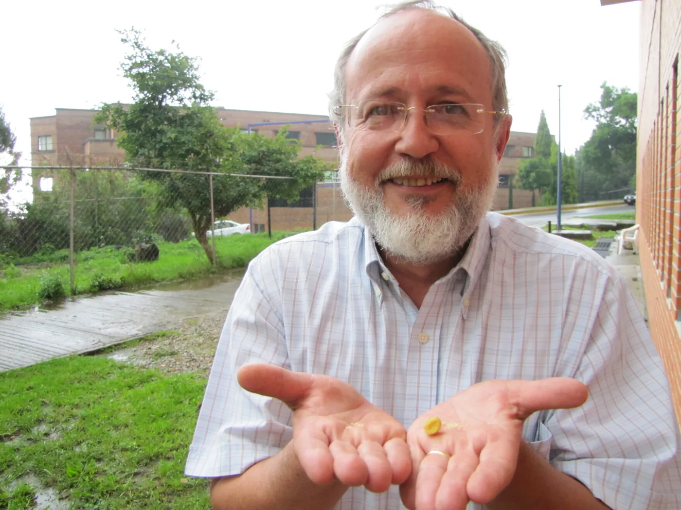
At Rancho Ojo de Agua, an 80-year-old ranch on 444 acres in Puebla, Mexico, 59 horses live under the care of pediatrician Dr. Andres Alagon. The ranch sends plasma to Instituto Bioclon in Mexico City, a pharmaceutical company that consults with biochemist Dr. Alejandro Alagon at the Autonomous National University of Mexico in Cuernavaca. The horses follow a careful immunization schedule lasting four to six months. Technicians start by injecting venom equal to half a scorpion sting [4]. Over six months, handlers increase the venom until horses easily tolerate the equivalent of 100 stings without getting sick. The horses produce abundant antibodies, showing how antivenom works inside living host animals. Donkeys, goats, rabbits, and camels also serve as donor hosts.
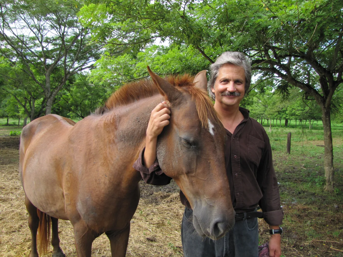
As the donor animal responds, its B cells make polyclonal antibodies against multiple venom targets. Once blood tests show high antibody levels, ranch workers collect blood using plasmapheresis, separating liquid plasma and returning red cells to the animal. Blood cell reinfusion protects horses from developing anemia during production cycles. In the United States, sheep provide the antibodies used in CroFab, which received approval from the Food and Drug Administration in October 2000 [1].
Enzymatic Cleavage into Active Fragments
Raw donor plasma contains thousands of animal proteins that humans cannot safely receive. Serum proteins like albumin cause allergic shocks without neutralizing venom. Chemists salt out unwanted proteins using ammonium sulfate or caprylic acid [1]. Laboratories then isolate immunoglobulin G, the main antibody type used to neutralize venoms.
A complete immunoglobulin G molecule has two arms called Fab fragments that bind venom, connected to a stem called the Fc region. The Fc stem engages human immune cells, triggering allergic reactions. To make safer medicine, biochemists cut off the Fc stem with enzymes. Treating immunoglobulin G with the enzyme pepsin cleaves the molecule below the hinge, leaving a double-armed F(ab’)2 fragment of 110 kilodaltons while degrading the constant region. The enzyme papain cuts above the hinge, creating two single Fab pieces of about 50 kilodaltons each [2]. Chromatography removes the cut stems, leaving only the binding segments needed to neutralize toxins. Doctors call this approach fabotherapeutics, which explains how antivenom works with fewer side effects in human patients [4].
Fragment size changes how the drug moves through human tissue compartments during therapy. Small 50-kilodalton Fab units travel quickly into deep muscle tissue, neutralizing local toxins that cause swelling [3]. But human kidneys filter small Fab units out of blood within hours. Larger 110-kilodalton F(ab’)2 fragments remain in the blood longer, maintaining protection over several days [2]. Longer circulation prevents recurrent hemotoxicity, which happens when short-lived antivenom leaves the body while leftover venom still leaks from the original bite wound into peripheral capillaries.

Cross-Neutralization Limits in Snake Antivenom
Product recipes depend on the target snake species chosen during donor priming. A monovalent antivenom uses venom from one species, creating antibodies calibrated for that single snake. A polyvalent recipe mixes venoms from several snakes, creating broad protection for rural clinics where patients cannot name the snake [2]. Over 600 snake species have dangerous venom, making broad coverage essential.
Cross-neutralization lets some serums protect against snakes never injected into donor horses. Closely related vipers share similar protein shapes in their venoms. Instituto Bioclon makes Antivipmyn using venom from South American rattlesnakes (Crotalus durissus) and fer-de-lance pit vipers (Bothrops asper) [2]. Even though it uses only two tropical venoms, Antivipmyn neutralizes venom from all North American pit vipers. Understanding cross-protection clarifies how antivenom works across related species.
Broad protection also applies to blood clotting disorders caused by snake venoms. In a 2018 report in Communications Biology, Ainsworth and colleagues examined 57 snake venoms, showing that some single-species antivenoms neutralized clotting toxins across unrelated species. The Ainsworth team showed that the metal binder EDTA also stopped venom toxicity in laboratory animals by blocking venom enzymes [5]. Preclinical evaluation now pairs animal tests with antivenomics, a mass spectrometry method described by José María Gutiérrez and colleagues in 2017 to map venom-antibody binding in laboratory settings [6]. Mass spectrometry reveals which venom proteins bind tightly to therapeutic antibodies, helping scientists refine antibody cocktails before beginning human trials.

Clinical Risks of Hypersensitivity
Giving horse or sheep proteins by vein carries known allergic risks for patients. Clinical pharmacologist H. Asita de Silva and colleagues wrote in the British Journal of Clinical Pharmacology that allergic reactions happen in many snakebite clinics [7]. Reactions occur in two main forms: immediate allergic shock and delayed serum sickness.
Early allergic symptoms appear within an hour of starting antivenom infusion [7]. Patients can develop hives, fever, wheezing, and falling blood pressure. Fragment antivenoms reduce these risks because they lack the constant animal stem [2]. But patients with alpha-gal allergies still face higher reaction rates when given F(ab’)2 products containing trace animal sugars. Doctors watch patients closely during infusion to treat allergic symptoms quickly.
Delayed reactions emerge five to 14 days after treatment as serum sickness [7]. Serum sickness occurs when human antibodies attack leftover animal antibodies still in the blood. Immune clusters settle in joints and kidneys, causing joint pain, high fever, and skin rashes. Even with these side effects, antivenom remains the only cure for snake envenomation. Doctors do not withhold treatment over allergic fears because untreated venom causes death and tissue loss [1]. Clinics treat allergic reactions with adrenaline, antihistamines, and steroids while keeping antivenom flowing [7]. In Europe, bites from native vipers cause limb swelling that requires hundreds of antivenom doses each year, even though products lack common European Medicines Agency approval [13].
Cold Chain Hurdles and Shortages
Envenomation remains a neglected health problem that hits poor tropical farmers hardest in agricultural regions worldwide. Snakebites cause between 80,000 and 100,000 deaths worldwide each year, leaving about 300,000 people with lasting disabilities or lost limbs [8]. Keeping antivenom stable presents big challenges in rural areas where power grids fail. Producers rely on lyophilization, a freeze-drying method that pulls water out under vacuum to leave dry powder [1]. Dry powder vials stay good for five years at room temperature. Liquid antivenom needs steady refrigeration below 8 degrees Celsius (46 degrees Fahrenheit). Without working refrigerators, liquid serums spoil before reaching remote clinics.
Market problems and distribution gaps make shortages worse in developing regions. In Southeast Asia, a 2022 study by Patikorn and colleagues in BMJ Global Health reviewed antivenom supplies across seven Association of Southeast Asian Nations members. Regional shortages persist because rural clinics lack tracking systems and imported vials often fail against local snake species [9]. Dr. Leslie Boyer of the VIPER Institute at the University of Arizona said that vials cost only $14 to $20 to make, but poor distribution networks make final costs unbearable [11].
Supply shortages also hit the United States healthcare system. Stock for American coral snake antivenom expired in 2008 and 2009, prompting the Food and Drug Administration to extend expiration dates through 2017 while Pfizer maintained limited supplies by 2021 [2]. To protect patients, Miami-Dade Fire Rescue runs 71 fire-rescue stations and manages the only public antivenom bank in the country from Paulison Fire Rescue Headquarters in Florida. The Miami team ships vials by air to out-of-state hospitals, offering round-the-clock doctor consultations that explain how antivenom works during critical envenomation emergencies [10].
Synthetic Alternatives to Traditional Antivenin
The cost of horse serum has led researchers to develop synthetic medicines. A single treatment course often demands $800 to $1,000 across rural African communities, whereas hospital charges in the United States can surpass $14,000. In San Francisco, former Celgene antibody researcher Daniel Dempsey co-founded Venomyx with Deepankar Roy and Alex Capovilla at the IndieBio accelerator to create horse-free treatments. The team injected a llama with venom, sequenced its B cells, and inserted protective genes into Escherichia coli bacteria. Bioreactors filled with modified bacteria make antibodies without keeping large horse herds. In Copenhagen, startup VenomAB at the Technical University of Denmark grows humanized antibodies in mammalian cells [11].
Natural immunity in small mammals also provides clues for new antivenoms. Opossums resist pit viper venoms naturally, a trait researcher Binie V. Lipps patented in 1996 after isolating a protective blood protein. Claire F. Komives of San Jose State University worked with the Indian Institute of Technology Delhi to engineer Escherichia coli that make the first 11 amino acids of that protein. Researchers cut the chains into single 11-mer peptides, costing about $1.00 per gram to produce. The National Natural Toxins Research Center at Texas A&M University in Kingsville confirmed this peptide protects mice against Russell’s viper (Daboia russelii) venom [12].
Human immunity may provide the clearest path toward universal treatments. Wisconsin resident Tim Friede took bites from venomous snakes for twenty years, building broad immunity. In a 2025 study in Cell, Centivax CEO Jacob Glanville and Peter Kwong of Columbia University isolated two antibodies, LNX-D09 and SNX-B03, from Friede’s blood. A mixture of these antibodies and varespladib protected mice against 13 snake species completely and six others partially [8]. Tests will soon begin on dogs in Australia. These discoveries show how antivenom works when engineered with modern biotechnology.
- WEBSITE Wikipedia. (2026). Antivenom. [Article Link]
- WEBSITE Wikipedia. (2026). Snake antivenom. [Article Link]
- ACADEMIC JOURNAL Gutiérrez, J. M., León, G., Lomonte, B., & Angulo, Y. (2011). Antivenoms for snakebite envenomings. Inflammation & Allergy – Drug Targets, 10(5), 369-380. [Article Link]
- ONLINE NEWS Ortiz Uribe, M. (2011, August 2). Horses key to making antivenom up for FDA approval. KJZZ. [Article Link]
- ACADEMIC JOURNAL Ainsworth, S., Slagboom, J., Alomran, N., Plá, D., Alhamdi, Y., King, S. I., Bolton, F. M. S., Gutiérrez, J. M., Vonk, F. J., Toh, C., Calvete, J. J., Kool, J., Harrison, R. A., & Casewell, N. R. (2018). The paraspecific neutralisation of snake venom induced coagulopathy by antivenoms. Communications Biology, 1(1), 34. [Article Link]
- ACADEMIC JOURNAL Gutiérrez, J. M., Solano, G., Plá, D., Herrera, M., Segura, Á., Vargas, M., Villalta, M., Sánchez, A., Sanz, L., Lomonte, B., León, G., & Calvete, J. J. (2017). Preclinical evaluation of the efficacy of antivenoms for snakebite envenoming: State-of-the-art and challenges ahead. Toxins, 9(5), 163. [Article Link]
- ACADEMIC JOURNAL Silva, H. A. d., Ryan, N. M., & Silva, H. J. d. (2015). Adverse reactions to snake antivenom, and their prevention and treatment. British Journal of Clinical Pharmacology, 81(3), 446-452. [Article Link]
- ONLINE NEWS Orf, D. (2026, April 1). The man who let deadly snakes bite him for 20 years—and the universal antivenom hiding in his blood. Popular Mechanics. [Article Link]
- ACADEMIC JOURNAL Patikorn, C., Ismail, A. K., Abidin, S. A. Z., Blanco, F. B., Blessmann, J., Choumlivong, K., Comandante, J. D., Doan, U. V., Ismail, Z. M., Khine, Y. Y., Maharani, T., Nwe, M. T., Qamruddin, R. M., Safferi, R. S., Santamaria, E. B., Tiglao, P. J., Trakulsrichai, S., Vasaruchapong, T., Chaiyakunapruk, N.,. Othman, I. (2022). Situation of snakebite, antivenom market and access to antivenoms in ASEAN countries. BMJ Global Health, 7(3), e007639. [Article Link]
- WEBSITE Miami-Dade County. (2025). Venom response and antivenom bank. Miami-Dade Fire Rescue. [Article Link]
- ONLINE NEWS Molteni, M. (2017, March 20). Bacteria are brewing up the next generation of antivenoms. WIRED. [Article Link]
- ONLINE NEWS Davenport, M. (2015, March 30). Opossum compounds isolated to help make antivenom. Scientific American. [Article Link]
- ACADEMIC JOURNAL Lamb, T. W., Haro, L. d., Lonati, D., Brvar, M., & Eddleston, M. (2017). Antivenom for European Vipera species envenoming. Clinical Toxicology, 55(6), 557-568. [Article Link]


