





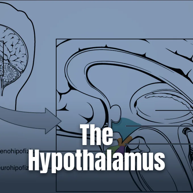
When asking what is the hypothalamus, neurobiologists describe an almond-sized brain region at the base of the forebrain that links the nervous system to the endocrine network through the pituitary gland to preserve bodily balance [4]. The cell cluster forms the floor of the forebrain and borders the third fluid chamber [3]. It weighs four grams in human adults. Yet it runs feedback loops that keep internal physiology balanced despite shifting conditions in the outside world. By tracking blood heat, fluid volume, and fuel levels, it coordinates involuntary nerve acts to defend overall health [1].
- What Is the Hypothalamus and How Does It Function?
- Core Anatomy of the Hypothalamus
- Autonomic Regulation Through Brainstem Networks
- Endocrine Signals to the Pituitary Gland
- Feeding Circuitry Driving Mammalian Appetite
- Circadian Rhythms and Core Temperature
- Developmental Influences on Adult Behavior
- Clinical Consequences of Hypothalamus Damage
What Is the Hypothalamus and How Does It Function?
The name hypothalamus comes from Greek words hypo, meaning under, and thalamos, meaning chamber, pointing to its spot right below the thalamus. All vertebrate brains possess this hub. In human adults, it fills four cubic centimeters and matches an almond in form and size [4]. By serving as a bridge between networks, it turns neural signals from sensory inputs into blood-borne chemical instructions to govern distant tissues [8].
George Boeree said that the organ acts like a household thermostat, keeping balance through steady negative feedback adjustments. When cold lowers body heat below its set point, brain circuits trigger shivering and narrow blood vessels until warmth returns. Heat prompts sweating to shed extra warmth. It also steers hunger, thirst, pain thresholds, and mating drives through similar negative feedback loops [1].
Control signals travel along two output paths to reach distant organs and govern whole-body tasks during daily activities. Downward nerve tracts pass through the brainstem into fight-or-flight and calming chains to alter heart rate, lung volume, and gut action within seconds during sudden shifts in activity or outside stress [1]. Hormonal paths connect with the pituitary gland, where neurosecretory cells release peptides that enter capillary loops to control energy pace and tissue growth across the body [4], [8]. Magnocellular cells send axons directly into the posterior gland, while parvocellular cells release regulatory peptides into portal blood to instruct anterior gland cells [4],. Both paths work in concert to protect internal stability during sudden environmental challenges [8].
Core Anatomy of the Hypothalamus
Under international anatomical standards, the structure bears the code A14.1.08.401 in TA98 and entity number FMA:62008 [3]. Neuroanatomists split it into four regions from front to back: the preoptic, supraoptic, tuberal, and mammillary areas. Across the coronal plane, it divides into periventricular, intermediate, and lateral zones [4]. Its medial wall lines the fluid chamber [3].
Specific cell groups make neurohypophysial peptides that travel down axons into the posterior gland to modulate fluid balance and reproduction. The paraventricular nucleus, or PVN, and the supraoptic nucleus, known as SON, contain magnocellular neurosecretory cells that manufacture oxytocin and vasopressin. Oxytocin directs uterine contractions during childbirth and milk ejection during nursing, whereas vasopressin promotes renal water retention to keep systemic blood volume and arterial pressure stable during dehydration. Parvocellular neurons in the PVN release corticotropin-releasing hormone, or CRH, into the hypophyseal portal system, allowing chemical signals to reach anterior gland cells and trigger broader systemic cascades [4], [8]. These dual cell types allow fast neural firing alongside sustained hormone output [4].
At the base of the third chamber, ependymoglial cells known as tanycytes form a flexible barrier between cerebrospinal fluid and brain parenchyma. F. Langlet reported in the Journal of Neuroendocrinology that tanycytes extend long processes that reach portal vessels and fuel-sensing neurons to regulate nutrient transport during fasting. Their tight junctions adjust permeability when circulating fuel levels shift [11]. Fenestrated capillaries lack an ordinary blood-brain barrier [8].
Autonomic Regulation Through Brainstem Networks
The region serves as the central control post for the autonomic nervous system, steering both its sympathetic and parasympathetic arms to preserve equilibrium. When threat or acute physical exertion demands immediate action, sympathetic pathways dilate pupils, raise heart rate, expand bronchial airways, and direct oxygenated blood straight into large skeletal muscle beds. After threat fades, parasympathetic fibers stimulate digestive glands, slow cardiac speed, and constrict airways to restore body energy stores. George Boeree said that an enteric nervous system also manages gastrointestinal movements under central guidance. Coordination between these arms keeps vital organs operating within safe boundaries across shifting environmental states [1].
Sensory inputs from visceral organs converge on the brain to guide autonomic decisions. The vagus nerve sends unbroken streams of data regarding stomach fullness, gut distension, and arterial blood pressure. Ascending tracts from the brainstem reticular formation deliver information about skin heat, while optic nerve fibers report daylight cycles to guide central autonomic decisions [1],. Projections from the locus coeruleus, solitary tract nucleus, and ventrolateral medulla carry feedback from thoracic organs [4].
Descending motor signals leave the forebrain along the hypothalamospinal tract to synapse on sympathetic neurons in the lateral horn of spinal cord segments T1 through L3 during systemic stress. Caudal pathways send signals into brainstem motor centers by running through the dorsal longitudinal fasciculus, while parallel axons course through mammillotegmental routes and the medial forebrain bundle. Such motor pathways link directly to the amygdala and emotional processing circuits to trigger coordinated defensive reactions across the organism. Brainstem loops adjust blood vessel diameter and visceral tone during times of sudden behavioral stress to ensure rapid survival responses [4].
Endocrine Signals to the Pituitary Gland
In a detailed synthesis in Comprehensive Physiology, researcher I. J. Clarke said that the organ functions as a neuroendocrine control center by releasing hypophysiotropic peptides into the hypophysial portal circulation. Small parvocellular neurons project axons into the median eminence, discharging pulses of releasing and inhibiting peptides into specialized capillary beds to regulate pituitary output with minimal total hormone volume. The peptides travel down portal venules straight to anterior pituitary cells without undergoing dilution in systemic blood. Target cells in the gland respond by releasing thyroid-stimulating hormone, adrenocorticotropic hormone, growth hormone, prolactin, or gonadotropins into general circulation to coordinate whole-body growth and energy balance [8].
The hypothalamus-pituitary-thyroid axis, or HPT axis, illustrates the feedback loops that sustain homeostatic control, as documented by T. M. Ortiga-Carvalho, M. I. Chiamolera, C. C. Pazos-Moura, and F. E. Wondisford in Comprehensive Physiology. Hypothalamic thyrotropin-releasing hormone prompts pituitary cells to release thyroid-stimulating hormone. Circulating thyroid hormones bind nuclear receptors in the brain and pituitary gland, creating a negative feedback loop that halts further hormone release [10].
Parallel feedback loops direct adrenal steroid output and reproductive roles across mammalian species. Parvocellular cells release CRH to trigger pituitary adrenocorticotropic hormone, which prompts adrenal glands to release cortisol into the bloodstream before cortisol binds glucocorticoid receptors in the PVN to shut down further discharge. Readers can examine feedback loops controlling stress responses in the HPA axis to understand how persistent disruption impacts metabolic balance. Self-limiting endocrine circuits prevent hormone toxicity while providing necessary adjustments during prolonged physical exertion. When systemic cortisol climbs, local receptors in the paraventricular nucleus halt further synthesis to protect downstream tissues [4].


Feeding Circuitry Driving Mammalian Appetite
Appetite control centers on the arcuate nucleus, or ARC, in the mediobasal hypothalamus, where two neuron classes exert opposing effects on hunger. One group releases neuropeptide Y, or NPY, and agouti-related peptide, or AgRP, to stimulate feeding when triggered by ghrelin. The other class produces POMC and CART to blunt appetite and suppress food intake after meals [6].
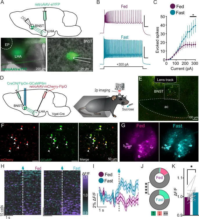
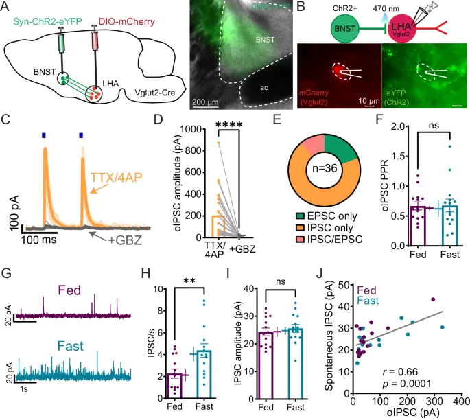
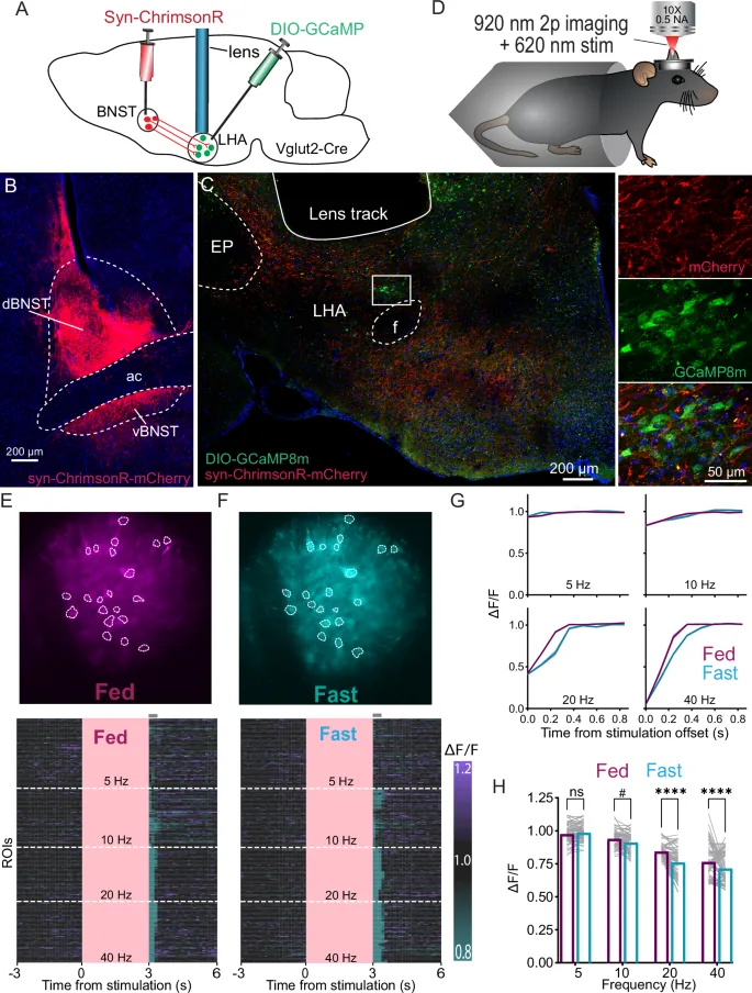
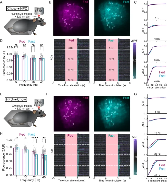
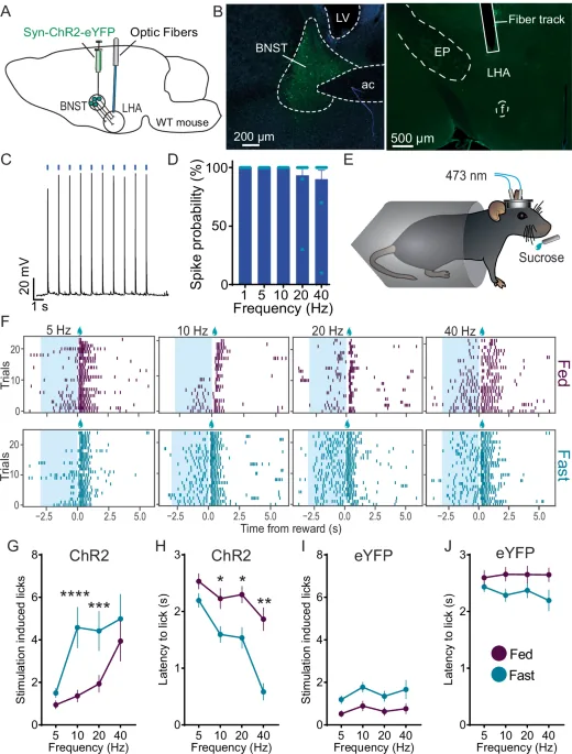
Arcuate neurons project to the lateral hypothalamic area, or LHA, a classical feeding center [5]. In a 2025 study in Nature Communications, Kuldeep Shrivastava, Vikshar Athreya, Yi Lu, Jorge Luis-Islas, Ashley Han, Tess F. Kowalski, and Mark A. Rossi traced synaptic pathways directing reward seeking in mice. They showed that glutamatergic neurons expressing vesicular glutamate transporter 2, or Slc17a6, act as an active brake on food consumption by receiving monosynaptic inhibitory GABAergic inputs from the bed nucleus of the stria terminalis. Optogenetic stimulation of BNST to LHA projections triggered sucrose reward seeking up to 40 hertz in fasted animals, directly increasing lick rates at the delivery spout [2].
Shrivastava and colleagues discovered that fasting increased the excitability of BNST neurons, enhancing inhibitory tone onto LHAVglut2 cells to promote food approach and guide motivated eating behavior in hungry mice. However, feeding mice a high-fat diet blunted this pathway and weakened neuron recruitment. Satiety cues also link with memory circuits linked to appetite and obesity to shape eating habits. Returning animals to standard chow restored normal synaptic sensitivity over time [2].
Circadian Rhythms and Core Temperature
The organ synchronizes daily bodily rhythms by matching cellular clocks to external light cycles across day and night [4]. Retinohypothalamic tracts from optic nerves send illumination signals to the suprachiasmatic nucleus, or SCN, resting right above the optic chiasm [1],. Daylight input tells the SCN to suppress pineal melatonin synthesis and adjust autonomic tone for daytime wakefulness. At night, falling light levels prompt the SCN to facilitate sleep onset, lower arterial blood pressure, and drop nocturnal body heat to match internal metabolic demand to circadian sleep cycles [4]. Daily timing programs let peripheral organs anticipate energetic needs before food arrives [7].
In a review in Science, A. Adamantidis and L. de Lecea evaluated how diencephalic circuits integrate sleep states with homeostatic demands. They showed that neural substrates for wakefulness, rapid eye movement sleep, and non-rapid eye movement sleep overlap anatomically and functionally with networks controlling thermoregulation and goal-oriented behaviors during waking hours. Sleep-active cells in the preoptic area cool the body during rest, whereas wake-active circuits support higher daytime temperatures to match behavioral demand [7].
Within the LHA, specialized orexin neurons synthesize orexin-A and orexin-B to sustain wakefulness throughout the day. These cells send axons to histaminergic cells in the tuberomammillary nucleus, dopaminergic groups in the ventral tegmental area, and noradrenergic neurons in the locus coeruleus to maintain alert states throughout the day. Cannabinoid receptor 1 joins physically with orexin receptor 1 to form a CB1-OX1 heterodimer that amplifies downstream signaling 100-fold. Degeneration of these orexin projection neurons causes narcolepsy, producing uncontrollable daytime sleep attacks and fragmented rest. When orexin input vanishes, brainstem targets lose excitatory drive and muscle tone collapses during strong emotional arousal [5].
Developmental Influences on Adult Behavior
Beyond sleep and metabolic balance, diencephalic nuclei coordinate basic defensive and social actions [4]. In Frontiers in Systems Neuroscience, Y. Hashikawa, K. Hashikawa, A. L. Falkner, and D. Lin showed that the ventrolateral part of the ventromedial hypothalamus, or VMHvl, governs attack behavior in rodents. Stimulating this nucleus triggers fighting in male mice, whereas silencing its ventrolateral neurons completely halts combative encounters. This conserved circuit allows animals to protect territory and secure mating access [9].
Structural divergence between male and female brains develops early in life through exposure to gonadal steroids. The sexually dimorphic nucleus of the preoptic area, or SDN-POA, shows distinct volume differences based on perinatal hormone exposure. In human male fetuses, testicular testosterone secretion surges from week eight of gestation through five to six months after birth to guide male neural development before peripheral steroid binding proteins become fully active. Brain cells aromatize testosterone into estradiol to guide male neural development. In adult rodents, neonatal testosterone shapes adult reproductive postures and hormone cycles [4].
Sensory pathways also relay pheromonal cues that influence maternal attachment and reproductive physiology across species. In female mice, exposure to urine from an unfamiliar male during early pregnancy causes implantation failure, a phenomenon known as the Bruce effect. Chemical cues received through olfactory nerves alter neuroendocrine secretion to alter reproductive timing [4]. In the arcuate nucleus, kisspeptin and neurokinin B neurons regulate gonadotropin-releasing hormone release to support fertility and ovulation [6].
Clinical Consequences of Hypothalamus Damage
Damage to diencephalic circuits contributes to complex metabolic, neuroendocrine, and mood disorders [8],. In IBRO Reports, A. Bao and D. F. Swaab showed that functional alterations in hypothalamic stress pathways represent a central feature of depressive illness by disturbing day-night rhythms, feeding patterns, and normal reward feelings. Chronic impairment harms bodily health and resilience [12].
Targeted injury to lateral pathways produces distinct clinical syndromes across human populations. Loss of orexin-producing neurons causes narcolepsy with cataplexy, a disorder marked by sudden loss of muscle tone during emotional arousal when patients experience laughter or surprise. Dysfunctional orexin signaling onto vagal brainstem centers has also been linked to visceral hypersensitivity, motility dysfunction, and irritable bowel syndrome [5]. Lesions in the ventromedial nucleus cause uninhibited eating and severe weight gain, whereas lateral damage abolishes hunger cues entirely [1], [4]. Opposing deficits show how separate nuclei balance daily energy intake. When the orexinergic brake on feeding fails, metabolic rate drops and patients struggle with persistent fatigue [5].
Endocrine deficits occur when hypothalamic communication with the pituitary gland fails [8]. In the arcuate nucleus, tuberoinfundibular dopamine, or TIDA, neurons secrete dopamine into the hypophysial portal blood to inhibit prolactin release from the anterior pituitary. Suckling or specific medications quiet these cells, allowing prolactin levels to climb and initiate milk production in nursing mothers. Damage to this inhibitory pathway causes hyperprolactinemia, leading to galactorrhea and infertility [6].
- WEBSITE Boeree, C. G. (2002). The Limbic System. [Article Link]
- ACADEMIC JOURNAL Shrivastava, K., Athreya, V., Lu, Y., Luis-Islas, J., Han, A., Kowalski, T. F., & Rossi, M. A. (2025). Energy state guides reward seeking via an extended amygdala to lateral hypothalamus pathway. Nature Communications, 16(1). [Article Link]
- WEBSITE Federative International Programme on Anatomical Terminologies. (2008). Hypothalamus (A14.1.08.401). [Article Link]
- WEBSITE Wikipedia contributors. (n.d.). Hypothalamus. [Article Link]
- WEBSITE Wikipedia contributors. (n.d.). Lateral hypothalamus. [Article Link]
- WEBSITE Wikipedia contributors. (n.d.). Arcuate nucleus (hypothalamus). [Article Link]
- ACADEMIC JOURNAL Adamantidis, A., & de Lecea, L. (2023). Sleep and the hypothalamus. Science, 382(6669), 405-412. [Article Link]
- ACADEMIC JOURNAL Clarke, I. J. (2014). Hypothalamus as an Endocrine Organ. Comprehensive Physiology, 5(1), 217-253. [Article Link]
- ACADEMIC JOURNAL Hashikawa, Y., Hashikawa, K., Falkner, A. L., & Lin, D. (2017). Ventromedial Hypothalamus and the Generation of Aggression. Frontiers in Systems Neuroscience, 11, 94. [Article Link]
- ACADEMIC JOURNAL Ortiga-Carvalho, T. M., Chiamolera, M. I., Pazos-Moura, C. C., & Wondisford, F. E. (2016). Hypothalamus‐Pituitary‐Thyroid Axis. Comprehensive Physiology, 6(3), 1387-1428. [Article Link]
- ACADEMIC JOURNAL Langlet, F. (2014). Tanycytes: A Gateway to the Metabolic Hypothalamus. Journal of Neuroendocrinology, 26(11), 753-760. [Article Link]
- ACADEMIC JOURNAL Bao, A., & Swaab, D. F. (2018). The human hypothalamus in mood disorders: The HPA axis in the center. IBRO Reports, 6, 45-53. [Article Link]
Cover image: Pharmattila / Wikimedia Commons, CC BY-SA 4.0


