





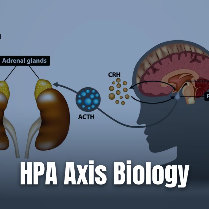
The Hypothalamic-Pituitary-Adrenal axis functions as the main hormone link between the human brain and the adrenal glands, directing how mammals adjust energy use, blood pressure, and immune defense during acute or ongoing threat. Neural inputs from central brain circuits join chemical signals from gland cells to keep the body stable when physical demands rise. Why does this hormonal circuit matter for daily health? With the HPA axis explained by medical teams at Cleveland Clinic and Simply Psychology, readers can see how three endocrine organs produce cortisol, guide energy shifts, and shield body tissues from harm [3].
Major Hypothalamic-Pituitary-Adrenal Axis Glands
Three primary organs build this endocrine network: the hypothalamus, the anterior pituitary gland, and the adrenal glands [4]. Clinical researchers at Cleveland Clinic show that coordinated hormonal signaling between these structures directs bodily adaptation during acute physical challenge [3].
Within the hypothalamus, the paraventricular nucleus, or PVN, acts as a primary receiving station for sensory signals from the amygdala. Author Shao Feng said in the ECPH Encyclopedia of Psychology from Springer Nature in Singapore and China Renmin University in Beijing that PVN cells make corticotropin-releasing hormone and AVP peptides. These nerve fibers project directly to the median eminence. CRH and AVP work together to stimulate pituitary ACTH output. Blood carries these chemical triggers downstream toward cells in the pituitary gland, forging a direct bridge between conscious threat appraisal in the forebrain and steroid release in body tissues [4].
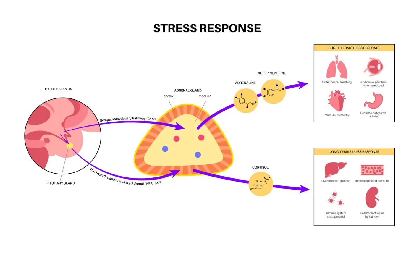
Each adrenal gland holds two functional units that divide fast and sustained chemical defense tasks during acute bodily threats. While the inner adrenal medulla releases adrenaline within seconds to drive fight-or-flight reactions, the outer adrenal cortex makes steroid hormones from stored cholesterol in three zones [8]. Authors Kishan Pankhania and Elly Voogt at TeachMePhysiology show that the outer zona glomerulosa makes aldosterone to balance salts, whereas the deeper zona fasciculata and zona reticularis secrete cortisol and androgens when triggered by pituitary ACTH. This structural division helps fast autonomic nerve signals pair with prolonged hormonal shifts throughout a physical ordeal [2].
First Hormone Signals in the Brain
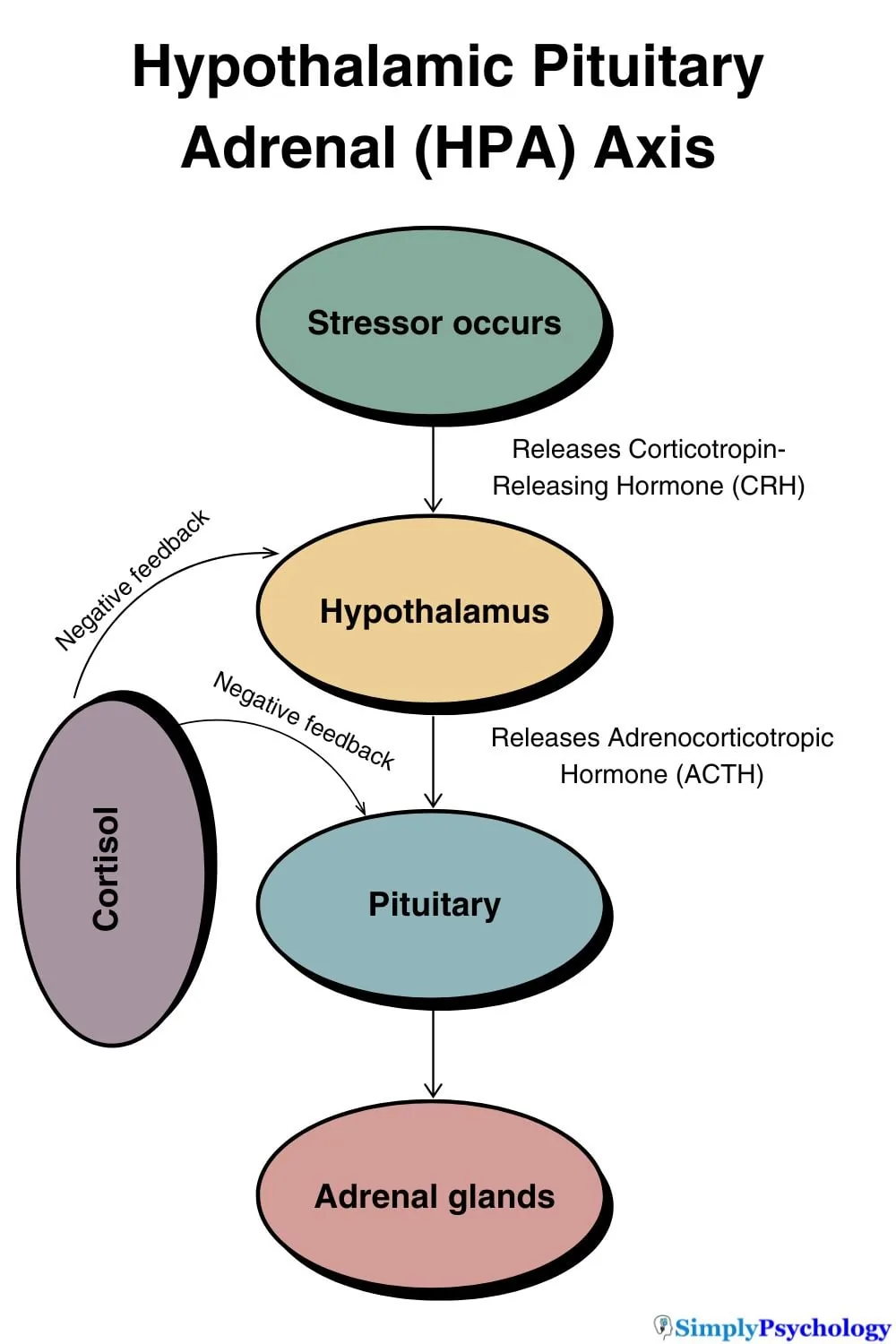
Brain firing begins when PVN neurons release corticotropin-releasing hormone, known as CRH, into portal blood vessels. Nerve terminals discharge CRH in short pulses rather than steady streams, preventing receptor dulling in target cells. The American Psychological Association records that brain cells also secrete arginine vasopressin, or AVP, with CRH during times of bodily demand. These two peptide signals travel through portal vessels to bind docking sites on pituitary cells, where their combined chemical presence drives far greater ACTH release than either hormone could achieve acting alone [5].
Hormone secretion follows a rhythmic cadence regulated by neural pacemakers, with the HPA axis explained by Mind-Body-Health researchers who track hourly pulses across the day. Tracking reviewed by Mind-Body-Health researchers confirms that PVN neurons fire chemical bursts roughly once per hour, with each discrete pulse sparking fifteen minutes of sustained adrenal cortisol production. Circulating cortisol has an elimination half-life of 100 minutes in blood [7].
Gland sensitivity dictates hormonal output. Physiologist Keller-Wood said in Comprehensive Physiology that pituitary corticotrope cells process incoming chemical cues through specialized surface receptors, stimulating intracellular messenger cascades that drive the transcriptional readout of POMC as a large precursor protein. AVP amplifies CRH drive. Gland cells package synthesized peptide hormones into secretory vesicles, discharging them directly into the bloodstream when incoming neural pulses trigger cell membrane depolarization during acute trauma, infection, or metabolic shock [12]. Without this tight neuroendocrine coupling, human body defenses fail.
Adrenal Cortex and Cortisol Output
Enzyme action splits the parent protein pro-opiomelanocortin into several active chemical signals. Authors Kishan Pankhania and Elly Voogt at TeachMePhysiology show that enzymatic cleavage yields ACTH, melanocyte-stimulating hormone, and pain-relieving beta-endorphins. Pituitary cells release ACTH into the bloodstream, where it travels toward the adrenal cortex to initiate steroid production, while circulating beta-endorphins blunt bodily pain during acute physical trauma [2].
Circulating ACTH binds tightly to the melanocortin-2 receptor, or MC2r, on cells within the adrenal zona fasciculata. This receptor activation triggers enzymatic pathways that convert stored cholesterol into active cortisol. Readers examining how cortisol hormone function in human metabolism operates can see how this steroid promotes liver gluconeogenesis, freeing glucose while breaking down muscle proteins into amino acids that supply the liver with raw fuel for energy synthesis. Zona reticularis cells also release DHEA [2].
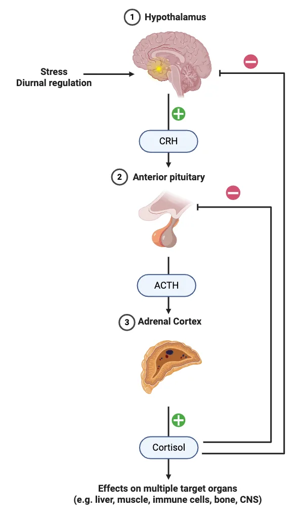
Cortisol shapes body cells by binding internal sensors that guide gene readout over hours. In target tissues, the glucocorticoid receptor, or GR, stays bound to helper proteins in cell fluid until cortisol crosses the lipid wall and binds its docking pocket. Once bound, the helper protein drops away, letting the cortisol-GR pair move into the cell nucleus, where it binds GRE sites on DNA to alter protein production. This genomic action guides prolonged bodily adaptation, while also dampening bone growth and reducing digestive calcium uptake during chronic steroid surges [2].
How the HPA System Uses Negative Feedback
Steroid hormones can cause tissue damage if their release continues without a shutoff. The negative feedback loop provides the main safety mechanism, allowing rising cortisol levels to signal the brain and pituitary gland to halt further hormone production [1]. Steroid molecules bind hypothalamic and pituitary receptors to suppress CRH and ACTH output [3].
Feedback control relies on two distinct sensor classes distributed across brain circuits. Physiologist Keller-Wood in Comprehensive Physiology reports that high-affinity MR sites bind basal cortisol, whereas lower-affinity glucocorticoid receptors engage during morning peaks and acute stress surges. Fast membrane actions suppress CRH release. Slower genomic pathways follow as steroid-receptor pairs silence POMC gene readout in pituitary cells, curbing hormone output at its genetic root [12].
Higher brain structures exert dual control over hypothalamic firing. The hippocampus contains a dense population of GR sites and acts as a firm brake on PVN neurons, while the amygdala delivers alarm signals that trigger hormone output [8]. Researchers Bodegom, Homberg, and Henckens in Frontiers in Cellular Neuroscience show that chronic stress causes dendritic loss in hippocampal neurons [13]. This structural damage weakens the brain’s braking mechanism, creating a harmful cycle where weakened hippocampal control permits cortisol levels to stay high, impairing memory storage and synaptic plasticity over extended periods. Immune proteins like IL-1 cross the blood-brain wall to trigger PVN firing [8].


HPA Axis Explained in Daily Rhythms
Daily biology dictates how steroid secretion shifts across day and night. The HPA axis explained through chronobiology shows that cortisol release follows a twenty-four-hour rhythm guided by master neural clocks in the suprachiasmatic nucleus, or SCN [7]. The SCN coordinates hypothalamic PVN activity, driving hourly pulses of CRH into portal vessels to match hormone supply with daily activity demands [8].
Blood cortisol drops to its lowest trough around midnight, allowing body tissues to focus on cellular repairs and immune healing during early deep sleep. Hormone production accelerates late at night, generating a morning surge termed the cortisol awakening response [8]. In healthy adults, cortisol levels peak thirty to forty-five minutes after waking. Nerve fibers from the SCN project straight to the adrenal cortex, boosting glandular sensitivity to ACTH during early morning hours [7].
The digestive tract maintains constant two-way signaling with brain circuits through the microbiota-gut-brain axis. Physiologist Ivan Pavlov first documented neural-digestive reflexes during his 1904 Nobel Prize research on digestive secretions [9]. Modern scientists Rusch, Layden, and Dugas in Frontiers in Endocrinology confirm that gut microbes produce over 90 percent of body serotonin and roughly 50 percent of dopamine [14]. Microbial compounds like butyrate nourish the gut wall, preventing luminal toxins from entering the bloodstream and sparking systemic inflammation. This gut barrier protects brain circuits from inflammatory stress [9].
How Does Chronic Stress Cause Harm?
Modern mental stressors trigger bodily pathways that evolved to handle acute physical threats. How does chronic worry transform this protective endocrine circuit into a cause of systemic tissue damage? Constant alarm keeps brain neurons firing continuously, denying tissues the calm intervals needed to restore homeostatic balance. Physiologist Hans Selye showed this progression in his 1956 study on the General Adaptation Syndrome, showing that sustained defensive exertion causes adrenal cortex enlargement, immune tissue shrinkage, and severe stomach ulcers in animal tests [1].
Persistent hormone surges blunt tissue sensitivity, producing glucocorticoid resistance [6]. Defensive chemical output continues unchecked despite high circulating cortisol levels [8]. Neuroscientist Sheng and colleagues in Frontiers in Behavioral Neuroscience show that early developmental disruption alters central neurotransmitter synthesis, fueling persistent low-grade inflammation that increases long-term risks for cardiovascular strain and metabolic fatigue across peripheral organ systems [15].
Hardship faced during early development leaves lasting marks on adult hormone regulation. Simply Psychology author Olivia Guy-Evans cited a meta-analysis led by Bunea that pooled thirty clinical datasets covering 4,292 people, revealing that childhood trauma produces a blunted adult cortisol response to social stress. While mild childhood tests can foster resilient coping, severe abuse alters brain receptor density, raising risks for depression and anxiety later in life. Authors Moylan and Zajkowska observed that recurrent depression relapses compound this hormone strain, making the underlying hormonal imbalance harder to reverse over time. Early hardships alter how the nervous system calculates threat thresholds throughout adult life [1].
Disorders Linked to Endocrine Breakdown
Clinical illnesses of the adrenal axis appear through two opposing extremes of hormone output. Doctors at Cleveland Clinic state that Cushing syndrome arises from sustained cortisol excess, often triggered by pituitary tumors that drive rapid central weight gain, muscle loss, and high blood pressure [3]. In contrast, Addison’s disease represents primary adrenal failure, where immune damage to the adrenal cortex halts cortisol making. Because low cortisol removes negative feedback, pituitary cells ramp up POMC production, yielding high ACTH and MSH that causes characteristic oral and palmar skin darkening in Addisonian patients [2].
Hormone systems cross-regulate each other to coordinate bodily balance. In women, pituitary hormones stimulate ovarian follicles to make sex steroids like estrogen and progesterone, which interact with adrenal hormones to guide tissue fat distribution, emotional stability, and vascular tone. Clinicians who track circulating estrogen levels in peripheral tissue observe these systemic shifts. High cortisol blunts hypothalamic GnRH release. This suppression disrupts reproductive cycles. In Metabolic Brain Disease, researchers Mbiydzenyuy and Qulu show that bidirectional crosstalk between adrenal and gonadal axes also modulates aggressive behavioral tendencies [11].

Psychiatric conditions display distinct hormone patterns that reflect contrasting types of axis trouble. While major depression often features high baseline cortisol and flat daily curves, patients with post-traumatic stress disorder show low baseline cortisol paired with overly sharp negative feedback braking. Meta-analyses led by Pan confirmed that twenty-four-hour urinary cortisol output is far lower in trauma survivors than in healthy controls. A companion analysis of morning saliva tests by Pan showed similar cortisol drops in PTSD groups, showing how trauma can recalibrate central hormone response [1].
Daily Habits and Clinical Care
Clinical nutritionist Maya Butti at BioMedica said that adopting steady daily routines helps stabilize adrenal hormone output across waking hours. Having the HPA axis explained in clinical care clarifies how steady bedtimes and balanced meals prevent sharp glucose swings that place emergency demands on adrenal tissues. Regular meals and sound sleep help brain pacemakers keep their hourly pulses stable [6].
Mental therapies offer clear biological benefits by calming overactive brain circuits. Cognitive behavioral therapy helps people reframe stressful thought patterns, calming amygdala alarms and reducing drive onto brain cells. Drug researchers have also evaluated targeted chemical compounds to treat severe endocrine dysregulation. In clinical trials led by Joseph Belanoff in 2002, the steroid receptor antagonist mifepristone lessened psychotic episodes among patients experiencing major depressive illness, while human investigations by Zobel demonstrated that synthetic CRH blockers alleviated clinical anxiety. Neither agent serves as a frontline treatment today, but both show that individual receptor sites within the endocrine cascade represent viable therapeutic targets [1].
Nutrient minerals like magnesium, zinc, and vitamin C support adrenal steroid production, while herbs such as Withania somnifera and Siberian ginseng help reduce bodily fatigue during recovery [6]. Doctors at Cleveland Clinic emphasize that people suspecting hormone trouble must seek testing from medical specialists. Blood, saliva, and twenty-four-hour urine tests allow doctors to tell lifestyle burnout apart from organic illnesses like Addisonian failure or pituitary tumors [3].
- WEBSITE Guy-Evans, O. (2026, September 17). Hypothalamic-Pituitary-Adrenal (HPA) Axis. Simply Psychology. [Article Link]
- WEBSITE Pankhania, K., & Voogt, E. (2025, November 28). Hypothalamic-Pituitary-Adrenal (HPA) Axis: ACTH, Cortisol, Regulation and Functions. TeachMePhysiology. [Article Link]
- WEBSITE Cleveland Clinic. (2024, April 15). HPA Axis: The Stress Response System. [Article Link]
- BOOK CHAPTER Feng, S. (2024). Hypothalamic-Pituitary-Adrenal Axis. In Z. Kan (Ed.), The ECPH Encyclopedia of Psychology (pp. 684-685). Springer. [Article Link]
- DICTIONARY ENTRY American Psychological Association. (2018, April 19). Hypothalamic–pituitary–adrenal axis (HPA axis). In APA Dictionary of Psychology. [Article Link]
- WEBSITE Butti, M. (2024, April 24). The HPA axis explained. BioMedica. [Article Link]
- WEBSITE Mind-Body-Health. (2013, March 7). Hypothalamic-Pituitary-Adrenal (HPA) Axis. [Article Link]
- WEBSITE Wikipedia. (n.d.). Hypothalamic–pituitary–adrenal axis. [Article Link]
- WEBSITE Wikipedia. (n.d.). Gut–brain axis. [Article Link]
- WEBSITE Wikipedia. (n.d.). Hypothalamic–pituitary–gonadal axis. [Article Link]
- ACADEMIC JOURNAL Mbiydzenyuy, N. E., & Qulu, L. (2024). Stress, hypothalamic-pituitary-adrenal axis, hypothalamic-pituitary-gonadal axis, and aggression. Metabolic Brain Disease, 39(8), 1613-1636. [Article Link]
- ACADEMIC JOURNAL Keller-Wood, M. E. (2015). Hypothalamic-Pituitary-Adrenal Axis-Feedback Control. Comprehensive Physiology, 5(3), 1161-1182. [Article Link]
- ACADEMIC JOURNAL Bodegom, M. v., Homberg, J. R., & Henckens, M. J. A. G. (2017). Modulation of the Hypothalamic-Pituitary-Adrenal Axis by Early Life Stress Exposure. Frontiers in Cellular Neuroscience, 11, 87. [Article Link]
- ACADEMIC JOURNAL Rusch, J. A., Layden, B. T., & Dugas, L. R. (2023). Signalling cognition: the gut microbiota and hypothalamic-pituitary-adrenal axis. Frontiers in Endocrinology, 14, 1130689. [Article Link]
- ACADEMIC JOURNAL Sheng, J. A., Bales, N. J., Myers, S. A., Bautista, A. I., Roueinfar, M., Hale, T. M., & Handa, R. J. (2021). The Hypothalamic-Pituitary-Adrenal Axis: Development, Programming Actions of Hormones, and Maternal-Fetal Interactions. Frontiers in Behavioral Neuroscience, 14, 601939. [Article Link]


