





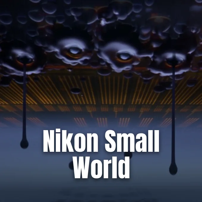
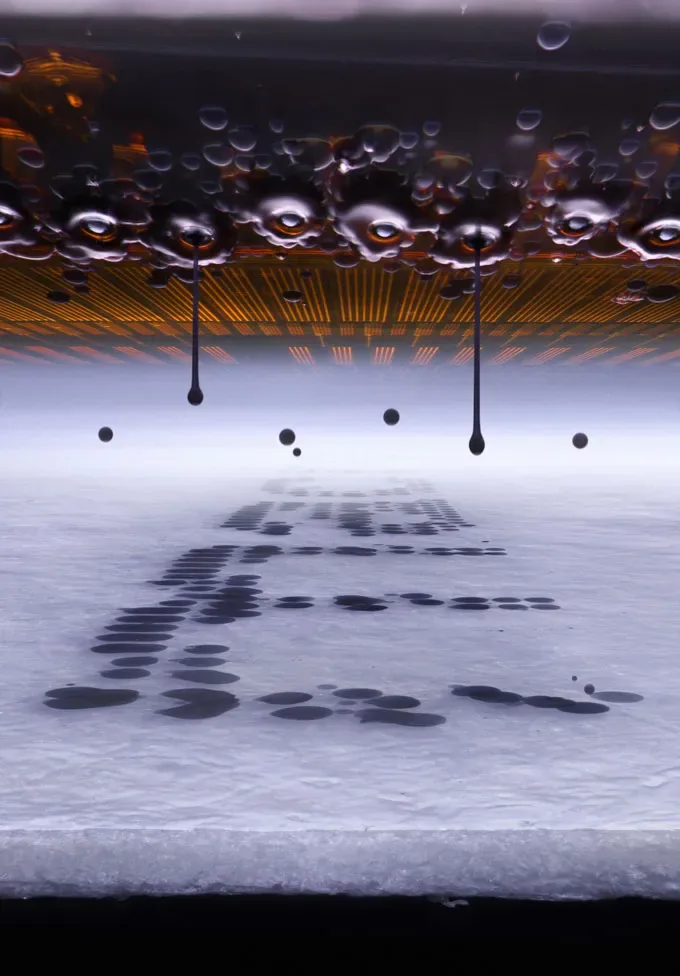
German experimental photographer Ole Bielfeldt won the 2026 Nikon Small World photomicrography competition on October 21 after engineering a custom optical setup to capture printer ink droplets suspended mid-air [1]. Bielfeldt, a 25-year-old design student living in Cologne, Germany, built the apparatus to freeze microscopic fluid motion that unfolds far too quickly for conventional mechanical camera shutters. His work led a field of 86 recognized images chosen from thousands of global entries, illuminating hidden biological forms alongside everyday mechanical processes documented across global research institutions [2].
Freezing Printer Ink Droplets
Bielfeldt first discovered his passion for microscopic imaging in 2021 during the COVID-19 lockdown after finding an old school microscope stored away in his parents’ attic. Because standard commercial camera adapters were not available for the antique instrument, the German photographer built his own coupler out of a tube and wood to attach his father’s camera to the eyepiece. Bielfeldt taught himself photomicrography through persistent trial and error, documenting behind-the-scenes experiments on social media under his creative brand macrofying. He avoided artificial intelligence tools to alter his imagery, relying strictly on traditional optical techniques and physical timing without investing in pricey commercial laboratory equipment. His curiosity about how standard desktop machines form crisp letters on paper inspired his winning project [1].
Microscopic ink droplets travel from printer nozzles at speeds approaching 20 meters per second, moving much faster than standard high-speed camera shutters can resolve. To freeze the fluid stream without blur, Bielfeldt disassembled a desktop printer and constructed a specialized rig only slightly larger than his head. He altered the mechanical assembly so that the printhead stayed stationary in sharp focus while motorized rollers pulled paper underneath. Working in complete darkness, Bielfeldt illuminated the flying droplets with a single brief flash while instructing the machine to repeatedly spell out the word “macro”. Printers seemed like magic before this project. “It’s so, so advanced technically, but at the same time it’s so, so common,” Bielfeldt said about modern printing hardware [2].
Evaluating Nikon Small World Submissions
The Nikon Small World competition is an annual showcase founded to recognize excellence in photography through the light microscope. In the 2026 contest, judges evaluated 1,802 entries submitted by photographers, researchers, and hobbyists hailing from 69 different countries [1]. The international judging panel selected 86 recognized photographs on October 21, awarding placements across biological, medical, and physical disciplines [2]. Bielfeldt earned first place ahead of experienced laboratory specialists from leading global universities. His victory shows that homemade garage engineering can compete with elite institutional equipment [1].
Submissions to Nikon Small World reveal intricate natural structures that remain completely invisible without high-magnification optical lenses. Across decades of competition, scientists and artists have relied on photomicrography to track cellular behaviors, study crystal formation, and observe living tissues [2]. Bielfeldt spent several weeks refining synchronization between the mechanical paper feed and his camera flash trigger in Cologne, Germany. Each successful frame required complete darkness and optical stillness [1]. By turning an ordinary desktop printer into an award-winning subject, Bielfeldt showed how microscopic photography bridges the gap between everyday engineering and popular scientific appreciation [2].
Pollen on a Jumping Spider
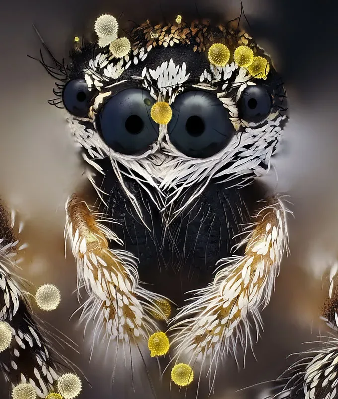
Second place in the competition went to German photographer John-Oliver Dum for an intimate macro portrait of a jumping spider covered in bright plant pollen. Dum developed his passion for arachnid photography after experiencing a painful spider bite during his childhood in Germany. Rather than avoiding arachnids as an adult, observing the graceful motion of a captive curly hair tarantula (Tliltocatl albopilosus) inspired Dum to document their anatomy up close. He spent several years studying regional seasonal rhythms in Europe to understand when specific spider species feed among blooming flora [2].
Dum captured the spider with the help of his wife, who cultivated blooming plants chosen specifically for the distinctive color and texture of their pollen grains. His macro lens revealed bristling body hairs and several reflective eyes among eight visual organs that normal human vision overlooks. “The biggest challenge was finding a spider like this on the right plants, with the right pollen attached to it, at exactly the right time of year and at exactly the right moment,” Dum said about his patient process. Dum said that close observation of delicate ecological interactions helps foster environmental conservation among the public. “We first have to see the beauty of this largely unknown world before we can truly understand what we may be destroying,” Dum said [2].


Vascular Networks in the Mouse Eye
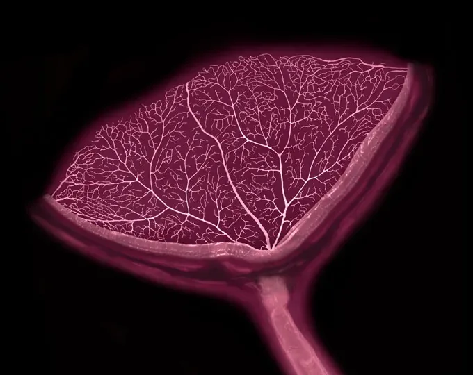
Cell biologists Dr. Sinem Karaman and Satu Paavonsalo from the University of Helsinki in Finland took 19th place with an image of ocular blood vessels. Instead of preparing a standard flat retina mount, Paavonsalo and Karaman created a three-dimensional view of an intact mouse eye, revealing a circular vascular network spreading outward from the central optic nerve. The branching capillaries resemble the petals of a flower, mapping how circulatory networks deliver oxygen to delicate ocular tissue layers [2].
Karaman and Paavonsalo study vascular systems at the University of Helsinki to understand how nutrient delivery breaks down in metabolic conditions such as obesity and diabetes. Mounting the ocular sample in three dimensions without damaging microscopic vessel walls required multiple delicate attempts in their Helsinki laboratory. “I had to hope that the sample was correctly positioned and that the staining was detected throughout the sample,” Paavonsalo said about the delicate process. Paavonsalo said that visualizing vascular networks in three dimensions provides structural insight that flat mounts cannot deliver. Fluorescent scans confirmed capillary density with high structural precision. Their Helsinki team hopes these structural findings will point to underlying mechanisms of vascular breakdown in human diseases [2].
Tardigrade Molting and Urban Slime Molds
Evolutionary developmental biologist Dr. Gonzalo Quiroga-Artigas from the University of Montpellier in France earned 12th place by photographing a molting water bear (Hypsibius exemplaris). The tiny creature, measuring only two-and-a-half times the width of a human hair, can enter suspended animation to survive harsh conditions including outer space. As the tardigrade pushed free from its outer covering, Quiroga-Artigas watched it leave behind an egg safely nestled inside the shed cuticle [2].
Quiroga-Artigas said that females of this species produce clutches containing twelve or fewer eggs during a single cycle, but the shedding sequence unfolds so swiftly that documenting the event under high magnification proves exceedingly rare. “Egg laying is closely synchronized with molting,” Quiroga-Artigas said about the timing. He chose this specific frame because “the egg looks almost like a precious gem being left behind by its mother”. Snapping the winning image demanded painstaking mounting and imaging of many living specimens at Montpellier. His work offers developmental biologists in France a clear visual timeline of how maternal coverings protect developing embryos [2].
Photographer Eric Epstein took sixth place with a detailed study of an urban slime mold (Hemitrichia sp.) collected from decaying logs in Prospect Park, Brooklyn, New York. To bring the delicate fruiting bodies into crisp focus, Epstein stacked 240 images taken across fine focal planes. “In this liquidy state, any touch would demolish” the fruiting bodies, Epstein said about harvesting the fragile woodland samples in Brooklyn. Certain slime mold networks mirror cosmic web structures of gas and dark matter connecting distant galaxies, while other varieties store bacteria to feed offspring. Epstein said he hopes the photograph hints “at [a] vast array of activity beyond our unaided perception” [2].
Stem Cell Research in Nikon Small World Portfolios
At the University of California, Berkeley, undergraduate researcher Faith Fernandes and structural cell biologist Dr. James Glover earned an Image of Distinction in Nikon Small World for photographing lab-grown neurons. Working at UC Berkeley, Fernandes and Glover reprogrammed adult human cells into induced pluripotent stem cells, returning them to an early developmental state capable of becoming any cell type in the human body. They subsequently guided those stem cells to mature into functioning human neurons, documenting the Berkeley team’s first attempt to cultivate neural networks with this technique [2].
Under the microscope, bright orange spheres mark cell nuclei where genomic DNA resides, while radiant blue neurotubules form structural pathways that transport intracellular molecules across the neural web. “I was so instantly enamored with this piece that I made it my computer wallpaper as soon as it was taken,” Fernandes said about the visual result. Pluripotent stem cells offer promising therapeutic pathways for regenerative medicine, allowing scientists at Berkeley and worldwide to investigate cellular treatments for degenerative conditions like Parkinson’s disease. Biomedical researchers have already used reprogrammed cells to regrow eye tissue, investigate premature aging in mouse studies, and culture miniature organ models for evaluating therapeutic compounds without relying on animal testing [2].
- ONLINE NEWS Ars Technica. (2026, October). Ink droplets in motion snag top prize in Nikon Small World contest. [Article Link]
- ONLINE NEWS Skalnik, A. (2026, October 21). Peek into hidden worlds revealed by the 2026 Nikon Small World contest. Science News. [Article Link]
APA 7: PerEXP Teamworks. (2026, October 23). Ole Bielfeldt Wins Nikon Small World with Suspended Ink Photo. PerEXP Teamworks. https://perexpteamworks.com/en/nikon-small-world-bielfeldt-ink-droplets/


