





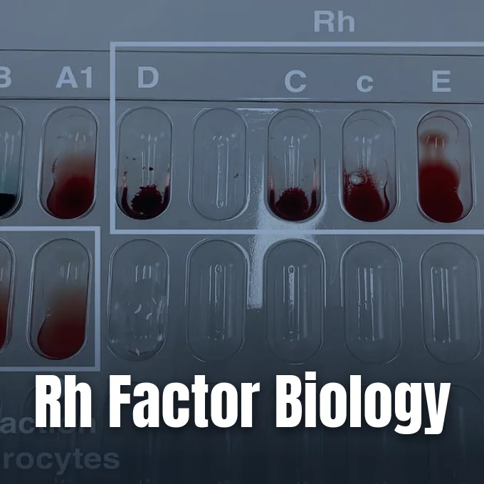
The Rh factor is an inherited protein on the surface of human red blood cells that defines whether a person’s blood type is positive or negative. While the ABO classification groups blood by carbohydrate markers, the Rh system depends on protein antigens embedded in the cell membrane [8]. When the primary protein known as the D antigen is present, the blood is Rh positive, and when it is absent, the blood is Rh negative [9]. Because the human immune system can recognize foreign red cell proteins during clinical care, identifying this marker helps medical teams prevent dangerous reactions during blood transfusions and pregnancy [10].
What Does the Rh Factor Mean?
The Rh factor tells doctors whether red cells carry the D antigen, which is the most immunogenic non-ABO protein found in human blood. A person with type A blood and the D antigen is A positive, while someone who lacks it is A negative [9]. Pairing this marker with the four basic ABO blood types yields eight common blood profiles used in clinics worldwide [11].
Unlike the ABO system, where healthy infants develop anti-A or anti-B antibodies through ordinary contact with environmental microbes, the Rh system does not generate natural defense antibodies against the D protein [9]. An Rh negative individual enters life without circulating anti-D antibodies in their plasma, so their immune system only builds these targeted defenses after direct exposure to positive red cells during an incompatible transfusion or childbirth [6]. Once this primary immune response occurs, memory B cells retain the ability to generate lasting IgG antibodies that stay in circulation for decades, which means that any later contact with positive blood can trigger fast destruction of donor or fetal cells without warning to the patient [10]. This fundamental biological distinction explains why blood banks track the Rh status of every unit with careful laboratory protocols [12].
For daily health, being Rh negative carries no disadvantage and causes no symptoms, because the missing protein does not weaken oxygen delivery or shorten ordinary life expectancy [9]. The trait functions as a benign genetic variation that only matters when blood from two distinct people mixes in medical care [10]. Doctors check this status routinely so they can supply compatible units during surgery [12].
Discovery of the Rh Blood Group System
Unexplained reactions puzzled early twentieth-century physicians when patients suffered chills and kidney failure despite receiving ABO-matched blood transfusions [11]. In 1939, Philip Levine and Rufus Stetson published the first clinical description of this condition after a woman suffered a severe reaction when transfused with blood from her husband following a stillbirth [5]. Their report showed that maternal serum contained an antibody that clumped the father’s red cells, providing the first clear evidence of an independent blood group factor [6].
In 1940, Karl Landsteiner and Alexander Wiener conducted laboratory experiments by injecting red blood cells from rhesus macaque monkeys into rabbits and guinea pigs. The animals developed antibodies that clumped rhesus monkey cells, but the researchers observed that this serum also clumped red cells from roughly 85% of human donors [11]. Believing they had found the same animal antigen in humans, they named the marker the Rhesus factor, though later testing proved the human protein was chemically distinct and led scientists to rename the original monkey antigen as LW [6]. The discovery reshaped transfusion medicine by explaining why prior crossmatches had failed, and it gave physicians their first biological framework for understanding newborn jaundice and hemolytic complications [5].
Subsequent investigations during the 1940s and 1950s uncovered additional related antigens on red cells, showing that human blood groups were far more diverse than Landsteiner’s initial three-type ABO model [8]. Early blood bankers quickly adopted animal and human serums to avoid hemolytic transfusion reactions during emergency surgery [12].
Antigens on the Red Cell Surface
The International Society of Blood Transfusion classifies the Rh system as system 004, recognizing 56 distinct antigens as of the May 2025 update [5]. Despite this diversity, five major antigens account for almost all clinical concerns: D, C, c, E, and e [4]. There is no lowercase d antigen in human biology, as lowercase d simply denotes the absence of the D protein in standard blood bank records [14].
Unlike ABO markers, which are carbohydrate chains attached to lipids or proteins, Rh antigens are pure transmembrane proteins that weave through the red cell lipid bilayer 12 times [6]. These proteins have six outward-facing loops that display antigenic sites and six inward-facing loops inside the cytoplasm. The D antigen sits on the first outer loop, making it readily accessible to circulating antibodies, while C and c occupy the second loop and E and e sit on the fourth loop [11]. Researchers have found that these membrane complexes help maintain the flexible biconcave shape of erythrocytes, while also functioning as transport channels for carbon dioxide and ammonium ions through the cell wall [4].

When all Rh antigens are missing from red blood cells, a rare condition called the Rh-null phenotype occurs [4]. Individuals with this condition have stomatocytic red cells that break down prematurely, causing chronic mild hemolytic anemia [7]. Because these patients lack all Rh surface antigens, they can only receive transfusions from other rare Rh-null donors [4].
Gene Structure on Chromosome 1
The genetic instructions for the Rh system reside on the short arm of chromosome 1, organized into two tightly linked genes designated RHD and RHCE. These two genes arose through an ancient duplication event, which explains why their coding sequences remain about 97% identical. Each gene contains 10 exons spanning approximately 75 kilobases of DNA, arranged in a tail-to-tail orientation with the small SMP1 gene located between them [4].
The RHD gene produces the RhD protein carrying the D antigen, while the neighboring RHCE gene encodes both the C or c and E or e antigens through alternative splicing and single-nucleotide polymorphisms. At amino acid position 103 of the RhCE protein, a single amino acid change determines antigen specificity: serine produces the C antigen, while proline yields the c antigen. Similarly, at position 226, alanine produces e while proline produces E [4]. In people of European descent, the Rh negative status usually results from a complete deletion of the RHD gene, whereas African and Asian populations often carry an inactive pseudogene with a premature stop codon or a hybrid gene that prevents functional protein expression [7].
Proper expression of these proteins on the cell surface requires a third gene, designated RHAG, located on chromosome 6. This gene produces the Rh-associated glycoprotein, which acts as a molecular chaperone to assemble Rh proteins into the membrane. If genetic mutations silence RHAG, no Rh antigens can reach the cell surface, producing the regulator form of the Rh-null condition despite normal genes on chromosome 1 [4].


The Rh Factor in Blood Transfusion
In transfusion medicine, matching the Rh factor prevents the recipient’s immune system from destroying donor red blood cells [12]. An Rh positive patient can safely receive red blood cells from either Rh positive or Rh negative donors because their body already tolerates the D antigen [9]. An Rh negative patient, however, should receive only Rh negative red cells to avoid sensitization and the formation of anti-D antibodies [10].
If an Rh negative individual who already carries anti-D receives Rh positive blood, preformed IgG antibodies quickly coat the transfused cells, marking them for destruction by macrophages in the spleen [7]. This extravascular hemolysis leads to fever, chills, flank pain, hemoglobin in the urine, and acute kidney failure [6]. According to clinical transfusion protocols from the University of Michigan Department of Pathology, red blood cell transfusions should begin at an initial rate no greater than 15 milliliters in the first 15 minutes, with the total unit transfused over two to four hours while staff monitor for adverse reactions. In contrast, platelets and cryoprecipitate components are typically administered at 10 milliliters per minute [12].
Because O negative red blood cells carry neither A nor B carbohydrate antigens nor the D protein, this blood type functions as the universal donor in medical emergencies. Trauma centers rely on O negative units when bleeding patients need immediate blood before typing tests can be completed [9]. Only about 6% to 8% of donors have O negative blood [4, 5].

Maternal Alloimmunization and Newborn Health
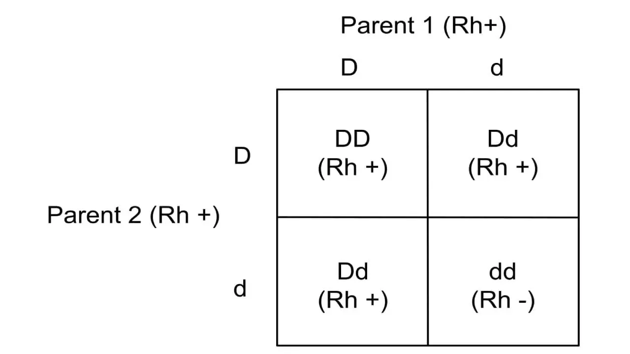
The most serious complication linked to the Rh factor occurs during pregnancy when an Rh negative mother carries an Rh positive fetus inherited from an Rh positive father. During delivery, or after events such as miscarriage or amniocentesis, fetal red cells can cross into maternal circulation [10]. The mother’s immune system recognizes the fetal D antigen as foreign and builds IgG anti-D antibodies, a process called alloimmunization [1].
While a first pregnancy usually delivers before maternal antibody levels rise significantly, subsequent pregnancies face severe risks [10]. Maternal IgG antibodies readily cross the placenta and bind to the red cells of an Rh positive fetus, causing progressive hemolysis known as hemolytic disease of the fetus and newborn or erythroblastosis fetalis. Severe fetal anemia can lead to hydrops fetalis, enlarged organs, and stillbirth, while newborn red cell breakdown produces high bilirubin levels that risk permanent brain damage without treatment [6, 14]. Non-invasive Doppler ultrasonography evaluating blood velocity in the fetal middle cerebral artery allows clinicians to detect fetal anemia early without invasive sampling risks [1].
Modern obstetric care prevents alloimmunization by administering Rh immune globulin, widely known as RhoGAM, to Rh negative mothers at 28 weeks of gestation and again within 72 hours after delivering an Rh positive infant [6]. This preventive treatment has lowered sensitization rates from 16% to under 1% in screened pregnancies [1, 9].
Clinical Impact of Variant D Antigens
Not every patient fits into a simple positive or negative category, as alterations in the RHD gene produce phenotypic variations known as weak D and partial D [2]. Weak D red cells express normal D antigens but at a much lower density, meaning they might test negative during routine room-temperature testing [5]. In contrast, partial D individuals lack specific epitopes of the D protein, which allows their immune systems to produce anti-D antibodies directed against the missing segments if exposed to complete D antigens [6].
Resolving these ambiguous phenotypes is important for both transfusion safety and maternal care [2]. Patients with weak D types 1, 2, and 3 can safely receive Rh positive blood and do not require immune globulin prophylaxis during pregnancy, while partial D patients must be treated as Rh negative to prevent sensitization [6]. Modern immunohematology laboratories use monoclonal antibodies with automated column agglutination and DNA-based testing to distinguish these alleles with high precision [3, 5]. Testing has progressed from simple immediate-spin tube methods to 37°C incubation and anti-human globulin assays, ensuring that weak reactions receive proper molecular confirmation before clinical decisions are made [5].

Recent genomic reviews in the literature show how DNA sequencing has uncovered novel antigens such as PARG, CEVF, CEWA, and CETW, numbered RH60 through RH63 by the International Society of Blood Transfusion [3]. Cell-free fetal DNA testing can now determine a baby’s Rh status from maternal plasma as early as 9 weeks of gestation using real-time PCR targeting exons 7 and 10 [6, 15].
Global Distribution of Blood Types
The frequency of the Rh factor and its underlying haplotypes varies widely in human populations as a consequence of ancient migrations, genetic drift, and natural selection [13, 14]. Worldwide, approximately 94% of people are Rh positive and only 6% are Rh negative, though regional proportions differ considerably. In Western Europe and the United States, roughly 15% to 17% of the population is Rh negative, with the highest concentration observed in the Basque population, where over 40% lack the D antigen [5, 14].
By contrast, Rh negative phenotypes are far less common in other parts of the world, appearing in about 3% to 5% of African populations and fewer than 1% of East Asian populations [5]. A genetic study of 2005 individuals across nine Arab-Berber populations in the Tlemcen region of Western Algeria demonstrated consistent regional homogeneity shaped by historical Mediterranean gene flow [13]. In sub-Saharan Africa, the R0 haplotype (Dce) predominates at frequencies above 40%, whereas European populations most frequently carry the R1 (DCe) and r (dce) haplotypes [5, 14]. In England, population studies show that R1 accounts for about 42.1% of haplotypes, followed by r at 38.9% and R2 (DcE) at 14.1% [5].
These demographic variations shape daily transfusion inventory management, requiring blood banks in Europe and North America to maintain larger stocks of Rh negative red cell units than facilities in East Asia [5]. Testing for Rh antigens beyond D remains standard practice when preparing chronically transfused patients to prevent delayed hemolytic reactions [6].
- ACADEMIC JOURNAL ACOG Practice Bulletin No. 192: Management of Alloimmunization During Pregnancy. (2018). Obstetrics & Gynecology, 131(3), e82-e90. [Article Link]
- ACADEMIC JOURNAL Ramsey, G. E. (2025). The Rh blood group system: RHD update. Immunohematology, 41(2), 31-48. [Article Link]
- ACADEMIC JOURNAL Floch, A., Tournamille, C., & Vege, S. (2026). The Rh blood group system: RHCE update. Immunohematology, 42(2), 52-65. [Article Link]
- WEBSITE Pan, S. (2026). Rh blood group system: Definition, antigens, antibody. Biology Notes Online. [Article Link]
- WEBSITE Edwards, T. (2025). Understanding rhesus blood groups: A complete guide. RhMPG – Rhesus Most Probable Genotype. [Article Link]
- WEBSITE MyH Editorial Team. (2024). Rh blood group: Types, negative, incompatibility & complication. My Hematology. [Article Link]
- WEBSITE News-Medical. (2017). What is Rh blood group? [Article Link]
- WEBSITE BloodBook. (2024). Blood typing systems: ABO, Rh, and the 26 ISBT blood group systems. [Article Link]
- WEBSITE BloodAnalysis. (2026). Rh factor: Rh positive vs Rh negative explained. [Article Link]
- ONLINE NEWS Morales-Brown, L. (2021). Rh factor: Definition and importance. Medical News Today. [Article Link]
- WEBSITE GeeksforGeeks. (2022). Rh blood group system: Overview, types, composition, importance and FAQs. [Article Link]
- WEBSITE University of Michigan Department of Pathology. (2013). Blood component and compatibility. [Article Link]
- ACADEMIC JOURNAL Aouar Metri, A., Sidi-Yakhlef, A., Biémont, C., Saïdi, M., Chaïf, O., & Ouraghi, S. A. (2011). A genetic study of nine populations from the region of Tlemcen in Western Algeria: A comparative analysis on the Mediterranean scale. Anthropological Science, 120(3), 209-216. [Article Link]
- WEBSITE Wikipedia contributors. (2026). Rh blood group system. Wikipedia. [Article Link]
- WEBSITE Wikipedia contributors. (2026). Rh factor testing. Wikipedia. [Article Link]


