





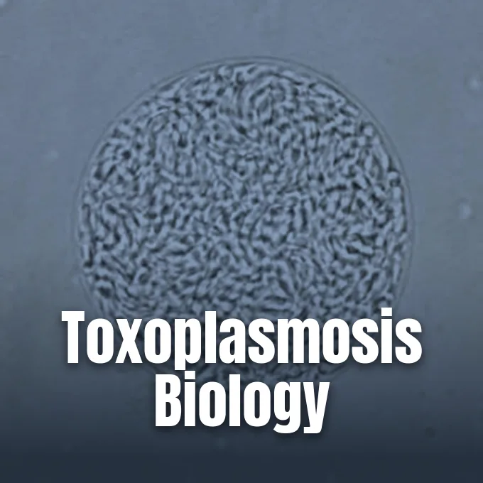
Toxoplasmosis is a common disease caused by the single-celled protozoan parasite Toxoplasma gondii, an organism that can invade any nucleated cell in warm-blooded animals. Research reviewed by N. C. Smith and colleagues indicates that about two billion people carry the parasite globally, with exposure rates topping 60% in some humid, low-altitude areas. Most healthy hosts experience no symptoms. But the parasite creates dormant cysts in muscle and brain tissue that persist for life, creating serious health risks if immune defenses drop or during pregnancy [11].
- Life Cycle of Toxoplasma gondii
- How Toxoplasmosis Spreads to Humans
- Cellular Invasion and Brain Cysts
- Clinical Signs in Healthy Hosts
- Why Is Toxoplasmosis Dangerous During Pregnancy?
- Central Nervous System Disease in Immunodeficiency
- Diagnostic Tests for Detecting Parasite Infection
- Medical Prevention and Toxoplasmosis Therapies
Life Cycle of Toxoplasma gondii
French scientists Charles Nicolle and Louis Manceaux first described the organism in 1908. It is an obligate intracellular apicomplexan pathogen that depends on host cell nutrients to replicate. Cats remain the definitive host. Domestic cats and wild felines serve as the only animals where the parasite can complete sexual reproduction. When felines ingest infected prey, sexual stages produce unsporulated oocysts in the feline gut lining that pass into the environment [6].
In intermediate hosts like humans, birds, and livestock, asexual replication proceeds through two distinct forms that shape the course of illness. Fast-dividing crescent-shaped tachyzoites drive acute illness by multiplying inside host cells until the host cell ruptures, allowing released parasites to invade nearby cells and travel through blood and lymph. When host immune defenses mount a response with specific antibodies and cellular mechanisms, tachyzoites convert into slow-dividing bradyzoites. These bradyzoites become sequestered within thick-walled tissue cysts that form in skeletal muscle, cardiac tissue, and the brain, where they evade antimicrobial drugs and remain dormant for the life of the host [6].
Cats shed millions of oocysts in their feces for up to three weeks after initial exposure. Freshly passed oocysts are not infectious. They need at least 24 hours in soil, water, or cat litter to sporulate into infective forms that can survive in moist outdoor soil for many months [1, 4].
How Toxoplasmosis Spreads to Humans
Foodborne spread is the most frequent route of human exposure. Eating raw or undercooked meat containing viable bradyzoite cysts introduces the organism into the human gut, with pork, lamb, and wild venison carrying notable cyst loads when animals graze outdoors [1, 8]. Coastal shellfish like oysters, clams, and mussels can also filter and retain oocysts from freshwater runoff [1].
Accidental swallowing of oocysts from contaminated soil, unwashed vegetables, or water supplies accounts for many non-meat cases. Intact skin does not absorb the parasite. But unwashed hands can transfer oocysts to food or mouth after gardening or cleaning cat litter boxes. In a study published in Clinical Infectious Diseases, J. L. Jones and J. P. Dubey pointed out that trends toward free-range and organically raised livestock can increase animal contact with outdoor cat feces, elevating cyst prevalence in farm meat [8]. Drinking unpasteurized goat’s milk or untreated municipal water has also caused documented community outbreaks [1].
Toxoplasmosis does not spread from person to person. Rare non-oral transfer happens during blood transfusions or solid organ transplants when an uninfected recipient receives organs from a Toxoplasma-positive donor [1, 4]. Laboratory technicians handling infected blood face accidental needle-stick contamination risks, but maternal-fetal spread across the placenta during pregnancy remains the chief non-oral danger [1].
Cellular Invasion and Brain Cysts
Cell entry is an active invasive process. When invading a host cell, Toxoplasma gondii forces its way inward and encloses itself in a specialized parasitophorous vacuole (PV) derived from the invaginated host plasma membrane. This vacuole resists endolysosomal fusion, shielding the parasite while it recruits host mitochondria and endoplasmic reticulum to scavenge lipids and amino acids [6].
During invasion, the parasite secretes rhoptry proteins designated ROP proteins from its apical complex. These proteins move to the host cell nucleus to modify cytokine expression while blocking host immunity-related GTPase proteins that would otherwise rupture the vacuole membrane. Strains carrying the GRA15 gene activate the nuclear factor kappa B (NF-κB) signaling pathway, stimulating host interleukin-12 release during early immune responses. The parasite also blocks programmed cell death by altering pro-apoptotic proteins BAX and BAK so they cannot migrate to mitochondria. Furthermore, experimental work by Wang and colleagues demonstrated that the parasite triggers host cell autophagy through a calcium-dependent signaling pathway, causing uninfected bystander cells to degrade while keeping infected host cells viable [6].
The GRA28 protein uses the MYR1 transport pathway to alter host gene regulation. Infected host cells become hyper-mobile. By mimicking dendritic cells, they ferry the parasite across the blood-brain barrier into neural structures, where host immune pressures force tachyzoites to turn into bradyzoites that form durable cysts in brain tissue [6].
Clinical Signs in Healthy Hosts
Most healthy people experience no symptoms. When clinical signs appear, they usually mimic mild influenza with fatigue, headache, low fevers, and diffuse muscle aches lasting several weeks. Swollen lymph nodes form the most common physical finding, appearing under the jawline or along the neck, although pediatric patients may develop lymph node swelling in the armpits or groin [6, 9].
Swollen lymph nodes resolve without treatment within one to two months in about 60% of cases. A quarter of patients take two to four months to recover, 8% require four to six months, and only 6% experience persistent swelling past six months. Eye damage can still occur. In a review in the Scandinavian Journal of Infectious Diseases, G. Saadatnia and M. Golkar emphasized that the parasite is a major cause of retinochoroiditis, producing inflammation in the retina and choroid that causes blurred vision, eye pain, and dark floaters [6, 9].
Skin lesions appear rarely in acquired cases, presenting as roseola-like spots, erythema multiforme eruptions, or prurigo nodules. Tachyzoites measure six by two micrometers. In PLoS ONE, J. Flegr, J. Prandota, M. Sovičková, and Z. H. Israili surveyed 88 countries, finding correlations between latent infection and subtle behavioral differences or road traffic accidents [6, 10].



Why Is Toxoplasmosis Dangerous During Pregnancy?
Primary maternal illness during pregnancy creates acute medical hazards because tachyzoites can cross the placenta to infect the fetus. Congenital transmission was first confirmed in 1941. Global health records document about 200,000 cases of congenital toxoplasmosis each year. If a woman had the parasite before conception, pre-existing antibodies protect the fetus, but new exposure during pregnancy allows tachyzoites to cross placental tissues into fetal circulation [3, 6].
Infected mothers rarely experience noticeable symptoms, yet the developing child can suffer major developmental injury. Up to half of infected fetuses are born prematurely, presenting with low birth weight, jaundice, enlarged liver and spleen, or purple blueberry muffin skin lesions. Fluid accumulation causes hydrocephalus. The classic presentation involves chorioretinitis, intracranial calcifications, and fluid buildup that leads to microcephaly, macrocephaly, or neonatal seizures. In chapters on pediatric infections by R. McLeod and K. M. Boyer, clinicians note that infants with mild congenital disease may appear normal at birth but later develop vision loss, hearing impairment, or learning disabilities during adolescence [3].
In an audiological study in Scientia Medica, Luciana Macedo de Resende, Gláucia Queiroz Manzan de Andrade, Marisa Frasson de Azevedo, Jacy Perissinoto, and Andrêza Batista Cheloni Vieira followed 106 infants diagnosed with congenital toxoplasmosis through the Minas Gerais State Neonatal Screening Program between September 2006 and March 2007. Every child began drug therapy before 2.5 months of age and received follow-up care for 12 months. Hearing impairment affected 46 infants. Testing showed normal hearing in 56.6% of children, while hearing deficits included conductive loss in 12.3%, sensory-neural loss in 3.8%, and central auditory dysfunction in 27.4% [5].
Central Nervous System Disease in Immunodeficiency
In people with advanced human immunodeficiency virus (HIV) or those receiving immunosuppressive chemotherapy, latent brain cysts can reactivate into proliferating tachyzoites. Reactivation causes toxoplasmic encephalitis. This focal neurological illness emerges when helper T-cell counts fall below 200 cells per microliter of blood, with risk peaking when CD4 counts drop below 50 cells per microliter. Patients develop subacute headaches, fever, confusion, hemiparesis, speech loss, cranial nerve palsies, and seizures [2, 4].
Medscape records show that clinical central nervous system toxoplasmosis developed in 3% to 15% of AIDS patients in the United States, causing 789 deaths between 2000 and 2010. In European and African centers before routine prophylaxis, cerebral reactivation occurred in 50% to 75% of seropositive patients with severe immune failure. A study in Mexico City evaluated 320 AIDS patients. Brain toxoplasmosis was the leading condition in 42% of the group, followed by cerebral cryptococcosis at 28%, tuberculous meningitis at 8.7%, and non-Hodgkin lymphoma at 3.75% [4].
Brain imaging reveals multiple ring-enhancing lesions in the basal ganglia with surrounding edema. T2-weighted magnetic resonance scans often show a concentric target sign. Antiretroviral therapy dropped annual toxoplasmosis hospital stays in the United States from over 10,000 in 1995 to 3,643 in 2001 and 2,985 by 2008 [4].
Diagnostic Tests for Detecting Parasite Infection
Diagnostic choices depend on immune status. Serological testing for immunoglobulin G (IgG) and immunoglobulin M (IgM) antibodies forms the first-line laboratory method, where rising IgG titers confirm active infection while IgM antibodies persist for up to 12 months [4, 7]. In people with advanced HIV, however, impaired antibody production causes false-negative serological tests in up to 22% of confirmed cases [4].
Molecular testing has improved parasite detection. In a review in Parasites & Vectors, Q. Liu, Z. Wang, S. Huang, and X. Zhu noted that PCR tools allow rapid amplification of parasite DNA from clinical samples [12]. Testing cerebrospinal fluid by PCR for the B1 gene offers 83.3% sensitivity and 95.7% specificity for diagnosing toxoplasmic encephalitis. In pregnant women with acute infection, testing amniotic fluid by PCR allows prenatal detection of fetal transmission without invasive fetal cord blood sampling [4, 12].
Serotyping methods help classify parasite strains in global research [12]. Lumbar puncture carries herniation risks when intracranial pressure is elevated. Doctors start empiric drug therapy based on imaging findings. If lesions show no improvement after 10 to 14 days, a stereotactic brain biopsy confirms tissue tachyzoites and rules out primary CNS lymphoma [4].
Medical Prevention and Toxoplasmosis Therapies
Healthy non-pregnant adults rarely need medication because their immune systems suppress tachyzoite replication [11]. When persistent symptoms or eye damage occur, the standard medical regimen combines pyrimethamine and sulfadiazine. Leucovorin protects bone marrow. Because pyrimethamine inhibits folic acid synthesis, doctors co-administer folinic acid, known as leucovorin, while R. McLeod and K. M. Boyer point out that therapy continues until active lesions resolve [3, 11].
In pregnant women with acute gestational infection, doctors prescribe the antibiotic spiramycin to reduce parasite passage across the placenta. Therapy changes after amniocentesis. If fetal infection is confirmed, treatment shifts to pyrimethamine, sulfadiazine, and leucovorin under maternal-fetal medical supervision. Infants receive therapy for 12 months. In MedlinePlus medical reviews updated by Neil K. Kaneshiro, David C. Dugdale, and Brenda Conaway, clinicians recommend adding corticosteroids if active eye inflammation threatens vision or if spinal fluid protein levels are high [3].
Prevention relies on simple kitchen precautions. Meat should be cooked to safe temperatures that kill bradyzoite cysts, and cutting boards should be washed with soap and hot water after preparing raw meats [1]. Pregnant women and immunocompromised patients should avoid cleaning cat litter boxes or wear gloves and wash hands thoroughly. Cat owners should keep cats indoors and feed them commercial dry or canned food rather than raw meat to stop cats from hunting infected prey [1, 2].
- WEBSITE Centers for Disease Control and Prevention. (2025, February 14). Toxoplasmosis: Causes and how it spreads. [Article Link]
- WEBSITE Centers for Disease Control and Prevention. (2025, February 14). People at increased risk for toxoplasmosis. [Article Link]
- WEBSITE MedlinePlus. (2025, June 4). Congenital toxoplasmosis: MedlinePlus Medical Encyclopedia. [Article Link]
- WEBSITE Medscape. (2026, June 9). CNS toxoplasmosis in HIV: Overview, pathophysiology, epidemiology. [Article Link]
- ACADEMIC JOURNAL De Resende, L. M., Andrade, G. Q. M. d., Azevedo, M. F. d., Perissinoto, J., & Vieira, A. B. C. (2010). Congenital toxoplasmosis: Auditory and language outcomes in early diagnosed and treated children. Scientia Medica, 20(1), 13-19. [Article Link]
- WEBSITE Wikipedia contributors. (2026). Toxoplasmosis. [Article Link]
- ACADEMIC JOURNAL Robert-Gangneux, F., & Dardé, M. (2012). Epidemiology of and diagnostic strategies for toxoplasmosis. Clinical Microbiology Reviews, 25(2), 264-296. [Article Link]
- ACADEMIC JOURNAL Jones, J. L., & Dubey, J. P. (2012). Foodborne toxoplasmosis. Clinical Infectious Diseases, 55(6), 845-851. [Article Link]
- ACADEMIC JOURNAL Saadatnia, G., & Golkar, M. (2012). A review on human toxoplasmosis. Scandinavian Journal of Infectious Diseases, 44(11), 805-814. [Article Link]
- ACADEMIC JOURNAL Flegr, J., Prandota, J., Sovičková, M., & Israili, Z. H. (2014). Toxoplasmosis – A global threat. Correlation of latent toxoplasmosis with specific disease burden in a set of 88 countries. PLoS ONE, 9(3), e90203. [Article Link]
- ACADEMIC JOURNAL Smith, N. C., Goulart, C., Hayward, J. A., Kupz, A., Miller, C., & Dooren, G. G. v. (2020). Control of human toxoplasmosis. International Journal for Parasitology, 51(2-3), 95-121. [Article Link]
- ACADEMIC JOURNAL Liu, Q., Wang, Z., Huang, S., & Zhu, X. (2015). Diagnosis of toxoplasmosis and typing of Toxoplasma gondii. Parasites & Vectors, 8(1), 292. [Article Link]
Cover image: United States Center for Disease Control and Prevention / Wikimedia Commons, Public domain


