



Tuberculosis is an airborne bacterial illness caused by Mycobacterium tuberculosis that primarily attacks human lung tissue when infected persons cough, speak, or sneeze [1]. Understanding what is tuberculosis begins by distinguishing between a silent, dormant carriage and an open sickness that destroys tissue. The germ can stay quiet inside human cells for years [2]. Without prompt medicine, lung illness spreads to close household contacts, causes breathlessness, and can prove fatal [1].
- What Is Tuberculosis and How It Begins?
- How Mycobacteria Evade Alveolar Macrophages
- Active Disease Versus Latent Infection
- Recognizing Signs of Pulmonary Disease
- Diagnostic Testing for Tuberculosis
- First-Line Antibiotics and Drug Resistance
- Global Burdens of Tuberculosis
- International Elimination Targets Amid Funding Shortfalls
What Is Tuberculosis and How It Begins?
The history of this wasting illness reaches back thousands of years to ancient Greece, where early doctors called it phthisis. By the eighteenth century, people across Europe called it consumption because of its steady physical wasting. German physician Robert Koch discovered the tubercle bacillus in Berlin in 1882. He proved that TB was a contagious germ rather than an inherited family curse [7].
Under a laboratory microscope, Mycobacterium tuberculosis appears as a thin rod measuring 0.2 to 0.6 micrometers wide and up to 10 micrometers long. Its outer envelope holds an unusually thick layer of fatty mycolic acids that shield the microbe from environmental desiccation, defend it against ordinary chemical disinfectants, and prevent conventional Gram stain dyes from penetrating the cellular boundary [8],. Diagnostic technicians use the Ziehl-Neelsen procedure instead [15]. When stained with red carbolfuchsin and washed with harsh acid and alcohol, the bacilli keep their bright crimson tone [7]. The cells form woven cords. These intertwined cord-like ropes are shaped by trehalose dimycolate glycolipids in the bacterial envelope, a distinctive structural characteristic that researchers directly associate with mycobacterial virulence inside host tissue [15].
Most gut bacteria multiply every twenty minutes, but the tubercle bacillus divides only once every 18 to 24 hours [15]. It needs abundant oxygen to survive. In human lungs, the germ grows fastest in the upper lobes where oxygen levels run highest [7]. Outside the body, it can survive for months on dry wood or dust. Because fewer than ten bacilli can cause sickness, inhaling a tiny airborne drop is enough to infect a person [8].

How Mycobacteria Evade Alveolar Macrophages
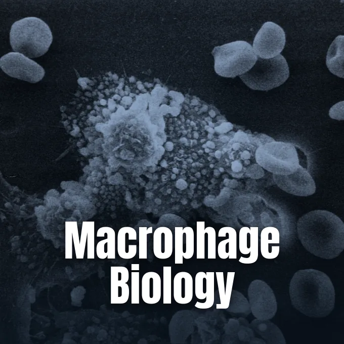
When a person inhales droplets from a coughing patient, the bacilli glide past bronchial airways into tiny alveolar air sacs [7]. White blood cells known as alveolar macrophages swallow the bugs inside digestive bubbles. Normal human cells merge these bubbles with acidic lysosomes to destroy foreign invaders. But the tubercle bacillus halts this merger by blocking early endosomal autoantigen 1 [15].
To thrive inside host defense cells, the bacterium turns off the chemical alarms that tell sick human cells to die. It secretes a lipid called isotuberculosinol to stall digestive bubble growth while pulling in cell nutrients through tiny transport vesicles. The bug also coats itself with 1-tuberculosinyladenosine (1-TbAd), a nucleoside that acts like an antacid to neutralize internal acid and swell host compartments. At the same time, the pathogen raises levels of the human enzyme PPM1A, which works through downstream JNK and AP-1 signaling pathways to shut down both intrinsic and extrinsic cell death programs and keep the infected macrophage alive. By blocking programmed cell suicide, the germ secures a safe shelter where it feeds and multiplies undisturbed [15].
Unable to clear the bugs, the immune system surrounds infected macrophages with T cells and fibrous tissue. This dense wall forms a granuloma with dead, cheesy tissue in the center and large Langhans cells around the border. Granulomas hold the germ in check, but they also protect dormant bacilli from destruction for decades [15].
Active Disease Versus Latent Infection
Doctors group TB into two states: latent carriage and active sickness [2]. In latent TB, host defenses trap the germs inside lung granulomas [7]. People with latent carriage feel fine, show no symptoms, and cannot pass the bug to others [2]. An estimated one-quarter of the global population carries latent TB, forming a vast human pool from which new cases emerge [1].
Most healthy people who breathe in the germ contain it for life [2]. Only about 5% to 10% of infected individuals ever develop active disease [1]. That breakdown happens most often in the first two years after exposure [9]. When immune defenses drop, dormant bugs wake up, break out of calcified scars, destroy lung tissue, and make the patient sick [7].
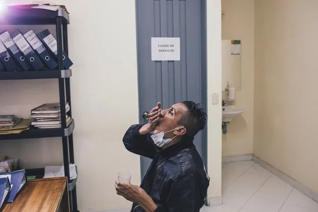
Underlying illnesses can speed the switch from a quiet carriage to active sickness. Co-infection with HIV is the biggest danger, raising the yearly risk of TB progression roughly twelvefold compared to people without the virus. Undernutrition also leaves the body weak, contributing to an estimated 0.97 million new cases globally in 2024. Without adequate nutrition and healthy CD4 T cells, the host immune barrier weakens until dormant mycobacteria escape from their fibrous prisons to invade surrounding healthy lung air spaces. Furthermore, poorly controlled diabetes, regular cigarette smoking, heavy alcohol use, and medical therapies (such as cancer chemotherapy or organ transplant drugs) weaken cellular defenses, disrupt lung granuloma stability, and allow quiet bacteria to resume rapid multiplication and cause open disease [1], [9].
Recognizing Signs of Pulmonary Disease
When Mycobacterium tuberculosis multiplies inside the lungs, it causes pulmonary TB, which makes up about 90% of all active cases [14]. The main warning sign is a persistent cough lasting more than three weeks, which brings up thick mucus that can turn bloody as tissue breaks down. Patients suffer chest pain, low fevers, heavy night sweats, and steady weight loss. People often feel worn out and lose their appetite for weeks. Because these signs start slowly, patients often mistake early illness for a winter chest cold, delaying doctor visits while spreading germs to family members. Without prompt antibiotic care, this ongoing lung decay causes shortness of breath, irreversible tissue scarring, and fatal lung collapse [5].
Unchecked lung damage brings sudden medical emergencies. Cavities can eat into bronchial arteries, forming weak vascular bulges called Rasmussen aneurysms that cause fatal bleeding. Germs can also break into the pleural chest cavity, causing painful pleurisy, fluid buildup, and hazardous collections of pus and air known as pyopneumothorax [14].
Although nine out of ten active illnesses remain centered inside pulmonary tissue, viable bacilli can travel through the bloodstream and lymphatic vessels to establish destructive extrapulmonary disease in virtually any organ of the human body. Scrofula inflames lymph nodes in the neck. Other vulnerable targets include the kidneys, the meninges surrounding the brain, and the musculoskeletal framework. Spinal bone loss produces Pott disease [14]. In vulnerable newborns, hematogenous dissemination produces miliary TB, scattering thousands of tiny millet-seed lesions across the liver, spleen, and brain tissues with devastating mortality if left untreated [8], [14].


Diagnostic Testing for Tuberculosis
Fast testing helps doctors start treatment early and stop airborne spread. To find latent carriage, clinics use skin tests and blood tests [2]. For the Mantoux skin test, a nurse injects five units of purified protein derivative under the skin of the forearm. The clinic checks the arm 48 to 72 hours later to measure any raised, firm swelling [7].
A well-recognized limitation of the traditional tuberculin skin test is that earlier childhood vaccination with Bacille Calmette-Guérin (BCG) frequently produces a confusing false-positive reaction that misleads physicians evaluating a patient’s true infection status [2]. Clinicians order blood assays to resolve ambiguity. Diagnostic blood tests like QuantiFERON-TB Gold and T-SPOT incubate patient blood samples with specific mycobacterial antigens and measure the release of interferon-gamma from sensitized T cells to confirm genuine exposure without interference from prior BCG vaccination [3], [7]. Testing requires only a single clinical visit [3]. Public health guidelines from NICE in England recommend these interferon-gamma release assays for confirming positive skin reactions among vulnerable populations and migrant new entrants [10], [13].
When people show lung symptoms, clinics collect morning sputum to look for acid-fast bacilli. A sputum smear gives quick hints in two days, but growing the bug on Lowenstein-Jensen egg media or Middlebrook 7H9 agar takes six to eight weeks [6], [8]. Rapid nucleic acid amplification tests (NAAT) can spot bacterial DNA and test for drug resistance in under two hours [6].
First-Line Antibiotics and Drug Resistance
Curing drug-susceptible TB takes a six-month course of multiple medicines taken daily. For the first two months, patients take four first-line oral drugs: isoniazid, rifampicin, pyrazinamide, and ethambutol. During the next four months, therapy continues with isoniazid and rifampicin to wipe out lingering bacteria [1]. These drugs strike different targets: isoniazid and ethambutol halt cell wall building, rifampicin stops RNA making, and pyrazinamide disrupts bacterial energy [12]. Because mycobacteria grow slowly and slip into sleeping states, patients must take every dose. Stopping pills early lets tough bacteria survive, multiply again, and gain drug resistance. Standard treatment protocols outlined by the World Health Organization require healthcare workers to monitor medication intake through directly observed therapy so that patients complete every dose and avoid developing resistant strains [1].
How do mycobacterial populations survive antibiotic exposure without immediate genetic mutation? In a 2024 preprint that has not undergone peer review, lead researcher Dániel Molnár, Éva Viola Surányi, and Judit Tóth at eLife grew Mycobacterium smegmatis under first-line antibiotics for 230 bacterial generations [18]. Gene sequencing showed no rise in mutation rates, proving that early survival came from non-genetic tolerance rather than drug-induced mutation. The bugs survived by switching metabolic paths and turning on DNA repair genes to keep their genomes stable [17].
When strains resist both isoniazid and rifampicin, the sickness is called multidrug-resistant TB (MDR-TB) [1]. Past treatments needed painful daily injections for two years [12]. In 2022, the World Health Organization recommended BPaLM, a six-month all-oral cure using bedaquiline, pretomanid, linezolid, and moxifloxacin. Globally in 2024, approximately 34 000 people with drug resistance started shorter oral cures, up from 5653 in 2023 [1].
Global Burdens of Tuberculosis
Even though effective medical therapies cure the condition, TB remains the deadliest single infectious agent on Earth. In 2024, an estimated 1.23 million deaths occurred worldwide, including 150 000 fatalities among people living with HIV. That same year, 10.7 million individuals contracted active TB across the globe, with men making up 5.8 million cases, women 3.7 million, and children 1.2 million. Eight nations carried two-thirds of all cases: India (25%), Indonesia (10%), the Philippines (6.8%), China, Pakistan, Nigeria, DR Congo, and Bangladesh. Global control initiatives averted roughly 83 million fatalities between 2000 and 2024 [1].
Co-infection with HIV creates a lethal partnership that accelerates clinical decline. In 2024, people living with HIV accounted for 5.6% of new TB infections, while global HIV testing among TB patients reached 82%. WHO collaborative interventions launched in 2004 and scaled up in 2005 averted an estimated 9.8 million deaths [1]. Analyzing 15,999 HIV patients in CCASAnet clinics across Brazil, Haiti, Mexico, and Peru, Harvard researcher Serena P. Koenig and Vanderbilt physician Catherine C. McGowan reported that an initial TB diagnosis doubled ten-year mortality to over 19% [20].

Social inequities drive sharp infection spikes among marginalized communities. In Canada, colonial harms and poor housing pushed the 2023 TB rate among Inuit to over 37 times that of the general population [11]. In England, UK Health Security Agency reports documented 4,855 notifications in 2023, with non-UK born residents accounting for 79.9% of cases. High urban rates clustered in Leicester at 40.7 per 100,000 and Newham at 40.6 [10].
International Elimination Targets Amid Funding Shortfalls
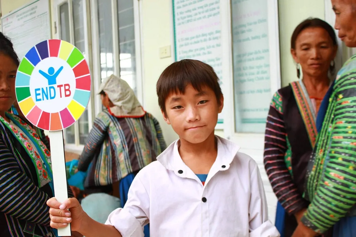
The World Health Organization End TB Strategy aims to cut TB deaths by 90% and new cases by 80% by 2030. Reaching these goals requires US$ 22 billion annually for care and prevention by 2027 [1]. But global spending reached only US$ 5.7 billion in 2023, covering just 26% of needed funds. Research funding also stalled at US$ 1.0 billion against a US$ 5 billion yearly goal [4].
Budget execution often lags approved allocations. In Delhi, official reports from the Directorate General of Health Services revealed that the National Tuberculosis Elimination Programme utilized only 47.4% of its approved budget in 2025, spending Rs 51.32 crore and leaving Rs 56.95 crore completely unspent. Beneficiary banking hurdles delayed direct benefit transfers. Under the Ni-kshay Poshan Yojana nutritional support initiative, which doubled monthly payments to Rs 1,000 in November 2024, administrative bottlenecks left over Rs 72 crore in food assistance unpaid to more than two hundred thousand eligible patients. Comptroller and Auditor General audits in Delhi flagged unpurchased autoclaves, staff shortages, and missed monitoring for 2,587 drug-resistant patients. Clinic infrastructure in Mehrauli remains severely strained [19].
Public advocacy helps push governments and drug companies to act. In his 2025 bestselling book Everything Is Tuberculosis, author John Green argued that the ongoing spread of this curable disease stems from human choices rather than nature. Green told the story of Henry, a young patient in Sierra Leone, and rallied his followers to petition drug makers for cheaper bedaquiline and rapid test cartridges [16]. Continued funding, modern tests, and strong community support are necessary to end this ancient killer for good [1], [16].
- WEBSITE World Health Organization. (2026). Tuberculosis (TB). [Article Link]
- WEBSITE Centers for Disease Control and Prevention. (2026). About Tuberculosis. [Article Link]
- WEBSITE Centers for Disease Control and Prevention. (2024). Testing for Tuberculosis. [Article Link]
- WEBSITE World Health Organization. (2025). 10 facts on tuberculosis. [Article Link]
- WEBSITE National Health Service. (2026). Tuberculosis (TB). [Article Link]
- WEBSITE MedlinePlus. (2025). Acid-Fast Bacillus (AFB) Tests. [Article Link]
- WEBSITE Schiffman, G. (2019). Tuberculosis: PPD Test, Causes, Symptoms, Treatment, and Prognosis. [Article Link]
- WEBSITE Public Health Agency of Canada. (2012). Pathogen Safety Data Sheets: Infectious Substances – Mycobacterium tuberculosis and Mycobacterium tuberculosis complex. [Article Link]
- WEBSITE Public Health Agency of Canada. (2023). Tuberculosis (TB): Prevention and risks. [Article Link]
- REPORT UK Health Security Agency. (2024). Tuberculosis incidence and epidemiology, England, 2023. [Article Link]
- WEBSITE Indigenous Services Canada. (2019). Tuberculosis in Indigenous communities. [Article Link]
- WEBSITE Immunopaedia. (2014). TB Drugs. [Article Link]
- REPORT National Institute for Health and Care Excellence. (2016). Recommendations: Tuberculosis. [Article Link]
- WEBSITE Wikipedia contributors. (n.d.). Tuberculosis. [Article Link]
- WEBSITE Wikipedia contributors. (n.d.). Mycobacterium tuberculosis. [Article Link]
- WEBSITE Wikipedia contributors. (n.d.). Everything Is Tuberculosis. [Article Link]
- ACADEMIC JOURNAL Molnár, D., Surányi, É. V., Trombitás, T., Füzesi, D., Hirmondó, R., & Toth, J. (2024). Genetic stability of Mycobacterium smegmatis under the stress of first-line antitubercular agents. eLife, 13. [Article Link]
- PREPRINT Molnár, D., Surányi, É. V., Trombitás, T., Füzesi, D., Hirmondó, R., & Tóth, J. (2024). Genetic Stability of Mycobacterium smegmatis under the Stress of First-Line Antitubercular Agents: Assessing Mutagenic Potential. [Article Link]
- ONLINE NEWS Shams, T. (2026). Rs 57 crore unspent: Delhi uses less than half of its tuberculosis budget in 2025. [Article Link]
- WEBSITE National Institute of Allergy and Infectious Diseases. (2019). Tuberculosis Diagnosis in People with HIV Increases Risk of Death Within 10 Years. [Article Link]


