





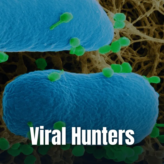
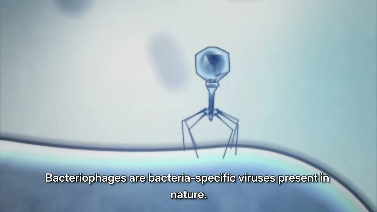
When curious observers ask what are bacteriophages, biologists define them as natural viruses that prey upon bacteria. These microscopic agents act as obligate intracellular parasites, multiplying only after penetrating and destroying a susceptible bacterial host. In marine surface waters, deep terrestrial soils, and human intestines, bacteriophages outnumber all other living organisms combined, shaping diverse microbial ecosystems across Earth through continuous viral predation. As science writer Melisse Lazarus notes, an estimated 10^31 bacteriophages populate the biosphere, far outnumbering global bacterial hosts [10]. Their precise host range lets them kill target pathogens without harming human cells [15].
What Are Bacteriophages?
The term bacteriophage means bacteria eater, an identity derived from the Greek root phagein to describe entities that devour microbial cells. Frederick Twort discovered them at the Brown Institution of London in 1915. Two years later, French-Canadian microbiologist Félix d’Hérelle at the Pasteur Institute in Paris independently isolated dysentery phages and realized they were viruses that attack bacteria. Félix d’Hérelle wrote: “In a flash I had understood: what caused my clear spots was in fact an invisible microbe… a virus parasitic on bacteria”. d’Hérelle introduced the concept of phage therapy in Paris in 1919 [15].
In Folia Microbiologica, researcher Sonika Sharma and colleagues note that bacteriophages are the most abundant viral entities on the planet [2]. In ocean surface waters, viral counts reach 9×10^8 virions per milliliter within dense microbial mats. Marine surveys show that bacteriophages infect up to 70% of marine prokaryotes, driving rapid nutrient turnover while recycling dissolved organic carbon back into marine food webs. Global phage counts exceed 10^31 virions [15].
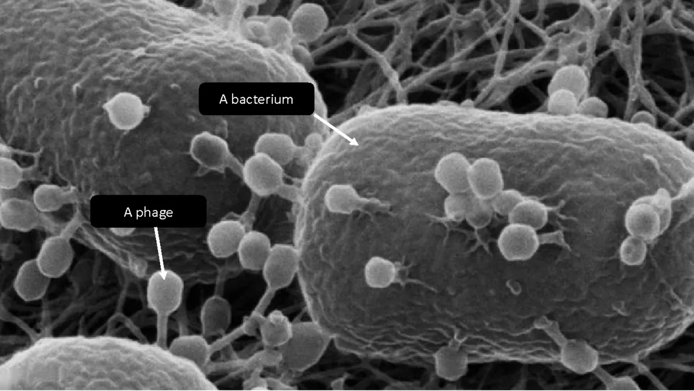
Why do bacteriophages target specific bacteria? Unlike broad chemical drugs that wipe out beneficial gut flora, each phage recognizes distinct docking molecules on host cell walls, with many phages killing only a single bacterial strain [1]. In News-Medical, Dr. Catherine Shaffer explains that phages bind surface markers like lipopolysaccharides, teichoic acids, or outer membrane proteins [6]. Because human tissues lack these bacterial receptors, bacteriophages cannot infect patient cells, allowing medical teams in the World Health Organization European Region to explore tailored clinical therapies [1].
Viral Anatomy
As microbiologists Sagar Aryal and Anupama Sapkota document, every phage consists of a nucleic acid genome packaged within a protective protein capsid shell [5]. Viral genomes range from four genes in simple viruses like MS2 to several hundred genes in complex tailed forms. The International Committee on Taxonomy of Viruses assigns tailed phages to the class Caudoviricetes, which includes podoviruses with short tails, myoviruses with contractile tails, and siphoviruses with long flexible tails [15]. Other families like Inoviridae form slender filaments [16].
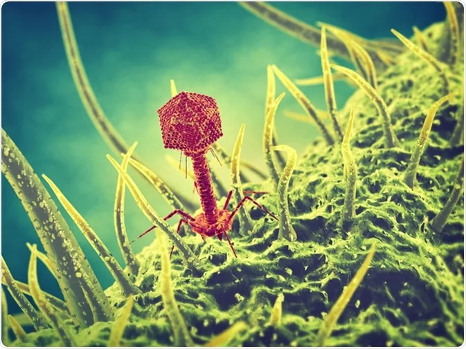
Escherichia virus T4, discovered by Max Delbrück and coworkers in 1944, provides a classic structural model of Caudovirales [5]. Its icosahedral head contains 2000 capsomeres enclosing linear double-stranded DNA [8]. Below the head, a short collar joins a contractile tail measuring 925 angstroms in length and 520 angstroms in diameter, wrapped in 24 annular rings. Six tail fibers extend from a hexameric baseplate to recognize outer surface receptors on Escherichia coli hosts [5].
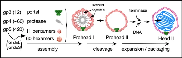
At the University of Pittsburgh, researcher Bob Duda demonstrated how bacteriophage HK97 capsids assemble through a defined multi-step pathway. The HK97 head builds from portal protein gp3 (47 kDa), protease gp4 (25 kDa), and major head protein gp5 (42 kDa), assisted by host chaperonins GroEL and GroES to form the precursor shell Prohead I. Protease gp4 cleaves gp5 from 42 kDa to 31 kDa, removing an internal delta scaffold domain to yield Prohead II before protein cross-linking creates a chain-mail capsid [4, 17]. In contrast, filamentous phages in the genus Inovirus, such as Ff phages fd, f1, and M13, assemble thousands of alpha-helical gene 8 coat proteins around circular single-stranded DNA, using membrane protein p1 to direct viral extrusion across host membranes without cell lysis [16].
The Lytic Infection Cycle
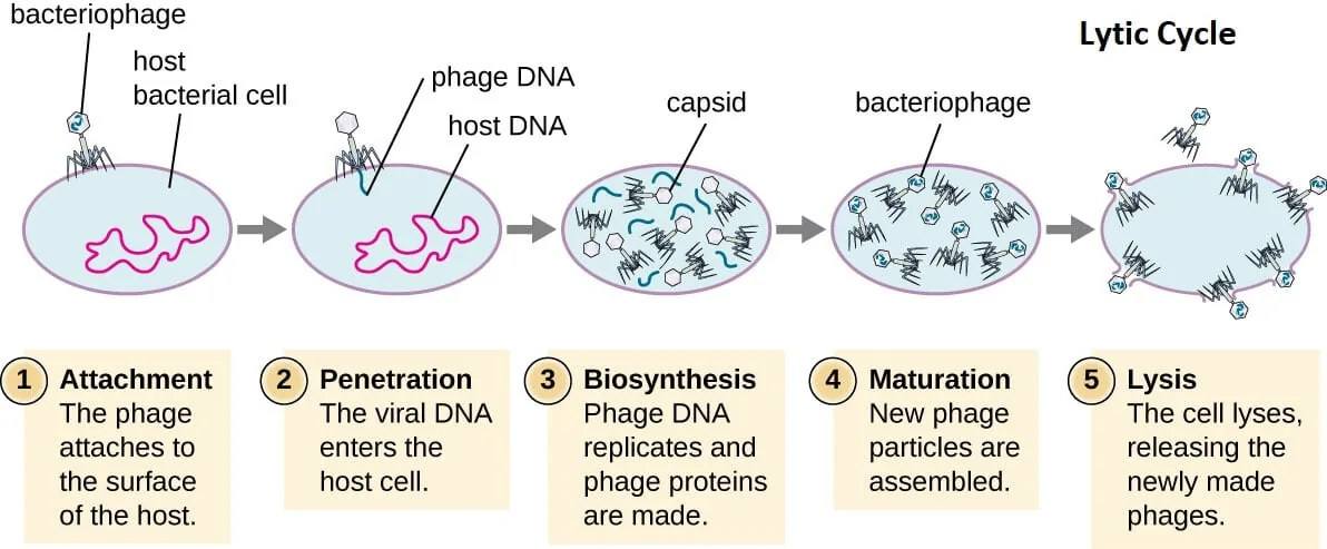
Virulent bacteriophages reproduce through the lytic cycle, a destructive pathway that terminates in host cell lysis [5]. Infection begins when tail fibers adsorb to host receptors such as outer membrane proteins, lipopolysaccharides, or cell wall teichoic acids [6, 8]. Once baseplate pins anchor against the bacterial surface, the phage discharges muramidase enzymes to weaken peptidoglycan bonds and drives a hollow core tube through the envelope to inject viral DNA into the cytoplasm [8]. The protein capsid stays outside [13].
Injected viral DNA seizes bacterial biosynthetic systems, stopping host transcription while recruiting host RNA polymerase to transcribe viral genes and replicate phage genomes at high speed [6]. Bacterial ribosomes translate early transcripts into replication enzymes and structural components, including capsid capsomeres, portal rings, and baseplate complexes [8]. In standard assays with coliphage T4, this latent period lasts about 20 to 25 minutes. During this window, the host bacterium operates as a viral factory, producing roughly 100 to 200 mature viral progeny that assemble piece by piece inside the cell [6, 12, 13].
Lysis completes the reproductive cycle when bacteriophages release specialized enzymes, including holins and endolysins, to breach the host envelope [5, 10]. Holins insert into the inner cell membrane to form pores, allowing endolysins to access and hydrolyze the outer peptidoglycan layer. Unable to withstand internal turgor pressure, the bacterial cell bursts in an explosive event that liberates hundreds of new infectious virions into the surrounding medium [5, 10]. These free virions immediately seek fresh bacterial hosts [10].
How Phages Establish Lysogeny
Temperate bacteriophages can choose an alternative developmental path known as the lysogenic cycle. Coliphage lambda, discovered in 1951 by Esther Lederberg in irradiated Escherichia coli cultures, serves as the premier molecular model for lysogeny [5]. As microbiologist Tankeshwar Acharya explains in Microbe Online, regulatory switches in lambda evaluate host physiology to decide between lytic development and dormant lysogeny. The viral protein CII helps initiate transcription of the CI repressor gene, producing CI proteins that silence lytic promoters and direct viral integration into the bacterial chromosome as a stable prophage. Integration creates a lysogenic bacterium [13].
An integrated prophage replicates passively alongside the bacterial chromosome during normal cell division, transmitting viral DNA to daughter cells without causing harm [5]. Continuous production of the CI repressor protein keeps the integrated prophage quiescent. In addition, free cytoplasmic CI repressor molecules bind incoming lambda genomes, conferring superinfection immunity that protects the lysogenized bacterial host from subsequent lethal attacks by matching phages. This immunity is strain-specific [13].
Environmental stress or ultraviolet radiation triggers the bacterial SOS response, ending prophage dormancy. Activated host RecA proteins stimulate autocatalytic cleavage of the CI repressor, relieving repression on the Cro promoter and prompting the prophage to excise and enter the lytic cycle. Prophages can also transfer novel virulence traits through lysogenic conversion. For instance, Corynebacterium diphtheriae produces diphtheria toxin only when carrying a tox-bearing prophage, while Shiga toxins in Escherichia coli and cholera toxins in Vibrio cholerae also originate from integrated phage genomes [13].

Bacterial Immune Defenses
Bacteria maintain diverse defense systems to counter persistent viral predation. Restriction-modification systems deploy restriction endonucleases that recognize and cut foreign viral DNA lacking host methylation marks. To prevent enzymatic digestion, bacteriophages like T4 substitute cytosine with glucosyl-hydroxymethylcytosine, shielding viral DNA from cleavage. Host bacteria also deploy adaptive immunity via CRISPR Cas9 genetic scissors, storing phage spacer sequences to guide Cas nuclease complexes against matching viral genomes. However, researchers note that covalent DNA modifications in T4 can block cleavage by wild-type Cas9 nucleases, requiring heavily engineered Cas9 variants to overcome these chemical shields. Phages and hosts co-evolve continuously [2].
In a 2026 report in Nature, Daniel S. Saxton, Michael T. Laub, and colleagues characterized SNIPE, a membrane-bound defense nuclease in Escherichia coli. Short for surface-associated nuclease inhibiting phage entry, SNIPE constitutively localizes to the bacterial inner membrane to protect against phage lambda and diverse siphoviruses. The protein features an amino-terminal transmembrane anchor, a central DUF4041 DNA-binding domain, and a carboxy-terminal GIY-YIG nuclease domain. Mutating catalytic residue E414A completely abolishes its antiviral activity [3].
Using radiolabeled 32P-DNA and fluorescent CFP-ParB tracking, the researchers showed that SNIPE associates with host entry proteins and the viral tape measure protein to cleave incoming phage DNA during injection. Membrane tethering keeps SNIPE auto-inhibited against host chromosomes, preventing autoimmune damage. Along with membrane nucleases, bacterial cells rely on enzymatic repair systems like photolyase repair of damaged DNA when environmental factors induce structural lesions in microbial chromosomes. These combined systems limit viral infection [3].
Historical Milestones in Research
In 1896, British chemist Ernest Hanbury Hankin documented that water from the Ganges and Yamuna rivers in India passed through fine porcelain Chamberland filters to kill cultures of Vibrio cholerae. In 1915, British pathologist Frederick Twort at the Brown Institution of London noticed glassy clearing in micrococcus colonies, proposing a filterable virus caused the destruction. Two years later in Paris, Félix d’Hérelle isolated dysentery-fighting viruses, coining the term bacteriophage and conducting the first human therapeutic trials in 1919. D’Hérelle treated dysentery patients successfully in France [15].
During the 1920s and 1930s, clinicians in France, Brazil, and the Soviet Union embraced bacteriophages for clinical treatments. In Georgia, Georgian researcher Giorgi Eliava worked closely with d’Hérelle to establish specialized institutes that supplied phage cocktails to Soviet soldiers [15]. In 1923, American firm Eli Lilly and Company began manufacturing biological phage products for commercial distribution. However, the discovery of penicillin in 1928, followed by industrial mass production of chemical antibiotics in the 1940s, led Western clinics to abandon phage therapy in favor of broad-spectrum pharmaceuticals [12, 15].
In 1944, Max Delbrück and coworkers organized the Phage Group, establishing T-even coliphages as standardized models to explore viral replication mechanisms [5]. In 1952, Alfred Hershey and Martha Chase used radiolabeled 32P-DNA and 35S-protein in phage T2 to demonstrate that nucleic acids, rather than proteins, carry hereditary information. Max Delbrück, Alfred Hershey, and Salvador Luria shared the 1969 Nobel Prize in Physiology or Medicine for these foundational discoveries. In 2018, George Smith and Gregory Winter won the Nobel Prize in Chemistry for phage display technology, using filamentous phages to evolve peptides and therapeutic antibodies. The 1969 and 2018 Nobel awards underscore the lasting value of phage research [15].
Bacteriophages in Modern Medicine
The worldwide rise of antimicrobial resistance has rekindled medical interest in phage therapy. As Vaishnavi Mishra and Nandkishor Bankar report in the Journal of Pure and Applied Microbiology, overusing antibiotics in humans and livestock has created dangerous multidrug-resistant pathogens. Health agencies warn that half of current antibiotics could lose clinical efficacy within years against threats like methicillin-resistant Staphylococcus aureus (MRSA), Pseudomonas aeruginosa, and extended-spectrum beta-lactamase producing Enterobacteriaceae [12]. To address this crisis, the World Health Organization coordinates with the Global AMR R&D Hub in Berlin to evaluate therapeutic phages as targeted alternatives [1].
Bacteriophages possess unique clinical traits that set them apart from traditional chemical drugs. They exhibit auto-dosing properties, multiplying specifically at infection sites where target bacteria concentrate and declining naturally once host pathogens are eliminated [12]. Phages also produce matrix-degrading enzymes that dissolve dense bacterial biofilms, clearing dormant persister cells that survive conventional antibiotic therapy [6]. In San Diego, California, physicians successfully treated a 68-year-old diabetic patient suffering from necrotizing pancreatitis caused by multidrug-resistant Acinetobacter baumannii using intravenous and percutaneous phage cocktails after three months of failed antibiotic treatment [11, 15]. Dr. Noreen Iftikhar highlights that phages provide targeted weapons against such untreatable superbugs [11].
Clinical application requires strict safety screening, as phages must infect the patient’s specific bacterial isolate without carrying toxin genes or mediating generalized transduction. Crude phage lysates must also undergo rigorous purification to remove bacterial endotoxins that could trigger dangerous inflammatory cytokine release or elevated tumor necrosis factor alpha in septic patients [6, 1]. In the United States and Western Europe, bacteriophages are not yet approved for routine prescription, remaining restricted to compassionate use protocols and investigational clinical trials [1]. The FDA approved early clinical trials in 2006 and 2019 [12].
Agricultural and Environmental Roles
Food safety agencies have approved biological biocontrol agents using bacteriophages to eliminate foodborne pathogens without altering the nutritional quality, flavor, or texture of commercial foods [10, 15]. In 2006, the United States Food and Drug Administration (FDA) and the USDA approved LMP-102, manufactured by Intralytix, to treat ready-to-eat poultry and meat products against contamination. That same year, the FDA approved LISTEX, developed by Micreos, to eliminate Listeria monocytogenes on cheese products. In 2011, the USDA recognized LISTEX as a clean label processing aid across all food products. These preparations target Salmonella, Listeria, and Escherichia coli [15].
Agricultural growers use phage formulations to control destructive crop diseases without accumulating toxic copper sprays or chemical residues in the environment. Phage sprays protect apple and pear orchards against Erwinia amylovora, the bacterium responsible for fire blight. Similar treatments target Xanthomonas and Erwinia on tomato, pepper, and potato crops, reducing bacterial spot and soft rot. In poultry and livestock farming, phages reduce intestinal colonization by Salmonella and Campylobacter, lowering disease transmission from farm settings to consumers. Phages thus offer sustainable agricultural biocontrol [1].
In marine and freshwater ecosystems, bacteriophages drive crucial planetary biogeochemical cycles. Marine viruses infect and lyse approximately 20% of microbial biomass daily, converting particulate cellular carbon into dissolved organic matter. This viral shunt accelerates nutrient cycling and supports planktonic food webs across global oceans [14]. Environmental bacteriophages also assist municipal wastewater treatment and hospital surface sanitization, supporting One Health strategies coordinated by the World Health Organization to limit the spread of drug-resistant microbes [1].
- REPORT World Health Organization. (2025). Bacteriophages and their use in combating antimicrobial resistance. World Health Organization Regional Office for Europe. [Article Link]
- ACADEMIC JOURNAL Sharma, S., Chatterjee, S., Datta, S., Prasad, R., Dubey, D., Kumar, R., & Vairale, M. G. (2016). Bacteriophages and its applications: an overview. Folia Microbiologica, 62(1), 17–55. [Article Link]
- ACADEMIC JOURNAL Saxton, D. S., DeWeirdt, P. C., Doering, C. R., Roney, I. J., & Laub, M. T. (2026). A membrane-bound nuclease directly cleaves phage DNA during genome injection. Nature, 627, 412–419. [Article Link]
- WEBSITE Duda, R. (n.d.). Bob Duda’s bacteriophage research. Department of Biological Sciences, University of Pittsburgh. [Article Link]
- WEBSITE Aryal, S., & Sapkota, A. (2022). Bacteriophage: Definition, Structure, Life Cycles, Applications, Phage Therapy. Microbe Notes. [Article Link]
- ONLINE NEWS Shaffer, C. (2018). What are Bacteriophages? News-Medical Life Sciences. [Article Link]
- WEBSITE Vajiram Content Team. (2025). What is Bacteriophage? Vajiram & Ravi. [Article Link]
- WEBSITE BYJU’S. (2019). Bacteriophage. BYJU’S Biology Learning. [Article Link]
- WEBSITE PrecisionPhage. (2025). What are Bacteriophages? PrecisionPhage Insights. [Article Link]
- WEBSITE Lazarus, M. (2025). 37 Facts About Bacteriophages. Facts.net Earth and Life Science. [Article Link]
- ONLINE NEWS Iftikhar, N. (2019). Phage Therapy: How It Works, Pros and Cons, Availability, and More. Healthline Media. [Article Link]
- ACADEMIC JOURNAL Mishra, V., Bankar, N., Tiwade, Y., & Ugemuge, S. (2024). How Phage Therapy Works, Its Advantages and Disadvantages: Mini Review. Journal of Pure and Applied Microbiology, 18(1), 58–69. [Article Link]
- WEBSITE Acharya, T. (2020). Bacteriophage: Structure, Replication (Lytic and Lysogenic), and Uses. Microbe Online. [Article Link]
- BOOK Mc Grath, S., & van Sinderen, D. (Eds.). (2007). Bacteriophage: Genetics and Molecular Biology. Caister Academic Press. [Article Link]
- WEBSITE Wikipedia contributors. (n.d.). Bacteriophage. Wikipedia, The Free Encyclopedia. [Article Link]
- WEBSITE Wikipedia contributors. (n.d.). Filamentous bacteriophage. Wikipedia, The Free Encyclopedia. [Article Link]
- WEBSITE Wikipedia contributors. (n.d.). Bacteriophage HK97. Wikipedia, The Free Encyclopedia. [Article Link]


