





Asking what are endorphins leads to the body’s built-in system for managing pain and body stress. Endorphins are natural opioid peptides produced mainly in the pituitary gland and hypothalamus that act as pain relievers throughout the human nervous system to maintain physiological balance. When tissues experience sudden injury or hard exertion, specialized cells release these neuropeptides to bind with opioid receptors and suppress discomfort across sensitive pathways. Such actions keep people functioning during demanding tasks and help restore chemical balance after stress [7].
What Are Endorphins?
The term endorphin comes from a blend of the Greek word endon, which means within, and morphine, named after Morpheus, the ancient Greek god of dreams [11]. In biological terms, it describes a natural pain reliever that mimics the pain-blunting effects of opiate medications without coming from an outside plant or synthetic compound [7]. Research into these natural opioids began in earnest during the early 1970s, as pharmacologists sought pain-relieving agents that lacked the addiction hazards and overdose dangers of traditional opium drugs. Scientists wanted to identify the specific compounds inside animal tissue that naturally docked into newly discovered opioid receptors to ease human suffering without producing destructive chemical dependency [11].
In 1973, researchers John Hughes and Hans Kosterlitz at the University of Aberdeen isolated enkephalin peptides from pig brain tissue. Soon afterward, Rabi Simantov and Solomon H. Snyder extracted similar peptides from calf brains. Pharmacologist Eric J. Simon coined the broad name endorphins to classify these natural opiate-like compounds [11].
Work continued rapidly as biochemists purified larger peptide fragments from pituitary tissue. In 1976, Choh Hao Li and David Chung sequenced camel pituitary endorphins, establishing that beta-endorphin has potent pain-relieving properties [11]. Wilhelm Feldberg and Derek George Smyth verified these findings in 1977, showing that beta-endorphin produces stronger pain relief than morphine and that naloxone cleanly reverses its calming effects. More than 20 separate endorphin forms exist within human biology, yet beta-endorphin is the main form studied by Cleveland Clinic specialists for pain control [7].
Molecular Structure of Opioid Peptides
All endorphins originate from a large precursor protein named proopiomelanocortin, commonly called POMC [1]. Inside the trans-Golgi network of pituitary cells, POMC attaches to carboxypeptidase E, which steers the prohormone into budding cellular vesicles. Cellular enzymes known as prohormone convertases then cut POMC into smaller active chains. Proprotein convertase 1 cleaves POMC to yield adrenocorticotropic hormone, known as ACTH, and beta-lipotropin, while proprotein convertase 2 trims beta-lipotropin into beta-endorphin and gamma-lipotropin [11].
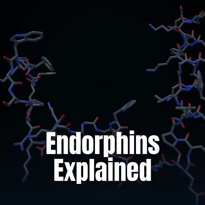
The complete human beta-endorphin molecule consists of a single polypeptide chain containing 31 amino acids [9]. Its primary chemical sequence begins with the conserved opioid pentapeptide motif Tyr-Gly-Gly-Phe-Met at the N-terminus and concludes with glutamic acid at the C-terminus [10]. The EMBL-EBI database catalogs it under CHEBI:10415, while the KEGG COMPOUND database records human entry C02210 for Homo sapiens with an average molecular mass of 3463.072 daltons [9], [10]. The 31-amino-acid chain forms an alpha-helical structure that resists quick breakdown by body enzymes, giving it a longer half-life than smaller enkephalins that circulate in nerve synapses. Because beta-endorphin keeps this durable structure, it can travel through biological fluids over longer paths and reach distant cell targets far better than shorter peptides that break down in seconds [12].
Body enzymes can split beta-endorphin into smaller peptide pieces. Cleaving the bond between threonine-16 and leucine-17 creates alpha-endorphin, while cleavage between leucine-17 and phenylalanine-18 yields gamma-endorphin. Early animal studies suggest alpha-endorphin acts somewhat like a mild psychostimulant, whereas gamma-endorphin displays actions comparable to neuroleptic drugs [11].

How Opioid Receptors Block Pain
Beta-endorphin relieves bodily distress by acting as a high-affinity agonist at mu-opioid receptors [1]. Opioid receptors belong to the large family of G-protein coupled receptors embedded in neuronal cell membranes across the human body. When an endorphin docks on the binding pocket of a mu-opioid receptor, the coupled G-protein activates and splits into Galpha and Gbeta-gamma subunits [12]. In a 2020 review in the International Journal of Molecular Sciences, authors A. R. Pilozzi, C. Carro, and X. Huang showed that this signaling chain alters how target neurons handle incoming impulses [1].
The freed Gbeta-gamma subunit binds to the inner loop of voltage-dependent calcium channels, blocking calcium entry during nerve signals. Because transmitter release needs calcium ions, this block stops presynaptic nerve ends from sending chemical messages to neighboring cells. At the same time, G-protein subunits bind to the tail of inward-rectifying potassium channels, driving potassium ions out of the cell. Potassium outflow causes membrane hyperpolarization, shifting the membrane potential to a negative state that keeps the neuron from firing action potentials during periods of severe stress [12].
In outer nerves, membrane hyperpolarization halts the release of substance P, a peptide that carries pain messages to spinal cord neurons [11]. In the brain, beta-endorphin produces pain relief while guiding emotional states through a two-step relief path [6]. It binds to opioid receptors on inhibitory cells, shutting down the release of gamma-aminobutyric acid, known as GABA [11]. With GABA turned off, nearby dopamine cells freely release dopamine into brain reward paths, creating calm and comfort that accompany pain relief during difficult recovery phases [6], [7].
Neural Pathways in the Brain
In the central nervous system, endorphin molecules work through synaptic contacts and broader fluid paths termed volume transmission. In a 2015 study, researchers J. G. Veening and H. P. Barendregt showed that beta-endorphin moves through cerebrospinal fluid, drawing on computational state models from Alan Turing [3].
Hypothalamic neurons make beta-endorphin and send axons into pain-control areas of the brainstem and limbic network. High numbers of mu-opioid receptors sit in the periaqueductal gray, the locus coeruleus, the amygdala, and the rostral ventromedial medulla. When endorphins bind receptors in the periaqueductal gray, they trigger descending pathways that travel down into the dorsal horn of the spinal cord. Descending pathways intercept incoming pain messages from injured tissues before signals reach the conscious brain cortex, keeping perception manageable during sudden trauma [12].
Not every endorphin fragment in brain tissue produces the same outcome. While full-length beta-endorphin(1-31) provides strong pain relief, body enzymes can cleave its tail to produce shorter forms like beta-endorphin(1-27) and beta-endorphin(1-26). Lab tests show that beta-endorphin(1-27) acts as a competitive blocker at mu receptors, lowering the painkilling power of the full peptide in animal models. Chemical modification through N-terminal acetylation also turns off beta-endorphin, stopping it from binding opioid receptors. Such enzymatic changes show that brain tissue does not rely on a single static chemical, but actively balances a family of related peptides whose competing actions shape fine sensory and emotional responses across diverse brain circuits [12].

Why Exercise Triggers Endorphins
Aerobic exercise gives one of the clearest natural triggers for releasing beta-endorphin from the pituitary gland into the bloodstream [7], [11]. As muscle contractions rise and metabolic products build up, nerves register the workload as body stress. The hypothalamus responds by coordinating hormone signals, prompting pituitary cells to send out endorphins to soothe tired muscle fibers. Hormone secretion raises pain thresholds, helping runners and athletes keep going through intense effort without stopping from early muscular fatigue [7].
For decades, popular culture credited the pleasant buzz known as the runner’s high to endorphins alone [7],. Modern neuroscience presents a more nuanced view. Because large peptides face hurdles crossing the blood-brain barrier, pituitary beta-endorphin in the blood cannot easily enter brain mood centers. Clinical tests using opioid blockers show that athletes still feel post-run joy even when their endorphin receptors are chemically blocked by pharmacology drugs [11]. Researchers now know that smaller fat-soluble endocannabinoids, which pass easily through the blood-brain barrier, generate the brain buzz, while blood endorphins focus on soothing sore muscles and easing fatigue during hard workouts [7], [11].
Beyond solo running, coordinated group movement stimulates the opioid network in special ways. In a 2017 double-blind study in Adaptive Human Behavior and Physiology, researchers B. Tarr, J. Launay, C. Benson, and R. Dunbar tested strangers dancing together in synchrony. Half of the dancers took 100 mg of naltrexone, an opioid blocker, while the rest received a placebo before dancing. Dancers on naltrexone showed clear hyperalgesic drops in pain tolerance compared to placebo controls, proving that synchronized dance triggers natural endorphin release that raises human pain thresholds [2].
Skin Synthesis After Sunlight Exposure
Endorphin synthesis also happens outside the brain. In a 2014 study published in Cell, science writer Sue McGreevey reported on work from Harvard Medical School and Massachusetts General Hospital showing that skin makes beta-endorphin under ultraviolet rays. Led by David E. Fisher at Mass General, researchers studied why people seek sun despite skin cancer warnings [8].
To test this reaction, scientists exposed mice to daily ultraviolet light equal to 20 to 30 minutes of midday Florida sun for six weeks. Within one week, beta-endorphin in animal blood rose and stayed high through the trial. The treated mice grew less sensitive to touch and heat than unexposed control mice. When researchers gave naloxone to block opioid receptors, normal skin feeling returned, confirming that skin-made beta-endorphin raised pain thresholds [8].
Naloxone in sun-exposed mice also caused classic opioid withdrawal symptoms, like shaking and teeth chattering. Mice lacking the beta-endorphin gene or lacking POMC in their skin showed no such reactions after ultraviolet light. David E. Fisher said: “Our study identified an organic pathway encoded in skin whereby UV radiation causes the synthesis and release of beta-endorphin and produces opiate-like effects, including addictive behavior.”. Backed by National Institutes of Health grants R01-AR043369-16 and R01-CA150226-03 and the Melanoma Research Alliance, the research showed that sun-seeking habits stem from a natural biological drive driven by skin endorphins [8].

Clinical Research on Stress and Headaches
Neurologists have tested how blood endorphin levels connect to chronic headaches. In a 2013 trial in Cephalalgia, U. K. Misra, J. Kalita, G. M. Tripathi, and S. K. Bhoi tested plasma beta-endorphin in 25 migraine patients diagnosed under International Headache Society rules and 20 matched controls. Of the 25 patients, 19 were female, with a median age of 35 years. The authors tested whether pain relief from repetitive transcranial magnetic stimulation, known as rTMS, matched changes in opioid levels [4].
Each patient received three rTMS sessions of 600 pulses at 10 Hz over the right abductor digiti minimi motor spot. Blood tests showed clear gains that tracked drops in migraine frequency over one month, confirming that magnetic pulses help restore low opioid levels in headache sufferers [4].
Beyond headaches, endorphins shape immune defenses and tumor stress paths. In a 2012 review in Cancer Research, D. K. Sarkar, S. Murugan, C. Zhang, and N. I. Boyadjieva showed how hypothalamic beta-endorphin cells slow tumor growth. In lab rat models of breast and prostate cancer, placing beta-endorphin neurons in the hypothalamus calmed sympathetic nerves. This shift raised natural killer cell and macrophage actions while lowering inflammatory cytokines, proving that brain endorphins alter tumor environments by curbing chronic stress [5].
Everyday Habits That Support Endorphin Release
The body uses simple daily habits to keep up healthy endorphin levels. Hearty laughter offers an easy trigger, since belly movements during laughing create muscle tone that prompts the pituitary gland to send out calming peptides [7], [11]. Quiet meditation, breath routines, and massage also bring relaxation by prompting opioid release [7], [11]. Food choices help too; eating spicy dishes with capsaicin creates a mild burning feeling on the tongue, prompting the brain to release endorphins to cool the heat [7].
People often confuse endorphins with dopamine, but the two chemicals serve different roles in the body [6],. Endorphins act as natural painkillers to ease strain during hard tasks [7]. Dopamine acts as a reward chemical, reinforcing good habits by giving a sense of pleasure upon reaching goals. In Highlights in Science Engineering and Technology, researcher R. Zheng showed how endorphins and dopamine work together to support wellbeing across daily life. When endorphins bind brain receptors, they quiet inhibitory GABA cells, letting dopamine flow into reward centers to provide both physical relief and a lingering boost of satisfaction [6], [11].
When someone’s body has an endorphin shortage, low dopamine activity can cause aches, worry, and low mood [7]. While synthetic opioid drugs bind the same receptors, they carry high addiction risks by flooding cells with artificial doses [6], [7]. In contrast, natural endorphin release is safely controlled by body feedback loops that keep healthy balance [1],. Regular exercise, laughing with friends, and good stress habits provide safe ways to support these natural painkillers [7].
- ACADEMIC JOURNAL Pilozzi, A. R., Carro, C., & Huang, X. (2020). Roles of β-Endorphin in Stress, Behavior, Neuroinflammation, and Brain Energy Metabolism. International Journal of Molecular Sciences, 22(1), 338. [Article Link]
- ACADEMIC JOURNAL Tarr, B., Launay, J., Benson, C., & Dunbar, R. (2017). Naltrexone Blocks Endorphins Released when Dancing in Synchrony. Adaptive Human Behavior and Physiology, 3(3), 241-254. [Article Link]
- ACADEMIC JOURNAL Veening, J. G., & Barendregt, H. P. (2015). The effects of Beta-Endorphin: state change modification. Fluids and Barriers of the CNS, 12(1), 3. [Article Link]
- ACADEMIC JOURNAL Misra, U. K., Kalita, J., Tripathi, G. M., & Bhoi, S. K. (2013). Is β endorphin related to migraine headache and its relief?. Cephalalgia, 33(5), 316-322. [Article Link]
- ACADEMIC JOURNAL Sarkar, D. K., Murugan, S., Zhang, C., & Boyadjieva, N. I. (2012). Regulation of Cancer Progression by β-Endorphin Neuron. Cancer Research, 72(4), 836-840. [Article Link]
- ACADEMIC JOURNAL Zheng, R. (2022). Pleasure and Achievement: Dopamine and Endorphins. Highlights in Science Engineering and Technology, 6, 83-89. [Article Link]
- WEBSITE Cleveland Clinic. (2023, September 6). Endorphins: What They Are and How to Boost Them. Health Library. [Article Link]
- WEBSITE McGreevey, S. (2014, June 19). Addicted to the Sun. Harvard Medical School. [Article Link]
- WEBSITE European Bioinformatics Institute. (2012, May 29). Beta-Endorphin (CHEBI:10415). Chemical Entities of Biological Interest. [Article Link]
- WEBSITE Kyoto Encyclopedia of Genes and Genomes. (2009, January 1). KEGG COMPOUND: C02210 beta-Endorphin. GenomeNet. [Article Link]
- WEBSITE Wikipedia contributors. (2026). Endorphins. Wikipedia, The Free Encyclopedia. [Article Link]
- WEBSITE Wikipedia contributors. (2026). Beta-Endorphin. Wikipedia, The Free Encyclopedia. [Article Link]
Cover image: Mplanine / Wikimedia Commons, CC BY-SA 4.0


