






Curious observers investigating unusual brain ailments often study the prion protein to understand how lethal particles spread without genetic code. Prions are toxic agents formed from an abnormally folded host protein that lacks any DNA or RNA genome [14]. Unlike bacteria or viruses, these agents multiply by forcing normal cellular units to take up their altered shape. The resulting protein clumps leave microscopic holes throughout brain tissue, triggering fatal neurological collapse [6].
What Is a Prion Protein?
Medical records indicate that fatal spongiform illnesses plagued European sheep herds long before modern lab teams isolated the causative agent. In 18th-century Europe, British shepherds documented scrapie as an incurable disorder driving sheep to scrape their fleeces against fences until they collapsed from exhaustion [12]. During the 1950s, medical teams observed kuru killing Fore tribespeople in Papua New Guinea [15].
Fore funeral customs involved preparing and eating tissue from deceased kin, which inadvertently transmitted the fatal agent across tribal villages. In the 1960s, American physician Carleton Gajdusek proved that brain extracts from deceased kuru patients transmitted spongiform disease into captive chimpanzees at the National Institutes of Health [9, 15]. The agent resisted ultraviolet rays and boiling water, confounding researchers who expected standard viral genomes [9, 12]. In 1967, British theorist J. S. Griffith suggested that an infectious agent could consist of an altered protein shape capable of converting normal counterparts [12]. American neurologist Stanley B. Prusiner isolated the scrapie agent at the University of California, San Francisco, confirming that it contained protein without nucleic acid [9]. Prusiner won the 1997 Nobel Prize in Physiology or Medicine for establishing the protein-only model [15].
Prusiner created the name prion in 1982 by joining parts of the phrase “proteinaceous infectious particle” [9, 15]. Skeptical virologists initially favored the virino model, suspecting a tiny nucleic acid core lay hidden inside [9]. Genetic tests in mice later confirmed that host protein alone sustains disease spread [14].
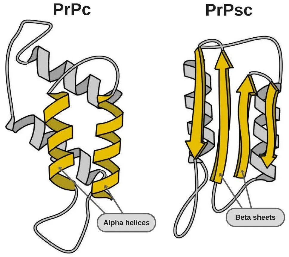
Molecular Architecture of PrP Isoforms
The seed of this neurodegenerative process is a normal cell-surface molecule known as cellular prion protein, or PrPC. In human cells, the PRNP gene on chromosome 20 directs the synthesis of a precursor chain spanning 253 amino acids [13, 15]. Cellular enzymes trim both ends, yielding a mature glycoprotein of 208 amino acids anchored to the outer cell membrane by a glycosylphosphatidylinositol anchor at serine residue 231. Structural data show that native PrPC features a loose amino-terminal tail and a stable carboxyl-terminal globular fold with three alpha helices and two small beta strands. A single disulfide bond links cysteine residues 179 and 214 to stabilize the normal shape [14, 15].
The lethal scrapie form, termed PrPSc, carries the exact same amino acid sequence as healthy PrPC. Protein folding accounts for the change. In 1993, K. M. Pan and colleagues published structural data in the Proceedings of the National Academy of Sciences. Infrared scans revealed that beta-sheet structure jumped from 3 percent in PrPC to 43 percent in PrPSc [1].
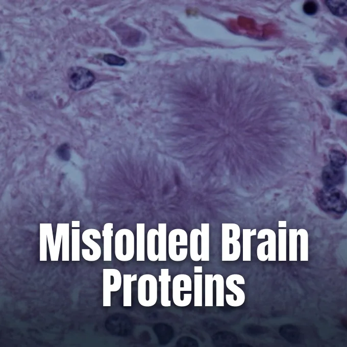
That structural flip turns flexible helical coils into flat beta sheets that stack into insoluble amyloid fibers [13, 14]. While normal PrPC dissolves in mild detergents and breaks down under proteinase K digestion, PrPSc resists proteases and clusters into dense brain plaques that damage surrounding neurons [14]. Measurements from circular dichroism and nuclear magnetic resonance indicate that the free-energy gap between both shapes is only 1 to 2 kcal mol-1 [13]. That small gap lets normal host units refold whenever they bump into an abnormal template. Readers tracing microscopic tissue damage can review how abnormal prions destroy brain cells during advanced neurodegenerative illness.
How Misfolded Chains Replicate Without Genes
Prion multiplication skips genetic copying and ribosomal translation entirely. Instead, replication follows template-directed conversion, in which a PrPSc aggregate binds a normal PrPC monomer and forces it to fold into the scrapie shape. The newly altered protein joins the growing fiber [14]. Each addition extends the amyloid chain and exposes fresh binding sites for nearby host proteins [11].
Early investigators debated whether replication relied on simple monomer pairs or larger aggregates. German biophysicist Manfred Eigen examined the reaction kinetics and showed that single-molecule conversion would need an impossible catalytic boost of 10 to the 15th power. Physical tests confirmed that active prions exist as ordered oligomers and elongated fibrils. As healthy PrPC units join the fibril tips, fluid shear forces inside lysosomes and endocytic vesicles snap the long amyloid fibers into smaller mobile fragments [14, 15]. Each fragment acts as an independent catalytic seed, producing exponential replication rates that scale directly with the square root of cellular PrPC concentration [15].
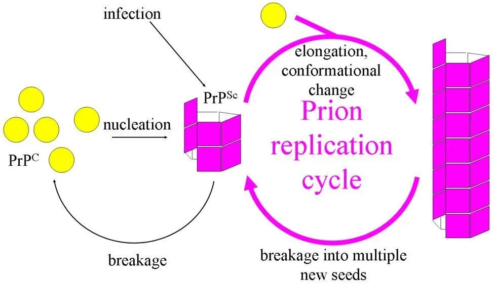
Host cell compartments tightly guide this replication cascade. Normal PrPC must reach cholesterol-rich lipid rafts on the plasma membrane before conversion can start. Depleting membrane cholesterol with filipin stops prion production in cell cultures. Surface receptors such as the LRP/LR laminin receptor and heparan sulfate proteoglycans help cells take up external prion seeds [11].
Normal Roles in Brain Function
Because PrPC appears across all mammal groups, biologists investigated its normal jobs in healthy tissue. Primate PrP sequences share between 92.9 percent and 99.6 percent amino acid identity, reflecting deep evolutionary roots. The amino-terminal segment holds five octapeptide repeats with the code PHGGGWGQ, which binds copper ions through histidine imidazole rings. High-resolution spectroscopic tests indicate that copper binding shifts the protein shape and helps shield vulnerable brain cells from heavy metal toxicity and free radical damage. PrPC also operates at synaptic junctions, where it supports cell adhesion and balances intracellular calcium levels during repetitive neural firing [14, 15].
Early gene knockout tests in mice gave unexpected clues. Rodents bred without the PRNP gene grew up looking healthy [9, 15]. But fine electrical recordings showed impaired hippocampal long-term potentiation, linking PrPC to synaptic memory formation. In peripheral nerves, regular enzymatic cleavage of PrPC tells Schwann cells to keep protective myelin sheaths intact around long axons [15].
Scientists eventually realized that related proteins back up PrPC. In 2009, developmental biologists discovered Shadoo, a mammal protein that carries out overlapping duties during embryo growth. Deleting either PrP or Shadoo yields living mice, but deleting both genes together kills the developing embryo [14]. PrPC also helps regulate circadian sleep cycles and blood stem cell health. Tests at the Whitehead Institute for Biomedical Research showed that long-term blood stem cells display PrPC on their membranes, and cells lacking the protein fail to sustain proper bone marrow renewal after transplantation [15]. That broad utility explains why PrPC remains active across mammalian biology [14].

Diseases Across Mammalian Hosts
Human transmissible spongiform encephalopathies divide into three clinical categories: sporadic, inherited, and acquired. The most common form is sporadic Creutzfeldt-Jakob disease, which makes up about 85 percent of all human diagnoses [2, 13]. Global clinics find 1 to 2 cases per million people each year, with symptoms usually starting between ages 55 and 75 [13].
Inherited disorders arise from more than 50 distinct PRNP gene variants passed down through autosomal dominant genetics [2, 13]. Fatal familial insomnia stems from an aspartic acid to asparagine swap at codon 178 alongside methionine at codon 129, wrecking thalamic sleep centers and causing intractable insomnia. Gerstmann-Sträussler-Scheinker disease causes unsteady walking and limb stiffness before memory loss sets in [2, 13]. Acquired cases include kuru from mortuary cannibalism, and iatrogenic CJD passed by neurosurgical tools or contaminated growth hormone batches prepared from 5,000 to 20,000 human pituitary glands before synthetic hormone debuted in 1987 [13]. All human prion diseases are uniformly fatal [6].
Animal herds face their own spongiform encephalopathies. Bovine spongiform encephalopathy struck British cattle in the 1980s and 1990s through meat-and-bone feed, eventually infecting consumers as fatal variant CJD across the United Kingdom. Today, chronic wasting disease spreads among deer, elk, and moose across 32 American states, Canada, and Scandinavia [6, 11]. The pathogen passes easily through saliva, urine, and contaminated range soil [8].

Are Prions Alive?
Can a tiny protein particle spread and cause fatal illness without being alive? Biologists classify prions as non-living biomolecules rather than living organisms. Unlike bacteria, fungi, or protozoa, prions lack cell membranes, cytoplasm, ribosomes, and metabolic energy production. Viruses carry genetic programs wrapped in protein coats, whereas viroids consist only of small circular RNA strands [12, 14]. Prions contain no nucleic acids whatsoever. They cannot divide, feed, or maintain internal balance. Instead, they act as rogue physical molds that spread by corrupting the shape of existing host proteins. Standard antibiotics and antiviral drugs have zero effect against them [12].
This absence of DNA and RNA forces scientists to rethink basic genetics. In prions, information is stored within the three-dimensional geometry of protein aggregates [4, 12]. Distinct prion strains cause different incubation windows and brain damage patterns while sharing the exact same amino acid sequence [10, 14]. Structural twists inside the amyloid core carry these distinct traits [12].
Prion mechanisms also appear in beneficial fungal biology. In 1965, British geneticist Brian Cox found a non-Mendelian trait in the baker’s yeast Saccharomyces cerevisiae called [PSI+]. In 1994, yeast researcher Reed Wickner showed that [PSI+] and another trait called [URE3] are prion forms of the normal proteins Sup35p and Ure2p. Fungal prions depend on the chaperone Hsp104 to trim amyloid fibers and pass daughter seeds to budding cells. In the filamentous fungus Podospora anserina, the HET-s prion governs cell compatibility so that only related colonies merge and share cytoplasmic nutrients during vegetative growth. These fungal models show that self-templating protein shapes can grant survival advantages under harsh conditions [4, 16].
Resistance to Medical Decontamination
Prions rank among the toughest disease agents known to medicine. Methods that kill regular bacteria and viruses leave abnormal PrPSc intact [8, 12]. Freezing does not hurt prions [8]. Alcohols and standard hospital washes cannot break down the dense beta sheets [12]. Reliable destruction requires exposure to dry heat of 900 degrees Fahrenheit for several hours [8].
Surgical clinics follow rigorous containment steps to prevent accidental contamination. Hospital instruments touching brain or eye tissue undergo specialized cleaning that pairs harsh chemicals with intense heat. Infection control guidelines mandate soaking surgical tools thoroughly in strong sodium hydroxide or concentrated sodium hypochlorite solutions for extended periods followed by high-pressure steam autoclaving. In nature, shed particles bind tightly to clay minerals like montmorillonite in topsoil, where they resist microbial breakdown and remain infectious to wildlife for many years [8, 12]. Grazing deer consume these mineral-bound prions, keeping woodland outbreaks alive [8].

Detecting prions in living patients once seemed nearly impossible. Traditional checks needed postmortem brain tissue showing sponge-like holes and swollen glial cells [13, 14]. Today, physicians rely on the Real-Time Quaking-Induced Conversion (RT-QuIC) test, which shakes spinal fluid to amplify and detect tiny traces of misfolded PrPSc [2, 12].
Mechanisms Shared with Other Brain Disorders
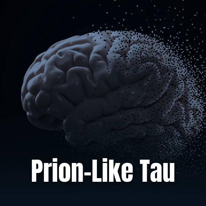
The discovery of protein-only spread has changed how neurologists study common brain illnesses. In a 2015 analysis published in the Annual Review of Neuroscience, researchers Lary Walker and colleagues showed that templated misfolding drives illnesses like Alzheimer’s, Parkinson’s, and Huntington’s. Because these conditions do not spread between people like infections, Walker suggested calling them “proteinaceous nucleating particles”. Under this molecular model, cellular proteins such as amyloid-beta, tau, and alpha-synuclein undergo initial misfolding events that seed further protein aggregation across interconnected neural circuits [3, 7]. This insight links many age-related dementias to a shared molecular pattern [3].
Direct laboratory confirmation came from Stanley Prusiner’s team in 2015. Neuroscientist Kurt Giles investigated multiple system atrophy, a movement illness resembling Parkinson’s. Transgenic mice injected with brain tissue from human patients developed fatal brain illness within 3.5 to 5 months, caused by self-propagating alpha-synuclein prions [10].
At the Paris Brain Institute, research directors Marie-Claude Potier and Stéphane Haïk track how amyloid plaques and tau tangles spread during Alzheimer’s disease. Pathological tau assemblies travel along nerve fibers to corrupt healthy proteins at neighboring synapses across interconnected brain networks [5]. Research models showing prion-like transmission of human tau in mice illustrate how seeded aggregates migrate through living neural tissue. By mapping the exact molecular steps of protein refolding, international lab teams hope to develop targeted therapies that block aggregate spread before extensive brain cell loss takes place.
- ACADEMIC JOURNAL Pan, K. M., Baldwin, M., Nguyen, J., Gasset, M., Serban, A., Groth, D., Mehlhorn, I., Huang, Z., Fletterick, R. J., & Cohen, F. E. (1993). Conversion of alpha-helices into beta-sheets features in the formation of the scrapie prion proteins. Proceedings of the National Academy of Sciences, 90(23), 10962-10966. [Article Link]
- WEBSITE Cleveland Clinic. (2024). Prion Disease: Causes, Symptoms, Diagnosis & Treatment. [Article Link]
- ACADEMIC JOURNAL Walker, L. C., & Jucker, M. (2015). Neurodegenerative Diseases: Expanding the Prion Concept. Annual Review of Neuroscience, 38(1), 87-103. [Article Link]
- ACADEMIC JOURNAL Halfmann, R., & Lindquist, S. (2010). Epigenetics in the Extreme: Prions and the Inheritance of Environmentally Acquired Traits. Science, 330(6004), 629-632. [Article Link]
- WEBSITE Paris Brain Institute. (2026). What are the Biological Mechanisms of Prion Diseases? [Article Link]
- REPORT Centers for Disease Control and Prevention. (2026). About Prion Diseases. U.S. Department of Health and Human Services. [Article Link]
- ACADEMIC JOURNAL Fraser, P. E. (2014). Prions and Prion-like Proteins. Journal of Biological Chemistry, 289(29), 19839-19840. [Article Link]
- REPORT Virginia Department of Wildlife Resources. (2024). What are Prions? [Article Link]
- ONLINE NEWS Lindquist, S., Rogers, M., & Heaphy, S. (1999). What Is a Prion? Scientific American. [Article Link]
- ONLINE NEWS Makin, S. (2015). A Red Flag for a Neurodegenerative Disease That May Be Transmissible. Scientific American. [Article Link]
- ONLINE NEWS Shetty, V. (2016). What Are Prions? ScienceABC. [Article Link]
- ONLINE NEWS Helmenstine, A. (2026). Prions – Definition, Structure, Diseases, and Facts. Science Notes and Projects. [Article Link]
- ONLINE NEWS Dha, N. (2021). What are Prions? News-Medical. [Article Link]
- WEBSITE Pan, S. (2024). Prions – Definition, Structure, Characteristics, Replication and Diseases. Biology Notes Online. [Article Link]
- WEBSITE Wikipedia contributors. (2026). Prion. Wikipedia, The Free Encyclopedia. [Article Link]
- WEBSITE Wikipedia contributors. (2026). Fungal prion. Wikipedia, The Free Encyclopedia. [Article Link]


