





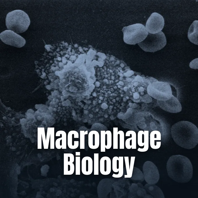
What is a macrophage? In the human body, a macrophage acts as a scavenger cell that detects, swallows, and breaks down worn cell parts, foreign debris, and invasive germs to protect nearby organs [1]. The cells arise from blood cells or emerge within embryonic tissues before birth, patrolling anatomical spaces through amoeboid motion and screening tissues for damage [2]. Beyond clearing debris, they alert local tissues to germ invasion by presenting antigens to helper T cells and sending chemical signals [1]. Without their vigilant activity, wounded organs fail to resolve swelling or heal after injury [4].
What Is a Macrophage?
At its core, the cell acts as a watchful guard inside living tissues. Nikita Verma and Swarnlata Saraf note that precursor cells leave the bloodstream to turn into resident macrophages in all vertebrates, measuring about 21 micrometres in diameter with CD14, CD68, and CD11b markers [2].
D. M. Mosser and co-authors describe these cells as body sensors, converting diverse mechanical, chemical, and metabolic inputs into distinct protective or restorative physical outcomes. In their review, they cite the definition that “A transducer is a device that converts energy from one form to another”. By sensing chemical inputs, the cells switch between defensive tasks and repair duties to sustain tissue stability [4].
Stationed groups take on local names throughout the body, such as Kupffer cells in the liver, alveolar cells in lung air sacs, and microglia in brain tissue. In the testes, the cells release 25-hydroxycholesterol to help nearby Leydig cells synthesize testosterone, building a protective zone that limits immune reactions in testicular tissue during health and disease. Cardiac cells connect straight to heart muscle myocytes through gap junctions, aiding steady electrical conduction across rhythmic cardiac tissue and regulating organ function during healthy resting states [2].
Discovery by Élie Metchnikoff
The formal identification of these eating guards dates back to 1884 in the Russian Empire. Zoologist Élie Metchnikoff observed mobile cells in starfish larvae gathering around inserted rose thorns, recognizing that internal cells could physically engulf foreign bodies to establish the foundation of cellular immunity [2]. He coined the term macrophage from Greek roots meaning large eater, later detailing these cellular actions in his 1905 book Immunity in the Infectious Diseases to prove that host defense relies on active cell ingestion [1]. His work founded cellular immunology as a scientific field [2].
Historically, anatomists grouped these widely distributed cells under the reticuloendothelial system, though medical texts now classify them as the mononuclear phagocyte system [2]. This framework links resident guards with blood precursors and dendritic cells [1]. The cells continuously guide host defense through dynamic chemical secretions rather than passive scavenging [4].
The word macrophage also appeared in early ecology literature with a completely separate biological meaning. In 1927, researchers Jordan and Hirsch used the labels macrophage and microphage to categorize distinct feeding strategies among heterotrophs. As cited by C. M. Yonge in 1928, an ecological macrophage “handles food items singly, while a microphage handles food items in bulk without manipulating them individually” during natural feeding behaviors in aquatic habitats. Although early ecology texts used the term into the twenty-first century, modern science reserves the name almost exclusively for white blood cells [5].
How Macrophages Clear Invading Pathogens
Phagocytosis is the main cellular method through which these white blood cells neutralize germ threats and clean up microscopic waste [1]. When an invading microbe or dying host cell enters tissue, the resident phagocyte extends cellular arms around the target until enclosing it within an internal vesicle called a phagosome to prepare for digestion. The newly formed vesicle fuses fast with lysosomes to create a digestive chamber known as a phagolysosome. Within this acidic compartment, potent enzymes and toxic reactive oxygen species break down proteins, lipids, and nucleic acids as the cell consumes high amounts of oxygen during the respiratory burst [2].
Target identification relies on surface pattern recognition sensors, including Toll-like receptors, scavenger sensors, and C-type lectins that recognize microbe wall components and initiate cellular engulfment. Phagocytic uptake rises when pathogens are coated with host opsonins like complement fragment C3b and immunoglobulin G antibodies. Complement docking sites CR1 and CR3 bind C3b, and Fc gamma proteins lock onto antibody tails [2].
Beyond germ invaders, the cells clear billions of aged apoptotic bodies through a silent disposal process called efferocytosis. When acute inflammation begins, short-lived neutrophils arrive first, expend their toxic granules, and undergo cell death within two days [2]. Monocytes and tissue cells enter the wound to ingest these dying neutrophils through efferocytosis, preventing toxic enzymes from spilling into nearby healthy matrix and triggering unwanted tissue damage [6]. Some clever pathogens, such as Mycobacterium tuberculosis and typhoidal Salmonella strains, resist phagosomal destruction to replicate inside host cells [2].
Macrophage Polarization and Tissue Repair
Rather than remaining locked in a single rigid behavioral state, these white blood cells display striking phenotypic plasticity across varying microenvironments [7]. Under classic immune models, microbe products like bacterial lipopolysaccharide and interferon-gamma activate cells toward the pro-inflammatory M1 phenotype to kill internal bacteria during acute infection [8]. These active cells produce nitric oxide by metabolizing arginine and secrete inflammatory mediators like tumor necrosis factor-alpha [2]. Their strong chemical output allows them to kill stubborn internal bacteria and eliminate malignant cells during the early acute stages of infection when pathogens invade host tissues [8].
In contrast, anti-inflammatory signals like interleukin-4 and interleukin-13 drive the alternative M2 phenotype, dedicated to resolving inflammation and encouraging wound healing [7]. Instead of synthesizing nitric oxide, active M2 cells metabolize arginine into ornithine, a chemical building block for collagen synthesis and tissue repair that supports clean wound healing after injury [2]. Polarization represents a fluid functional spectrum [8].
During wound recovery, timely transitions between these activation states determine whether an injured organ restores normal structure or develops disfiguring fibrotic scars that compromise long-term organ health. T. J. Koh and L. A. DiPietro point out that persistent inflammatory phenotypes stall healing in chronic ulcers and diabetic wounds [9]. Conversely, balanced shifts toward tissue-repair programs support blood vessel growth, fibroblast spread, and clean matrix deposition [6]. The cells coordinate this repair sequence by removing cellular debris and secreting growth factors that instruct neighboring structural cells. When regulation fails, prolonged swelling promotes pathological fibrosis and tissue scarring after severe injury [9].

Communication With Helper T Cells
As professional antigen-presenting cells, macrophages build a bridge connecting innate defenses with adaptive immunity [1]. After digesting a captured pathogen inside a phagolysosome, the cell processes microbe proteins into small peptide fragments. It displays these foreign peptides on major histocompatibility complex class II units positioned across its external plasma membrane [2].
Because tissue cells remain primarily within peripheral organs rather than migrating to T cell zones in lymph nodes, they interact mainly with previously active effector T cells or local memory cells. When pattern recognition sensors detect microbe structures in vivo, the cell activates the transcription factor NF-kappa-B to initiate broad immune defense. This trigger unleashes pro-inflammatory cytokines, including IL-1β, IL-6, and TNF-α. Released cytokines induce systemic fever, dilate local blood vessels, and upregulate adhesion units on vessel cells to recruit white blood cells from the bloodstream to the injury site [2]. CD4-positive helper T cells recognize displayed antigens, releasing activating cytokines that further stimulate local phagocytic destruction [1].
The cells release specific chemokines like CXCL8 to attract neutrophils, and produce CCL2, CCL5, and CXCL10 to draw incoming monocytes and lymphocytes toward the infection focus in infected tissues. Together with dendritic cells, they activate natural killer cells by sending IL-12, IL-18, and type I interferons. In modern medicine, understanding how spatiotemporal drug delivery directs natural killer cells shows how engineered therapeutics can complement innate immune signaling during therapy. Stimulated natural killer cells then release interferon-gamma, which feeds back onto local cells to boost their pathogen-killing capabilities [2].
Colony Stimulating Factors and Growth Signals
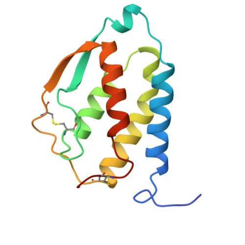
Survival and tissue growth of these cells rely on granulocyte-macrophage colony-stimulating factor, a cytokine secreted by T cells and fibroblasts. Known as GM-CSF or CSF2, it prompts bone marrow progenitors to produce white cells that migrate into tissues to mature, signaling through STAT5 and STAT3 transcription factors to boost defense [12].
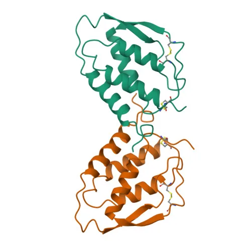
In 1992, M. R. Walter, W. J. Cook, and colleagues at the University of Alabama at Birmingham determined the crystal structure of rhGM-CSF at 2.8 angstrom resolution, reporting a compact four-helix bundle [10]. Later in 1996, D. A. Rozwarski and co-workers at Cornell University in Ithaca, New York refined the model to 2.4 angstroms in Proteins: Structure, Function, and Genetics, pinpointing critical residues on helix A and helix C. Their models revealed hydrophobic core clusters and buried hydrogen bonds that stabilize cytokine conformation [11].
Recombinant drugs soon emerged, with Genetics Institute making molgramostim, Immunex producing sargramostim, and Sandoz testing regramostim. The US FDA approved sargramostim under the brand Leukine in 1991 for bone marrow transplants, passing through Genzyme and Sanofi to Partner Therapeutics, and Amgen Inc gained approval for Imlygic in October 2015 [12].
Lessons From Salamander Regeneration
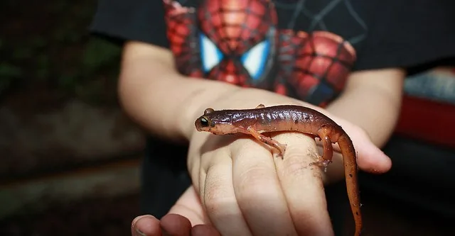
Human tissue repair often ends in fibrous scarring, but certain amphibians exhibit an astonishing capacity for complete structural regeneration. Salamanders can regrow complex body parts, including lost limbs, tails, spinal cord segments, retinas, and damaged heart muscle. Research teams at Monash University examined the role of immune cells in regenerative healing [3].
Dr. James Godwin and his colleagues at the Australian Regenerative Medicine Institute at Monash University examined whether macrophages drive scar-free regeneration in salamanders. When Godwin and his team selectively depleted macrophages from salamanders prior to limb amputation, the animals completely lost their ability to regenerate lost limbs and formed permanent scar tissue. Instead of forming a healthy blastema capable of rebuilding bone and muscle, the wounded stumps developed scar tissue reminiscent of adult mammalian wounds. Godwin believes these cells release chemical signals that suppress fibrosis and instruct surviving progenitor cells to rebuild complex tissues. In an interview with ABC News, Godwin remarked: “This really gives us somewhere to look for what might be secreted into the wound environment that allows for regeneration” [3].
The long-term objective of this comparative biology research is to uncover biochemical cocktails that stimulate human tissues to regenerate without permanent scar formation. Godwin explained to reporters: “The long-term plan is that we’ll know exactly what cocktail to add to a wound site to allow salamander-like regeneration under hospital conditions” during acute clinical treatment [3]. Research examining how broken bones heal and why cartilage repairs poorly illustrates the clinical challenge of guiding mammalian repair cells toward scarless tissue restoration. Finding the exact molecular signals secreted by salamander cells could pave the way for novel regenerative treatments in human orthopedic surgery [9].
Therapeutic Targets in Chronic Disease
Because of their central position in both driving and resolving inflammation, these phagocytes represent attractive targets for novel medical therapies [8]. In cardiovascular medicine, T. J. Barrett notes in Arteriosclerosis, Thrombosis, and Vascular Biology that plaque macrophages dictate the progression, stability, and regression of atherosclerosis in arterial beds. Classically active cells sustain vascular swelling and promote arterial plaque rupture, but alternative repair types clear necrotic lipid cores to foster lesion stabilization. Modulating cell types within arterial walls offers exciting clinical avenues to reduce heart attack and stroke risks. Treatments that shift plaque cells toward pro-resolving states could halt disease progression and promote vascular healing in vulnerable patients [13].
In oncology, solid tumors frequently recruit circulating monocytes and educate them into immunosuppressive tumor-associated macrophages. S. Chen and co-authors describe how these corrupted cells promote blood vessel growth and suppress cytotoxic T cells [8]. Investigating cellular cannibalism in cancer therapies reveals innovative strategies to reprogram these phagocytes into active antitumor fighters that eliminate resistant neoplastic growths.
Pharmacologists are also designing innovative targeted drug delivery platforms that leverage the natural phagocytic appetite of these defensive cells. By packing drug compounds inside biodegradable lipid carriers or engineered liposomes, doctors can deliver concentrated payloads directly into tissue cells to reduce harmful systemic toxicities [1]. This selective delivery approach enhances treatments for internal bacterial infections like tuberculosis, suppresses localized joint inflammation in rheumatoid arthritis, and reduces systemic toxicities [8]. Continued insights into cell signaling pathways ensure these ancient cellular defenders will remain at the forefront of modern therapeutic development [6].
- ACADEMIC JOURNAL Verma, N., & Saraf, S. (2017). A role of macrophages: An overview. Journal of Drug Delivery and Therapeutics, 7(6). [Article Link]
- WEBSITE Wikipedia contributors. (n.d.). Macrophage. Wikipedia, The Free Encyclopedia. [Article Link]
- ONLINE NEWS Souppouris, A. (2013). Scientists identify cell that could hold the secret to limb regeneration. The Verge. [Article Link]
- ACADEMIC JOURNAL Mosser, D. M., Hamidzadeh, K., & Gonçalves, R. (2020). Macrophages and the maintenance of homeostasis. Cellular & Molecular Immunology, 18(3), 579-587. [Article Link]
- WEBSITE Wikipedia contributors. (n.d.). Macrophage (ecology). Wikipedia, The Free Encyclopedia. [Article Link]
- ACADEMIC JOURNAL Watanabe, S., Alexander, M. J., Misharin, A. V., & Budinger, G. R. S. (2019). The role of macrophages in the resolution of inflammation. Journal of Clinical Investigation, 129(7), 2619-2628. [Article Link]
- ACADEMIC JOURNAL Murray, P. J. (2016). Macrophage polarization. Annual Review of Physiology, 79(1), 541-566. [Article Link]
- ACADEMIC JOURNAL Chen, S., Saeed, A. F. U. H., Liu, Q., Jiang, Q., Xu, H., Xiao, G. G., Rao, L., & Duo, Y. (2023). Macrophages in immunoregulation and therapeutics. Signal Transduction and Targeted Therapy, 8(1), 207. [Article Link]
- ACADEMIC JOURNAL Koh, T. J., & DiPietro, L. A. (2011). Inflammation and wound healing: The role of the macrophage. Expert Reviews in Molecular Medicine, 13, e23. [Article Link]
- ACADEMIC JOURNAL Walter, M. R., Cook, W. J., Ealick, S. E., Nagabhushan, T. L., Trotta, P. P., & Bugg, C. E. (1992). Three-dimensional structure of recombinant human granulocyte-macrophage colony-stimulating factor. Journal of Molecular Biology, 224(4), 1075-1085. [Article Link]
- ACADEMIC JOURNAL Rozwarski, D. A., Diederichs, K., Hecht, R., Boone, T., & Karplus, P. A. (1996). Refined crystal structure and mutagenesis of human granulocyte-macrophage colony-stimulating factor. Proteins: Structure, Function, and Genetics, 26(3), 304-313. [Article Link]
- WEBSITE Wikipedia contributors. (n.d.). Granulocyte-macrophage colony-stimulating factor. Wikipedia, The Free Encyclopedia. [Article Link]
- ACADEMIC JOURNAL Barrett, T. J. (2019). Macrophages in atherosclerosis regression. Arteriosclerosis, Thrombosis, and Vascular Biology, 40(1), 20-33. [Article Link]
Cover image: Susan Arnold / Wikimedia Commons, Public domain


