





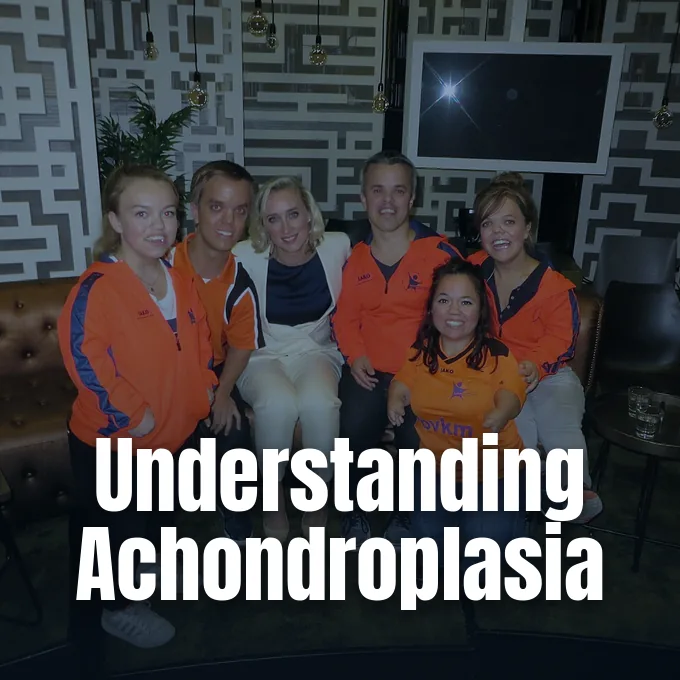
For families asking what is achondroplasia, the condition is a genetic disorder characterized by disproportionate short stature, where the limbs are noticeably shorter than the trunk because cartilage fails to convert into bone during development. The medical term translates literally into without cartilage formation, yet the underlying biological issue rests in how bone forms from cartilage rather than cartilage creation itself [1]. It is the most common form of short-limb dwarfism worldwide, affecting approximately one in every 25,000 infants born each year [3].
What Is Achondroplasia?
Achondroplasia accounts for roughly 70 percent of all dwarfism cases, shaping the lives of more than 250,000 people worldwide who navigate daily life with typical cognitive abilities [12]. Without medical treatment, adult men with the condition reach an average height of 131 centimeters, or four feet four inches, while adult women average around 124 centimeters [1]. Although their limbs stay short, people with the condition have a trunk of relatively normal length. Their mental development is normal [3].
The condition belongs to a larger family of skeletal disorders known as osteochondrodysplasias, which disrupt normal bone and cartilage development throughout childhood [6]. Medical experts have documented more than 100 separate conditions that alter bone growth, while organizations like Nemours KidsHealth note that the broader medical spectrum contains over 700 skeletal dysplasia variations. In dwarfism, clinicians distinguish between short-trunk and short-limb conditions [7]. Achondroplasia is a short-limb condition where bone shortening appears most noticeably in the upper arms and thighs, an anatomical pattern known as rhizomelic shortening [11]. This pattern differs from diastrophic dysplasia, which features mesomelic shortening in the forearms and calves [7]. Despite these physical variations, individuals build rewarding careers and participate fully in their communities [3].
Advocacy groups help clarify terminology surrounding short stature. According to Little People of America, the diagnosis of dwarfism applies when an adult reaches a final height of four feet ten inches or shorter. The organization encourages families to treat skeletal dysplasia as an anatomical difference rather than a personal flaw [7].

The Role of FGFR3 Mutations
At the biological core of the condition is a specific change within the fibroblast growth factor receptor 3 gene, commonly abbreviated as FGFR3. Located on human chromosome 4, this gene provides explicit instructions for producing a receptor protein that helps oversee bone development [1]. In healthy skeletal development, the normal FGFR3 protein serves as a natural brake on bone growth, regulating how quickly cartilage cells multiply before converting into hard bone [11].
In more than 97 percent of individuals with achondroplasia, the disorder stems from a single recurring mutation known as Gly380Arg in the transmembrane domain of the receptor. Researchers Zsolt Vajo, Clair A. Francomano, and Douglas J. Wilkin explained in Endocrine Reviews that this specific amino acid substitution causes the receptor to become constitutively active without needing its usual chemical trigger [9]. The receptor protein sends an unyielding inhibitory signal to growth plate chondrocytes in developing limbs [8]. Because the brake never releases, chondrocyte proliferation drops significantly, preventing long bones from expanding to typical lengths [11]. The resulting disruption explains why limb elongation slows while trunk growth proceeds along a more standard trajectory [1].
Mutations in the same receptor gene can produce other skeletal conditions. A milder mutation causes hypochondroplasia, which presents with less pronounced limb shortening and subtle facial differences [1]. Distinct alleles that cause stronger receptor activation result in thanatophoric dysplasia, a lethal condition characterized by severe ribcage underdevelopment. Another variant causes SADDAN dysplasia [9].
Genetic Inheritance Patterns
Achondroplasia follows an autosomal dominant inheritance pattern, meaning that a child needs only one altered gene copy from a single parent to develop the condition [1]. Despite this dominant transmission, about 80 percent of children with achondroplasia are born to average-height parents who carry no mutation in their own body cells. These occurrences arise from spontaneous, or de novo, genetic changes that occur during the formation of parental reproductive cells [4].
Scientific studies have shown that new spontaneous mutations in the FGFR3 gene originate almost exclusively during spermatogenesis in the father. In 2013, urologist Harry Fisch from the Male Reproductive Center at Columbia Presbyterian Hospital pointed out that the likelihood of this genetic alteration increases with advanced paternal age, particularly for men conceiving after age 35. For fathers over age 50, the incidence rate rises to approximately one in 1,875 births, compared to one in 15,000 in the general population. Biologists theorize that sperm cells carrying this specific FGFR3 mutation might possess a subtle selective survival advantage that allows them to multiply more effectively during sperm production [11]. This selective advantage may explain why the same base change recurs so consistently in unrelated families in many different populations [9].

When one parent has achondroplasia and the other does not, each pregnancy carries a 50 percent chance of passing on the condition. When both parents have achondroplasia, genetic odds divide: there is a 50 percent chance of having a child with achondroplasia, a 25 percent chance of average stature, and a 25 percent risk of homozygous achondroplasia [4]. Children with two copies usually die shortly after birth [1].
Physical Characteristics of Achondroplasia
The physical signs of achondroplasia are visible at birth and show how the underlying gene mutation alters bone development [3]. Infants typically present with a comparatively large head, a condition known medically as macrocephaly, which is accompanied by a prominent forehead called frontal bossing and a flattened nasal bridge [1]. Because the upper jaw is small, children often experience crowded teeth as their baby teeth emerge. Pediatric dentists can help manage this crowding with early orthodontic evaluations [3].
Limb proportions provide the most recognizable aspect of the condition, with shortening concentrated in the humerus and femur bones. Elbow movement is often limited, preventing individuals from straightening their arms completely at the joint [1]. Hand anatomy also exhibits a unique shape known as a trident hand, where the fingers are short and an extra gap separates the middle finger from the ring finger [11]. In contrast to the limbs, the spine and trunk are of normal length, which creates an apparent imbalance in overall body proportions. Joints may appear unusually flexible, leading to loose ligaments in the hands and feet during early childhood [3].
Spinal alignment changes as children grow and learn to support their own body weight. Infants often develop an outward curve in the upper spine known as thoracolumbar kyphosis, which pediatric orthopedists monitor carefully [8]. Once children begin walking, this curve gives way to an exaggerated inward sway in the lower back called lumbar lordosis [1]. Bowed legs, known clinically as genu varum, can also emerge [8].



Health Complications in Infancy
While most people with achondroplasia enjoy healthy lifespans, early infancy brings distinct medical vulnerabilities that need attentive clinical oversight [4]. The base of the skull forms through endochondral bone growth, which causes the opening where the spinal cord exits the brain, called the foramen magnum, to remain smaller than normal. If this narrow passage compresses the brainstem, infants can experience central sleep apnea, weakness, or sudden pauses in breathing [1]. The American Academy of Pediatrics recommends routine neurological evaluations to identify babies who might need surgical decompression to protect their brainstem [6].
Fluid circulation within the brain is another serious childhood concern [1]. About one in every 100 infants develops hydrocephalus, a condition where cerebrospinal fluid accumulates inside the skull because venous drainage pathways narrow [3]. In addition, altered facial anatomy narrows the Eustachian tubes, leaving children prone to recurrent middle-ear infections that can cause conductive hearing loss if left untreated. Audiologists at Children’s Hospital in Missouri emphasize testing young children early to preserve language skills [8].
As individuals enter adolescence and adulthood, anatomical narrowing in the spinal column can create progressive orthopedic challenges. Lumbar spinal stenosis occurs when the bony spinal canal pinches the spinal cord or peripheral nerve roots, resulting in pain, numbness, or tingling sensations in the legs that make walking difficult [1]. Walking long distances can become tiring when lower spinal nerves lack adequate space, prompting some adults to lean forward to relieve pressure on their lower back [6]. Weight management becomes an important health priority because extra body weight places excess mechanical strain on the spine and lower joints. Nutritionists advise tailored calorie intake to prevent obesity while preserving muscle strength. Regular low-impact exercise helps maintain core stability [12].

Diagnostic Methods and Imaging
Doctors often detect signs of achondroplasia during the third trimester of pregnancy through routine prenatal ultrasound examinations. While early fetal scans at 20 weeks appear normal, scans after 24 to 26 weeks may show a noticeable discordance between shortened femur length and a larger biparietal head diameter [11]. Obstetricians also look for excessive amniotic fluid, known as polyhydramnios, which can accompany skeletal dysplasias during late gestation [6]. When prenatal ultrasound raises clinical suspicion, maternal blood testing or amniocentesis can evaluate fetal DNA to confirm the specific FGFR3 variant [8].
After birth, pediatricians confirm the diagnosis through physical examination and a radiographic skeletal survey. X-rays reveal short, thick tubular bones with characteristic metaphyseal flaring and irregular growth plates in the arms and legs. The pelvic bones show small, squared iliac wings, a narrow sciatic notch, and a flat horizontal acetabular roof. Radiologists also examine the ribs, which appear shortened with cupped anterior ends [11].
While radiographic imaging provides clear evidence in most cases, molecular genetic testing offers definitive confirmation by identifying mutations in 99 percent of tested individuals. Testing is particularly useful when symptoms appear unusually mild or when clinical findings overlap with other skeletal dysplasias [4]. Clinical geneticists must differentiate achondroplasia from conditions like hypochondroplasia, thanatophoric dysplasia, or spondyloepiphyseal dysplasia [9]. Genetic counseling helps parents understand recurrence risks, clarify inheritance patterns, and plan future family decisions with accurate medical guidance. Counselors explain that having one child with a spontaneous mutation does not raise the odds for future pregnancies [3].
Managing Achondroplastic Dwarfism
Medical management of achondroplasia focuses on preventing complications, optimizing mobility, and preserving neurological function throughout a person’s life [8]. When neuroimaging detects brainstem compression at the base of the skull, neurosurgeons perform suboccipital decompression to widen the foramen magnum and relieve pressure on delicate neural tissue [4]. Children who develop hydrocephalus may receive a ventriculoperitoneal shunt or an endoscopic third ventriculostomy to redirect excess cerebrospinal fluid [11]. For respiratory obstruction, surgical removal of the adenoids and tonsils often helps open narrowed airways [4].
For decades, families asked whether human growth hormone could treat short stature, but clinical trials showed it does not provide lasting benefits because achondroplasia does not stem from hormone deficiency [3]. Modern medical research has shifted toward therapies that target the downstream signaling pathways of the mutant FGFR3 receptor [5]. In 2021, regulatory agencies approved vosoritide, a daily subcutaneous injection of a C-type natriuretic peptide analog that helps stimulate endochondral bone growth [6]. In March 2026, the U.S. Food and Drug Administration granted accelerated approval to Yuviwel, also known as navepegritide, for pediatric patients aged two years and older with open growth plates. This approval marked a new option for children seeking medical growth support [5].

Approval of Yuviwel was supported by a 52-week randomized, double-blind clinical trial designated Trial 1 (NCT05598320) involving 84 pediatric patients who had never received prior treatment. In the study, 57 children received weekly subcutaneous injections of Yuviwel at 0.1 mg/kg, while 27 received a placebo. Children receiving Yuviwel grew an average of 1.5 centimeters taller over 52 weeks compared to the placebo group, maintaining that growth rate across a subsequent open-label extension. The FDA reported that common side effects included injection-site reactions, vomiting, and temporary low blood pressure [5].
Daily Life and Long-Term Outlook
Adults with achondroplasia generally enjoy good health and an average lifespan when early childhood neurological and respiratory issues are properly managed [6]. However, physical changes in adulthood need ongoing attention, particularly regarding joint pain, lumbar spinal stenosis, and weight management [3]. For women with achondroplasia who become pregnant, obstetricians recommend delivery by Cesarean section because the small maternal pelvic canal prevents safe vaginal childbirth. Psychosocial support also plays an important role in helping individuals navigate public misperceptions and environmental barriers [11].
Environmental modifications can make daily living far more accessible for people of short stature. Simple adaptations like step-stools, lowered light switches, and pedal extenders for cars help foster personal independence from early childhood [7]. Physical activity is encouraged, particularly low-impact sports like swimming and bicycling that strengthen muscles without stressing vulnerable spinal joints [12]. A notable example is Australian Paralympic swimmer Kate Wilson, who was born with achondroplasia and took up swimming upon her pediatrician’s recommendation. Wilson advanced to compete in five swimming events at the Rio 2016 Paralympic Games, reaching the finals of the 100m breaststroke. Her career illustrates how athletic participation flourishes when supported by proper medical guidance [10].
Support networks offer practical guidance for individuals and their families. Organizations like Little People of America and the Genetic Support Network of Victoria connect parents with specialized medical centers, including the Victorian Clinical Genetics Services and the Southern Cross Bone Dysplasia Centre directed by Dr. Ravi Savarirayan [3]. With informed pediatric surveillance, modern pharmacological treatments, and adaptive community support, children born with achondroplasia continue to lead fulfilling, independent, and productive lives [12].
- WEBSITE National Library of Medicine. (2023). Achondroplasia. MedlinePlus Genetics. [Article Link]
- WEBSITE Genetic and Rare Diseases Information Center. (2013). Achondroplasia. National Institutes of Health. [Article Link]
- WEBSITE Department of Health & Human Services. (2001). Dwarfism. Better Health Channel, State Government of Victoria. [Article Link]
- WEBSITE National Human Genome Research Institute. (2019). About Achondroplasia. National Institutes of Health. [Article Link]
- PRESS RELEASE U.S. Food and Drug Administration. (2026). FDA approves drug for pediatric patients with most common form of dwarfism. [Article Link]
- WEBSITE Schwartz, C. I., Dugdale, D. C., & Conaway, B. (2025). Achondroplasia. MedlinePlus Medical Encyclopedia. [Article Link]
- WEBSITE Nemours KidsHealth. (2026). Dwarfism. [Article Link]
- WEBSITE University of Missouri Health Care. (2025). Achondroplasia. Children’s Hospital. [Article Link]
- ACADEMIC JOURNAL Vajo, Z., Francomano, C. A., & Wilkin, D. J. (2000). The molecular and genetic basis of fibroblast growth factor receptor 3 disorders: the achondroplasia family of skeletal dysplasias, Muenke craniosynostosis, and Crouzon syndrome with acanthosis nigricans. Endocrine Reviews, 21(1), 23-39. [Article Link]
- WEBSITE Paralympics Australia. (2016). Kate Wilson: Australian Para Swimming Athlete. [Article Link]
- WEBSITE Wikipedia contributors. Achondroplasia. Wikipedia, The Free Encyclopedia. [Article Link]
- WEBSITE Wikipedia contributors. Achondroplasia in children. Wikipedia, The Free Encyclopedia. [Article Link]
Cover image: Xavier020 / Wikimedia Commons, CC BY-SA 4.0


