





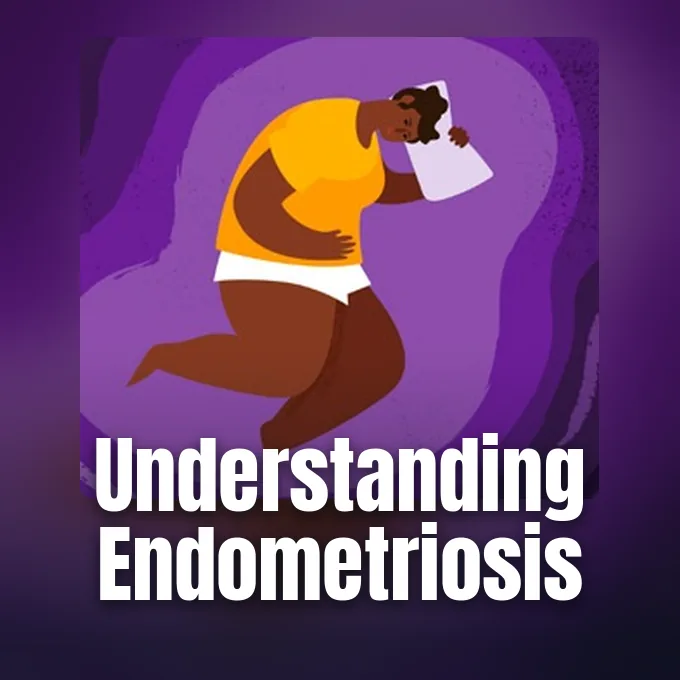
Understanding what is endometriosis requires looking beyond ordinary period cramps to examine how womb-like tissue grows in other parts of the body. The estrogen-driven illness affects about 10% of women worldwide, representing roughly 190 million people who deal with chronic pain or difficulty conceiving during their reproductive years [1, 2]. Displaced cells implant on pelvic organs, sparking localized swelling and monthly internal bleeding that cannot exit the body [4, 9]. Dr. Stacey Missmer and Dr. Andrew Horne observed in BMJ that the illness damages daily routines and emotional wellbeing across multiple life stages [2].
What Is Endometriosis?
In clinical medicine, endometriosis is defined as the growth of endometrial-like tissue outside the inner uterine cavity, where it forms lesions on pelvic organs and surrounding peritoneal membranes [1, 7]. The term stems from Greek words endo, meaning inside, and metrium, meaning uterus. When functioning inside the womb, the endometrium thickens each cycle under estrogen stimulation and sheds as menstrual blood through the cervix and vagina during normal monthly menses [10, 15]. Ectopic lesions react to these same monthly hormonal surges [4, 15]. In JAMA, Dr. Sawsan As-Sanie, Dr. Krina T. Zondervan, and Dr. Stacey A. Missmer reported that 9 million women in the United States currently live with this condition [1].
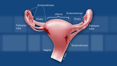
Trapped menstrual blood and shed cells irritate nearby tissue [4, 10]. Over time, recurring internal bleeding triggers chronic swelling, leading to dense fibrous scar bands called adhesions that stick adjacent reproductive organs and bowel loops firmly together [4, 15]. Clinicians classify these abnormal growths as endometrial implants, plaques, or nodules [10, 15].
At the University of Edinburgh, researchers at the MRC Centre for Reproductive Health led by Professor J. Pollard investigate how ectopic lesions establish blood supplies and survive [2]. Patients consult three or more clinicians before receiving an explanation [1]. Dr. Andrew Horne said that presentation patterns vary widely between individuals [2].
Primary Symptoms of Pelvic Pain
Pain is the main symptom of endometriosis [1, 4]. In JAMA, Dr. Sawsan As-Sanie reported that 90% of patients experience chronic pelvic pain, including severe cramps during menstrual bleeding and deep discomfort during sexual intercourse. Patients frequently endure severe dysmenorrhea, an intense period cramping that begins days before bleeding and resists standard analgesics [1, 4]. Many women also experience deep dyspareunia, meaning sharp pain during intercourse, alongside constant lower back aches [1, 15].
Beyond reproductive discomfort, displaced tissue on the bladder or bowel causes painful urination known as dysuria and severe distress during bowel movements known clinically as dyschezia during monthly periods [1, 15]. Patients also suffer from nausea, diarrhea, and abdominal bloating [4, 15]. Writing in The Guardian, journalist Saeed Kamali Dehghan documented how patient Ruhi Singh in Delhi suffered excruciating abdominal pain for a decade before receiving an accurate medical assessment. Dr. Preety R. Rajbangshi at the George Institute for Global Health India explained that cultural normalization of pain frequently delays clinical care for young women [14]. Symptom severity does not match the physical size of lesions [4, 10]. A woman with small superficial spots can endure agonizing cramps, whereas another with extensive ovarian cysts may experience almost no discomfort during monthly cycles [10, 15].
Sensory nerves often grow inside these lesions. Sensitized sensory nerve endings connect directly to the central nervous system, releasing neurochemicals that amplify pain signals whenever regional inflammation flares up within pelvic tissue [8, 10]. Over time, the nervous system becomes sensitized, producing persistent pain across the lower abdomen [2, 8].
How Ectopic Lesions Form in the Pelvis
Doctors have debated the origins of endometriosis for decades [10, 15]. In 1940, American gynecologist Dr. John Sampson formulated the theory of retrograde menstruation to explain how uterine lining escapes into the abdominal space [13, 15]. Sampson posited that during normal menses, fragments of shed endometrium flow backward through the fallopian tubes into the peritoneal cavity instead of passing out through the cervix and vagina [10, 15]. Once inside the pelvis, viable cells adhere to the peritoneum, build blood vessels, and form implants [7, 15].
Retrograde menstruation occurs in 90% of women who have periods [4, 13]. Another hypothesis involves coelomic metaplasia, where primitive cells lining the pelvic cavity transform into endometrial tissue under the influence of chronic inflammatory stimuli or hormonal signals. Displaced cells can also travel through lymphatic channels or blood vessels, reaching distant sites like the lungs [10, 16]. In Endocrine Reviews, Dr. S. E. Bulun reported that defectively programmed progenitor stem cells in the uterine lining contribute directly to abnormal extrauterine growth [7]. Inherited vulnerability is also documented, with family studies indicating that having an affected mother or sister increases personal risk several-fold [4, 15].
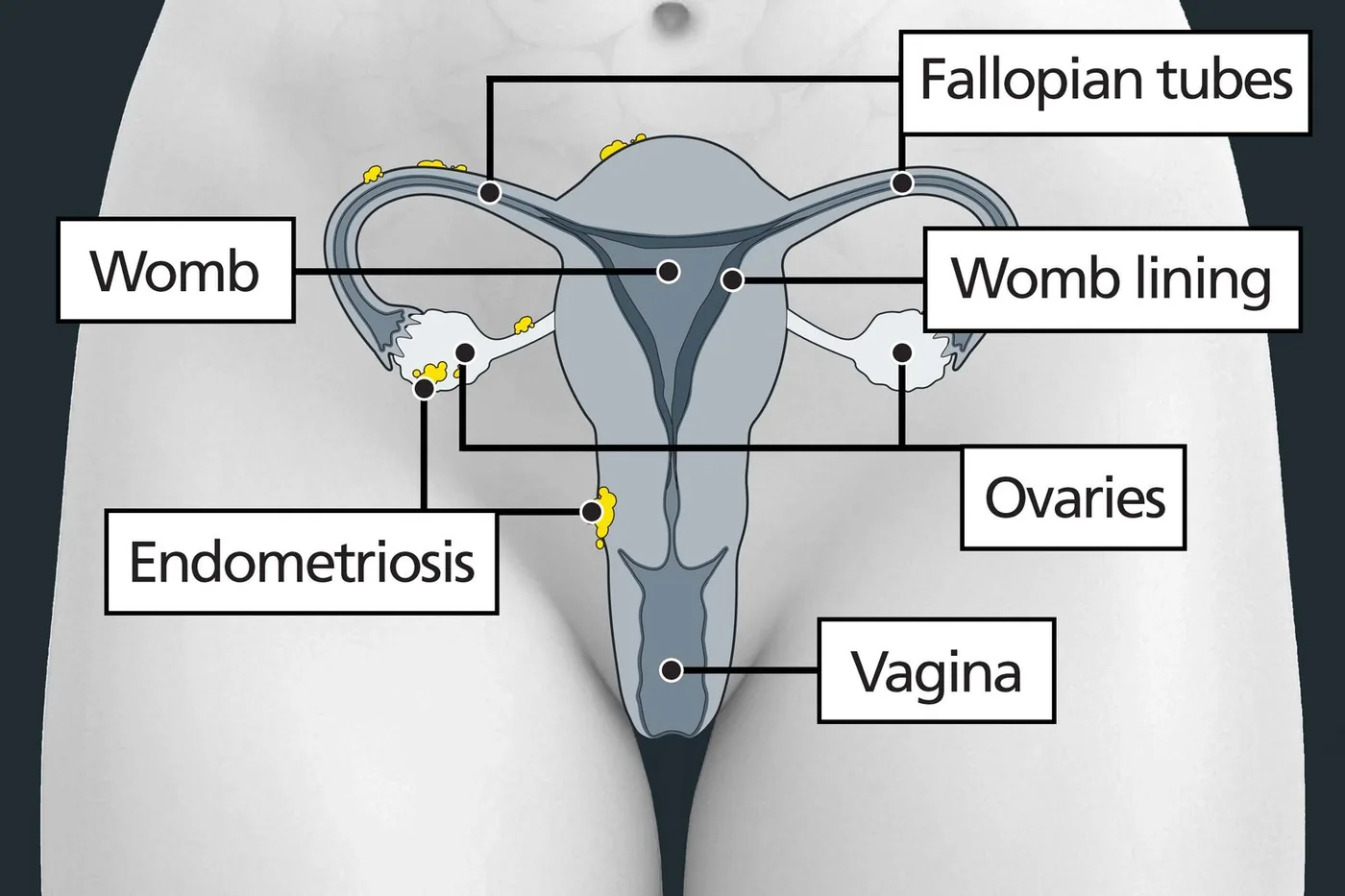
Hormones fuel ectopic cell survival [1, 7]. Bulun showed that endometriotic stromal cells overexpress the transcription factor steroidogenic factor-1, which drives high local estrogen output and sustains regional inflammatory signaling cascades. These cells also display progesterone resistance, lacking receptors that normally stop tissue from growing outside the womb [7, 11].
Anatomic Subtypes of Tissue Spread
Clinicians divide the disease into distinct anatomical subtypes based on lesion depth and location in the pelvis. The most common variety is superficial peritoneal endometriosis, consisting of flat patches on the peritoneal membrane. These implants appear red, black, or white under surgical inspection. Although superficial lesions do not penetrate deeply into underlying structures, they secrete active cytokines that irritate nearby pelvic nerves and cause persistent abdominal aching [7, 15].
A deeper subtype is deep infiltrating endometriosis, defined as lesions that penetrate more than 5 millimeters beneath the peritoneal surface into muscular organ walls. These implants invade the rectum, bladder, bowels, and uterosacral ligaments, forming firm nodules associated with extensive scarring and pain [4, 15]. When tissue affects the ovaries, cysts called endometriomas often develop. Clinicians call these “chocolate cysts” because they fill with thick, dark brown altered blood [11, 15]. In The Guardian, clinical nutritionist Apoorva Joshi described how deep rectovaginal lesions caused her eight years of severe pain before surgeons identified the disease [14].
Rarely, tissue travels outside the pelvis to cause thoracic endometriosis [15, 16]. This condition can trigger catamenial pneumothorax, where a lung collapses during menses, usually on the right side within 72 hours of menstrual bleeding. Patients experience sharp chest pain, coughing, and shortness of breath [16]. Cesarean scars can also harbor implants, forming tender lumps that swell during monthly periods [4, 15].

Genetic Links Between Endometriosis and Immunity
Endometriosis frequently co-occurs with other chronic inflammatory and autoimmune conditions [2, 13]. In a 2025 study published in Human Reproduction, geneticist Dr. Nilufer Rahmioglu and colleagues at Oxford University analyzed UK Biobank records from 8,000 endometriosis cases and 65,000 women with immune conditions. The analysis revealed a 14% increased likelihood of developing an immune disorder among patients, with the risk rising to 30% for those with three or more conditions [4, 13].
The Oxford team identified shared DNA regions that simultaneously influence endometriosis and autoimmune diseases like rheumatoid arthritis, multiple sclerosis, coeliac disease, and osteoarthritis [4, 13]. In London, patient Cherelle Gardiner described how her monthly period flares coincided with multiple sclerosis pain in her legs before doctors linked both conditions at a biological level. One shared DNA region regulates cellular growth and tissue repair, while another controls white blood cell movement during inflammation. Statistical tests confirmed a causal link with rheumatoid arthritis [4, 13].
Cardiovascular vulnerabilities are also tied to the illness. In a 2025 meta-analysis in BMC Public Health, Dr. S. Parsa demonstrated that women with endometriosis have a 23% higher risk of cardiovascular disease and a 13% higher risk of hypertension. Sustained vascular inflammation likely contributes to this elevated long-term arterial stiffness [5].
The Long Road to Clinical Diagnosis
Securing an accurate diagnosis takes 5 to 12 years [1, 9]. Most women consult three or more doctors before receiving a diagnosis, frequently hearing that intense period cramping is merely a normal part of womanhood [1, 14]. Cultural taboos and normalization of pain discourage young girls from seeking early medical help [9, 14]. In India, Dr. Rahul Gajbhiye at the National Institute for Research in Reproductive and Child Health found that diagnostic delays remain common across both rural and urban clinics [14].
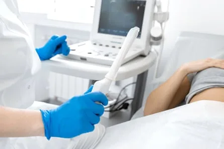
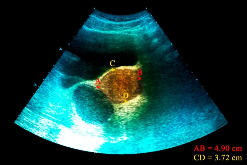
Clinical evaluation begins with a medical history and a pelvic exam, where a doctor checks for tender nodules along the uterosacral ligaments or behind the cervix [1, 3, 11]. Clinicians use transvaginal ultrasonography or pelvic magnetic resonance imaging to detect ovarian cysts and deep nodules in the bladder or bowel. However, a normal scan does not exclude endometriosis. Flat superficial peritoneal lesions cannot be detected through standard ultrasound or magnetic imaging [1, 11]. In New York, patient advocate Jill Fuersich spent a decade undergoing normal gastrointestinal scans before a specialist identified severe disease during surgery [4].
Laparoscopy with a tissue biopsy remains the definitive confirmation test [1, 11]. During keyhole surgery, a doctor passes a small camera through an incision below the navel to inspect organs and remove tissue samples [11, 12]. Guidelines from the European Society of Human Reproduction and Embryology emphasize that clinicians can begin medical therapies based on symptoms alone, without requiring patients to undergo diagnostic surgery first [1, 9].
Medical and Surgical Endometriosis Treatments
Treatment focuses on symptom relief and organ protection. First-line medical therapy combines over-the-counter pain medications like ibuprofen or naproxen with hormonal drugs that suppress ovarian estrogen production and quiet active lesion growth [1, 6, 9]. Physicians frequently prescribe combined contraceptive pills, the levonorgestrel intrauterine device, or depot medroxyprogesterone acetate injections [1, 9]. In JAMA, Dr. Sawsan As-Sanie evaluated 15 clinical trials involving 1,680 participants, confirming that hormonal suppression reduces pelvic pain scores by 13 to 17 points compared with placebo [1].
Roughly 11% to 19% of women get no relief from contraceptives. When first-line drugs fail, doctors prescribe gonadotropin-releasing hormone agonists (GnRH agonists) such as leuprolide or oral GnRH antagonists like elagolix and linzagolix [1, 13]. In the United Kingdom, the National Institute for Health and Care Excellence approved Ryeqo and linzagolix to lower circulating estrogen and control severe bleeding [13]. These drugs create a temporary, reversible menopausal state. To prevent hot flashes and bone mineral loss, physicians provide low-dose estrogen or progestin as add-back therapy during extended courses of GnRH treatment [4, 11]. Dr. Thomas Bainton at Chelsea and Westminster Hospital NHS Foundation Trust said that medical therapy controls symptoms only as long as patients continue taking the drugs [12, 13].
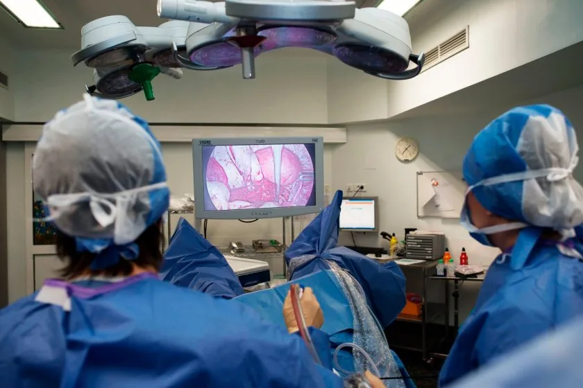
Surgery is recommended when medications fail [1, 12]. Gynecologic surgeons use keyhole laparoscopy to cut out deep nodules and divide scar adhesions that bind internal organs together in the pelvis [6, 12]. However, symptoms recur in up to 50% of patients within five years of surgery [12]. Hysterectomy does not guarantee complete relief if residual nerve pathways remain active [1, 8].
Fertility Challenges in Long-Term Care
Infertility affects 26% to 50% of women with endometriosis [1, 9]. Pelvic adhesions can distort fallopian tubes and block egg transport, while chronic inflammation releases toxic cytokines that harm sperm, eggs, and normal embryo implantation [4, 9]. For couples facing reproductive hurdles, assisted reproductive technologies such as in vitro fertilization (IVF) provide the highest likelihood of achieving a successful pregnancy [9, 10].
Pregnancy does not cure endometriosis. High progesterone levels during gestation temporarily ease pain by suppressing ovulation, but symptoms frequently return after childbirth once regular menstrual cycles resume [4, 10]. In Milwaukee, reproductive surgeon Dr. Grace Janik said that clinical care must adapt as an individual’s personal life goals and health needs evolve. Similarly, although menopause lowers estrogen levels, some postmenopausal women experience persistent pain, especially if they take menopausal hormone replacement therapy [4, 11]. Patient advocate Senie Byrne in Leesburg, Va., founded support networks after enduring years of dismissal before her diagnosis [4].
Care extends beyond medication and surgery [2, 9]. Pelvic physical therapy, dietary changes, and cognitive behavioral therapy help reduce muscle guarding and relieve the emotional toll of chronic pelvic pain [8, 9]. Support groups like Endometriosis UK and the Worldwide EndoMarch movement founded by Dr. Camran Nezhat in Palo Alto provide education and help patients overcome social isolation [12, 17]. Ongoing genetic research points toward better targeted therapies ahead [2, 13].
- ACADEMIC JOURNAL As-Sanie, S., Mackenzie, S. C., Morrison, L., Schrepf, A., Zondervan, K. T., Horne, A. W., & Missmer, S. A. (2025). Endometriosis. JAMA, 334(1), 64–78. [Article Link]
- ACADEMIC JOURNAL Horne, A. W., & Missmer, S. A. (2022). Pathophysiology, diagnosis, and management of endometriosis. BMJ, 379, e070750. [Article Link]
- ACADEMIC JOURNAL Solnik, M. J., Sanders, A., Sanfilippo, J. S., & Konje, J. C. (2025). Endometriosis. BMJ Best Practice. [Article Link]
- REPORT Office on Women’s Health. (2025). Endometriosis. [Article Link]
- ACADEMIC JOURNAL Parsa, S., Noroozpoor, R., Dehghanbanadaki, H., Khateri, S., & Moradi, Y. (2025). Endometriosis and risk of cardiovascular disease: a systematic review and meta-analysis. BMC Public Health, 25(1), 1–12. [Article Link]
- ACADEMIC JOURNAL Kalaitzopoulos, D. R., Samartzis, N., Kolovos, G. N., Mareti, E., Samartzis, E. P., Eberhard, M., Dinas, K., & Daniilidis, A. (2021). Treatment of endometriosis: a review with comparison of 8 guidelines. BMC Women’s Health, 21(1), 397. [Article Link]
- ACADEMIC JOURNAL Bulun, S. E., Yilmaz, B. D., Sison, C., Miyazaki, K., Bernardi, L. A., Liu, S., Kohlmeier, A., Yin, P., Milad, M. P., & Wei, J. (2019). Endometriosis. Endocrine Reviews, 40(4), 1048–1079. [Article Link]
- ACADEMIC JOURNAL Maddern, J., Grundy, L., Castro, J., & Brierley, S. M. (2020). Pain in Endometriosis. Frontiers in Cellular Neuroscience, 14, 590823. [Article Link]
- REPORT World Health Organization. (2025). Endometriosis fact sheet. [Article Link]
- REPORT Eunice Kennedy Shriver National Institute of Child Health and Human Development. (2020). About Endometriosis. [Article Link]
- WEBSITE Institute for Quality and Efficiency in Health Care. (2025). Endometriosis. [Article Link]
- REPORT Gloucestershire Hospitals NHS Foundation Trust. (2026). Laparoscopic (keyhole) surgery for endometriosis. [Article Link]
- ONLINE NEWS Clark, L. (2025). A deeper understanding of endometriosis is suggesting new treatments. New Scientist. [Article Link]
- ONLINE NEWS Dehghan, S. K. (2023). ‘Hell on earth’: India’s taboos around women’s pain leave endometriosis sufferers in agony. The Guardian. [Article Link]
- WEBSITE Wikipedia contributors. (2026). Endometriosis. Wikipedia, The Free Encyclopedia. [Article Link]
- WEBSITE Wikipedia contributors. (2026). Thoracic endometriosis. Wikipedia, The Free Encyclopedia. [Article Link]
- WEBSITE Wikipedia contributors. (2026). Worldwide Endometriosis March. Wikipedia, The Free Encyclopedia. [Article Link]


