



Glucagon is a 29-amino acid peptide hormone made by alpha cells in the islets of Langerhans, serving as the main opposing partner to insulin in human glucose control [1]. When blood sugar drops during fasting, physical exercise, or body stress, the pancreas releases this signal to raise blood glucose and fatty acids [2]. Readers asking what is glucagon find that its chief job involves prompting the liver to break down stored glycogen and generate new sugar molecules, shielding the brain from low blood sugar [3]. Working with other local signals, this ancient hormone keeps body energy in balance through precise cell interactions [4].
What Is Glucagon?
Glucagon is a single-chain peptide with a molecular mass of 3485 daltons, grouped inside the secretin family of hormones [1]. In healthy humans, the pancreas releases it when blood glucose levels fall, prompting liver tissue to turn stored glycogen into free glucose [2]. While insulin promotes fuel storage by helping muscle and fat cells take up sugar, this hormone starts catabolic paths that move fuel into the bloodstream [3]. This balance creates a continuous feedback loop that prevents severe low blood sugar and high blood sugar, preserving steady fuel delivery to glucose-dependent organs like the brain [1].
The chemical makeup of human glucagon has twenty-nine amino acid residues in a fixed chain: NH2-His-Ser-Gln-Gly-Thr-Phe-Thr-Ser-Asp-Tyr-Ser-Lys-Tyr-Leu-Asp-Ser-Arg-Arg-Ala-Gln-Asp-Phe-Val-Gln-Trp-Leu-Met-Asn-Thr-COOH [1]. Unlike steroid hormones, it cannot pass through cell walls [3]. The peptide travels through the portal vein and binds surface GCGR docking sites on liver cells [2].
Meals exert an immediate effect on hormone levels in the blood. When a person eats food rich in carbohydrates, blood glucagon levels drop to prevent an excessive postprandial rise in circulating sugar. Consuming a protein-rich meal prompts alpha cells to secrete the peptide, which prevents low blood sugar while helping the liver convert amino acids into new glucose [2]. Through these steps, the hormone adapts fuel flow to daily food intake [1].

Synthesis from the Proglucagon Precursor
The making of glucagon begins with the transcription of the preproglucagon gene, named Gcg, which yields matching mRNA copies in the pancreas, gut, and brainstem [1]. Translation builds an initial 180-amino acid precursor called preproglucagon [4]. Signal peptidase enzymes remove the leader segment, leaving a 160-amino acid proglucagon protein that serves as the base for several signals [1]. Although the amino acid chain is identical across tissues, proglucagon undergoes tissue-specific cleavage by local enzymes, yielding distinct peptide sets with divergent jobs [4].
Inside the pancreas, alpha cells express proprotein convertase 2, an enzyme that cuts proglucagon into mature glucagon between amino acids 33 and 61 [1]. This cleavage also yields glicentin-related pancreatic polypeptide from residues 1 to 30, intervening peptide-1, and the major proglucagon fragment [4]. Stomach alpha cells also build the peptide [1].
In contrast to pancreatic processing, enteroendocrine L cells of the gut express prohormone convertase 1/3 rather than proprotein convertase 2. This path makes glicentin, oxyntomodulin, glucagon-like peptide-1, and glucagon-like peptide-2. Active forms of GLP-1, like GLP-1 (7–36) amide and GLP-1 (7–37), act as incretin signals that spur insulin release and promote fullness. Natural GLP-1 lasts only two minutes in the blood because dipeptidyl peptidase-4, known as DPP-4, cleaves alanine 8 and glutamic acid 9, so only ten to fifteen percent reaches target tissues intact [4].
What Triggers Glucagon Release?
Alpha cells keep a freerunning basal output that is shaped by nutrient levels, nerve inputs, and local signals within each islet of Langerhans [1]. Low blood sugar is the chief physical trigger for release, prompting alpha cells to discharge their secretory granules into blood vessels [2]. When blood glucose drops, suppression from neighboring beta cells lifts. Beta cells co-secrete amylin and gamma-aminobutyric acid, known as GABA, with insulin to quiet alpha cells; when insulin and amylin output falls, alpha cells become derepressed and release the hormone [1].
Blood amino acids provide another strong stimulus for alpha cell release, with arginine and alanine triggering robust hormone output. Alanine often arrives from muscle transamination reactions involving pyruvate and glutamate, signaling protein breakdown and the need for hepatic fuel conversion. Autonomic nerves also steer secretion: epinephrine binds to beta-2, alpha-2, and alpha-1 adrenergic receptors to drive hormone output during sudden physical stress or vigorous exercise. Gut signals like acetylcholine, cholecystokinin, gastrin, and gastric inhibitory polypeptide, or GIP, spur secretion during digestion to balance fuel handling [1].
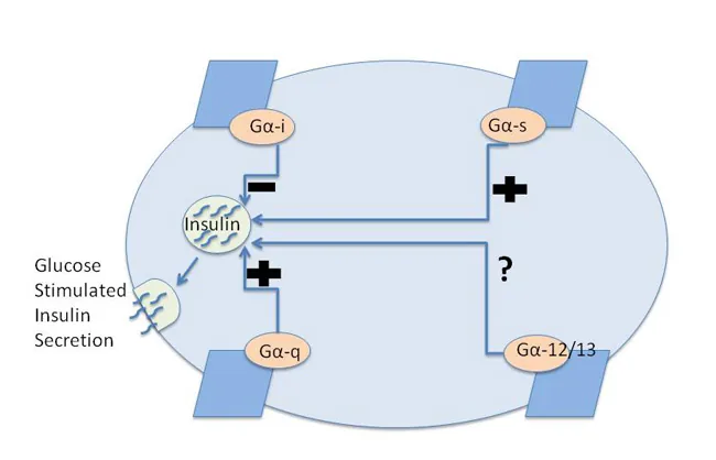
Inhibiting mechanisms balance these inputs to prevent excess glucose output. Somatostatin from pancreatic delta cells shuts down release. High levels of free fatty acids and ketone bodies also curb output [1]. Shaun Coughlin and his group reported that mouse islets express at least twenty-eight GPCRs, including ADRB2, ADRA2A, and SSTR2 [3].
Glucagon Actions on Liver Metabolism
The liver is the main target organ for the hormone, holding dense groups of surface GCGR proteins that direct hepatic glucose output [2]. When it binds to hepatocytes, it spurs glucose release through two paths: glycogenolysis, which breaks down stored glycogen, and gluconeogenesis, which builds new glucose from lactate, glycerol, and amino acids [5]. At the same time, it halts glycogen synthesis and slows glycolysis [2].
Lab studies led by C. J. Ramnanan, D. S. Edgerton, G. Kraft, and A. D. Cherrington showed that an acute rise in the hormone spurs hepatic glucose output almost entirely through an immediate enhancement of glycogenolysis, with little acute help from gluconeogenesis. Yet this glycogenolytic surge is self-limiting and fades over time, a waning effect called tachyphylaxis that occurs even without high blood sugar. In the overnight fasted state, basal glucagon secretion balances basal insulin action, maintaining the steady rate of glycogen breakdown needed to preserve normal fasting blood glucose [5].
At the enzyme level, the hormone starts a phosphate cascade that switches key control points [2]. Activated protein kinase A, or PKA, phosphorylates phosphorylase kinase, which turns inactive glycogen phosphorylase b into active glycogen phosphorylase a, the rate-limiting enzyme that clips glucose 1-phosphate from glycogen [1]. PKA also phosphorylates and inactivates glycogen synthase, halting glycogen assembly. To block glycolysis, PKA phosphorylates the bifunctional enzyme PFKFBPase, shutting off its 6-phosphofructo-2-kinase arm while activating its fructose-2,6-bisphosphatase arm. This enzymatic shift lowers fructose-2,6-bisphosphate, or F2,6P, which turns into F6P, relieving inhibition on fructose bisphosphatase so gluconeogenesis outpaces glycolysis [2].

Receptor Signaling Through Cyclic AMP
The actions of the hormone depend on the glucagon receptor, a 62-kilodalton glycoprotein in the class B secretin family of GPCRs, encoded by the GCGR gene [8]. Alfred Gilman and Martin Rodbell, who shared the Nobel Prize in 1994, proved that heterotrimeric G proteins pass signals from cell surface receptors to internal enzymes. Human cells hold sixteen alpha subunit genes yielding twenty-three isoforms, five beta subunit genes, and twelve gamma subunit genes [3].
When the hormone binds its receptor, the heterotrimeric G protein swaps GDP for GTP on the Gs alpha subunit [3]. The activated Gs alpha subunit separates from the beta-gamma complex and turns on adenylyl cyclase, which converts ATP into cyclic adenosine monophosphate, or cAMP [1]. Rising cAMP activates PKA, which phosphorylates downstream metabolic enzymes [2]. To a lesser degree, the receptor engages Gq proteins, activating phospholipase C to cleave PIP2 into DAG and IP3, which prompts calcium release from internal stores [8].
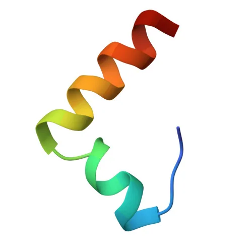
Structural work has mapped the chemical basis of receptor binding. At the University of Arizona in Tucson, Arizona 85721 and Howard Hughes Medical Institute, N. S. Sturm, Y. Lin, S. K. Burley, J. L. Krstenansky, J. M. Ahn, B. Y. Azizeh, D. Trivedi, and V. J. Hruby synthesized peptide analogues modified at positions 17, 18, and 21. They showed that a salt bridge between lysine 18 and glutamic acid 21 is vital for receptor binding, with analogues reaching 78 to 100 percent adenylate cyclase activation compared to natural glucagon at 1.5 nM [6]. Later, J. Ying, J. Ahn, N. E. Jacobsen, M. F. Brown, and V. J. Hruby ran two-dimensional NMR tests at 37 degrees Celsius and pH 6.0 in dodecylphosphocholine micelles to map antagonist [desHis1, desPhe6, Glu9]glucagon amide, revealing an L-shaped fold with two helices at a 90-degree angle stabilized by D15-R18 and R17-D21 salt bridges [7].

Lipid Oxidation and Energy Expenditure
Beyond controlling sugar, the hormone acts as a chief catabolic driver of fat breakdown and energy use [9]. In liver cells, it stops new fat creation by shutting down acetyl-CoA carboxylase, or ACC, through PKA phosphorylation. ACC normally turns acetyl-CoA into malonyl-CoA; when the hormone blocks ACC, malonyl-CoA levels fall. Because malonyl-CoA is an allosteric blocker of carnitine palmitoyltransferase I, or CPT1, its loss removes the barrier at the outer mitochondrial wall. This shift lets fatty acids enter mitochondria for beta-oxidation, fueling ketone body creation like acetoacetate and beta-hydroxybutyrate during fasting [1].
In body fat stores, the hormone aids lipolysis when insulin is low, activating hormone-sensitive lipase to split fat into glycerol and free fatty acids [1]. K. D. Galsgaard, J. Pedersen, F. K. Knop, J. J. Holst, and N. J. W. Albrechtsen showed that hepatic receptor signaling cuts liver fat accumulation [10]. Blocking the receptor with drugs causes dyslipidemia and raises liver fat, showing that basal tone protects against hepatic steatosis [9].
The hormone also spurs body heat production and lifts daily energy burn. M. Kleinert, S. Sachs, K. M. Habegger, S. M. Hofmann, and T. D. Müller noted that its thermogenic action has been known for six decades [12]. In a blinded human crossover trial, T. Tan, B. C. T. Field, K. McCullough, and S. R. Bloom found that glucagon infusion significantly increases resting energy expenditure measured by indirect calorimetry in ten volunteers. Giving it with GLP-1 stopped glucose spikes while keeping high energy burn, supporting dual-agonist therapies [11].
Clinical Manifestations in Diabetes
Flaws in hormone control sit at the center of metabolic disease. In type 2 diabetes, alpha cells resist insulin suppression, producing high glucagon levels that worsen hyperglycemia. This finding supports the bihormonal hypothesis, which holds that diabetes stems from an altered ratio of insulin to glucagon signaling. L. Janah and co-workers also showed that long-term metabolic strain causes liver glucagon resistance, harming amino acid clearance and urea synthesis [9].
In type 1 diabetes, loss of beta cells eliminates insulin as well as local amylin and GABA signals. Without suppression, alpha cells pour out the hormone in an unrestrained flow. This unchecked output drives rapid glycogen breakdown and fast ketogenesis, triggering diabetic ketoacidosis. Clinical tests showed that giving somatostatin to adults with type 1 diabetes delayed ketoacidosis from an average of ten hours to eighteen hours, proving the direct role of the hormone in acute metabolic crisis [1].
Rare clinical conditions also stem from gene mutations or tumors. Glucagonoma, a rare alpha cell tumor of the pancreas, causes severe hyperglucagonemia marked by weight loss, low blood amino acids, diabetes, and a rash called necrolytic migratory erythema [1]. Missense mutations at chromosome 17q25 in the GCGR gene cause Mahvash disease, an inherited disorder with glucagon resistance, alpha cell growth, and neuroendocrine tumors [8]. In clinical care, emergency glucagon kits and injections serve as rescue tools for acute severe low blood sugar in patients using insulin [1].
History of Isolation and Structural Mapping
The discovery of the hormone dates to the early 1920s during experiments on pancreatic secretions. After Frederick Banting and Charles Best isolated insulin in Toronto, scientists saw that crude pancreas extracts caused a brief blood sugar rise before the insulin drop. In 1922, C. Kimball and John R. Murlin isolated this substance and named it “glucagon”, joining “glucose” and “agonist” to reflect its glucose-raising power. In the 1950s, chemists at Eli Lilly purified the hormone and sequenced its 29 amino acids, which allowed Roger Unger to build the first radioimmunoassay in 1959 [1].
Molecular cloning in the late twentieth century uncovered the multi-hormone layout of the proglucagon gene. In 1979, Richard Goodman in Joel Habener’s laboratory at Massachusetts General Hospital harvested islet tissue from the Brockman bodies of American anglerfish. Goodman cloned anglerfish DNA into bacteria, creating a library that P. Kay Lund used to locate the glucagon sequence. In 1982, Lund and Goodman reported that the proglucagon gene codes for three distinct peptides: glucagon and two new companion hormones. Graeme Bell at Chiron Corporation led the team that isolated these peptides, naming them GLP-1 and GLP-2 [1].
Comparative biology shows the deep roots of these peptides across animal species. In young crayfish, excising the eyestalk produces hyperglycemia driven by endogenous glucagon-like molecules [1]. Over the past century, research by T. D. Müller, B. Finan, C. Clemmensen, R. D. DiMarchi, and M. Tschöp has turned this peptide from an obscure extract into a cornerstone of dual and triple agonist drugs that treat obesity and metabolic illness [13].
- WEBSITE Wikipedia contributors. (2026). Glucagon. Wikipedia, The Free Encyclopedia. [Article Link]
- WEBSITE Robertson, S. (2018). Glucagon Signaling Pathway. News-Medical. [Article Link]
- ACADEMIC JOURNAL Layden, B. T., & Lowe, W. L. (2010). G-Protein Coupled Receptors, Pancreatic Islets, and Diabetes. Nature Education. [Article Link]
- WEBSITE Wikipedia contributors. (2026). Glucagon-like peptide-1. Wikipedia, The Free Encyclopedia. [Article Link]
- ACADEMIC JOURNAL Ramnanan, C. J., Edgerton, D. S., Kraft, G., & Cherrington, A. D. (2011). Physiologic action of glucagon on liver glucose metabolism. Diabetes, Obesity and Metabolism, 13(s1), 118-125. [Article Link]
- ACADEMIC JOURNAL Sturm, N. S., Lin, Y., Burley, S. K., Krstenansky, J. L., Ahn, J. M., Azizeh, B. Y., Trivedi, D., & Hruby, V. J. (1998). Structure-Function Studies on Positions 17, 18, and 21 Replacement Analogues of Glucagon: The Importance of Charged Residues and Salt Bridges in Glucagon Biological Activity. Journal of Medicinal Chemistry, 41(15), 2693-2700. [Article Link]
- ACADEMIC JOURNAL Ying, J., Ahn, J. M., Jacobsen, N. E., Brown, M. F., & Hruby, V. J. (2003). NMR Solution Structure of the Glucagon Antagonist [desHis1, desPhe6, Glu9]Glucagon Amide in the Presence of Perdeuterated Dodecylphosphocholine Micelles. Biochemistry, 42(10), 2825-2835. [Article Link]
- WEBSITE Wikipedia contributors. (2026). Glucagon receptor. Wikipedia, The Free Encyclopedia. [Article Link]
- ACADEMIC JOURNAL Janah, L., Kjeldsen, S. A. S., Galsgaard, K. D., Winther-Sørensen, M., Stojanovska, E., Pedersen, J., Knop, F. K., Holst, J. J., & Albrechtsen, N. J. W. (2019). Glucagon Receptor Signaling and Glucagon Resistance. International Journal of Molecular Sciences, 20(13), 3314. [Article Link]
- ACADEMIC JOURNAL Galsgaard, K. D., Pedersen, J., Knop, F. K., Holst, J. J., & Albrechtsen, N. J. W. (2019). Glucagon Receptor Signaling and Lipid Metabolism. Frontiers in Physiology, 10, 413. [Article Link]
- ACADEMIC JOURNAL Tan, T., Field, B. C. T., McCullough, K., Troke, R., Chambers, E. S., Salem, V., Maffe, J. G., Baynes, K., Silva, A. D., Viardot, A., Alsafi, A., Frost, G. S., Ghatei, M. A., & Bloom, S. R. (2012). Coadministration of Glucagon-Like Peptide-1 During Glucagon Infusion in Humans Results in Increased Energy Expenditure and Amelioration of Hyperglycemia. Diabetes, 62(4), 1131-1138. [Article Link]
- ACADEMIC JOURNAL Kleinert, M., Sachs, S., Habegger, K. M., Hofmann, S. M., & Müller, T. D. (2019). Glucagon Regulation of Energy Expenditure. International Journal of Molecular Sciences, 20(21), 5407. [Article Link]
- ACADEMIC JOURNAL Müller, T. D., Finan, B., Clemmensen, C., DiMarchi, R. D., & Tschöp, M. (2017). The New Biology and Pharmacology of Glucagon. Physiological Reviews, 97(2), 721-766. [Article Link]


