





Histamine is a biogenic monoamine that coordinates immune signals, triggers stomach acid release, and regulates daily waking in the human central nervous system. First identified in 1910, the molecule acts locally in surrounding tissues rather than traveling as an endocrine hormone from specialized glands. Understanding histamine function reveals how a single small chemical messenger can stimulate digestion, provoke allergic inflammation, and maintain mental alertness across varied bodily systems [10, 12].
- What Is Histamine?
- Granule Storage Driving Histamine Function
- How Four Receptors Direct Histamine Function
- Vascular Leakage During Allergic Reactions
- Histamine Function in Stomach Acid Secretion
- Brain Neurotransmission Governing Daily Wakefulness
- Scombroid Toxicity in Dietary Fish
- Enzymatic Clearance of Ingested Amine
What Is Histamine?
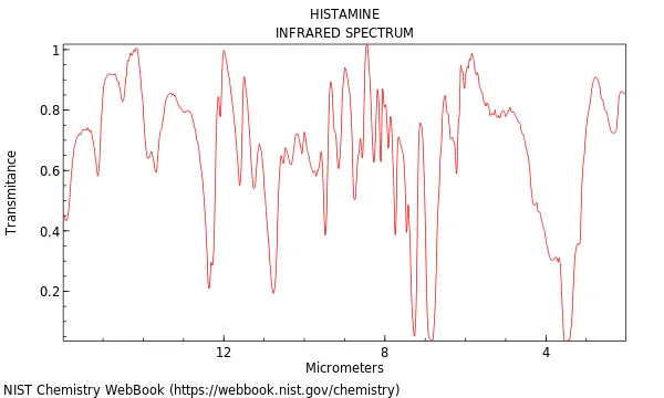
Histamine is an organic amine with the chemical formula C5H9N3, an average mass of 111.148, and the IUPAC standard InChIKey NTYJJOPFIAHURM-UHFFFAOYSA-N [1, 2]. It dissolves easily in water [10]. Early chemical literature recorded the compound under names like Ergamine and Ergotidine, while Coblentz Society, Inc. and Wyandotte Chemicals Corp. in Wyandotte, Michigan documented its infrared profile for standard databases. The National Institute of Standards and Technology (NIST) indexes the molecule under CAS Number 51-45-6 and code NSC 33792 in the NIST Chemistry WebBook, providing computed Mol files [1]. In ChEBI entry 18295 maintained by the European Bioinformatics Institute, it is classified as a human metabolite [2]. Because human tissues produce it locally without classical endocrine glands, early investigators described it as an autocoid [10].
Solid histamine base melts at 83–84 °C (181–183 °F) [1, 10]. In water, the molecule carries two basic centers with distinct traits. Under human bodily conditions, where blood pH stays between 7.35 and 7.45, the side chain amino group with a pKa near 9.4 takes up a proton, while the ring nitrogen with a pKa of roughly 5.8 stays unprotonated. As a result, histamine in human blood exists mostly as a singly charged cation. In liquid solution, the ring adopts two forms, with the tele tautomer being preferred over the pros tautomer because the proton sits farther from the side chain [10].
The amine contains only 17 atoms and has an exact molecular weight of 111.15 [6, 10]. Cells build the molecule through decarboxylation of L-histidine using the enzyme L-histidine decarboxylase, allowing cellular histamine function to coordinate at least 23 separate biological roles across human organ systems. It is tiny. Yet its impact spans the whole body [10].
Granule Storage Driving Histamine Function
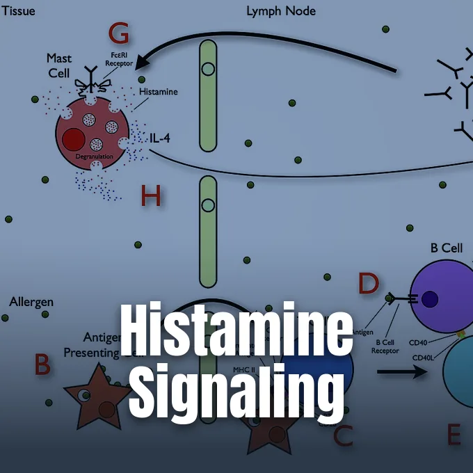
Tissue mast cells and circulating basophils store over 90% of body histamine inside cytoplasmic granules. Mast cells reside heavily at physical borders exposed to the outside world, including the nose, mouth, feet, gut lining, and blood vessel walls, where they act as rapid sentinels against foreign antigens. They sound the alarm. When micro-organisms break through outer skin or mucosal layers, these sentinels release chemical mediators to initiate tissue defense [10].
The main trigger for cellular degranulation is an immune pathway mediated by allergen-specific immunoglobulin E antibodies. When an allergen enters the tissues of a sensitized person, it binds and cross-links adjacent IgE molecules anchored to high-affinity receptors on the mast cell surface. Receptor cross-linking prompts storage granules to move toward the outer cell wall. Degranulation occurs within seconds as the granules fuse with the plasma membrane, discharging dense histamine, proteases, and cytokines into the nearby fluid. Non-immunologic agents can also induce degranulation; clinical drugs, such as morphine, curare alkaloids, and polymyxin antibiotics, displace histamine from internal binding sites in granules and provoke release without requiring any antibody interaction [10].
Gastric enterochromaffin-like cells in the stomach fundus store large pools of non-mast cell histamine, releasing the compound when triggered by hormonal gastrin and vagal acetylcholine cues. This local flow proves that histamine function directs routine digestion. Without this trigger, gastric acid output falls sharply [10].
How Four Receptors Direct Histamine Function
Mammalian tissues express four distinct histamine receptors, known as H1, H2, H3, and H4, all belonging to the rhodopsin-like family of seven-span G protein-coupled receptors [6, 12]. Ligand binding activates specific G proteins that regulate second messengers such as cyclic AMP or inositol trisphosphate to modulate histamine activity [10, 12]. Through these diverse G protein couplings, histamine function generates contrasting outcomes across target organs [12].
The H1 receptor sits mainly on vascular endothelial cells, smooth muscle fibers, and local sensory neurons. Stimulation of H1 receptors spurs phospholipase C, generating inositol trisphosphate and diacylglycerol to free internal calcium stores. In the airway tree, this calcium influx triggers smooth muscle contraction that narrows the bronchial lumen, while in skin vessels it prompts cell contraction that promotes edema and fluid leakage [10, 12]. In contrast, H2 receptors localize heavily on the basolateral surface of gastric parietal cells and cardiac myocytes. When histamine binds to H2 receptors, it couples to Gs proteins and turns on adenylate cyclase, raising internal cyclic AMP levels and pumping hydrochloric acid into the stomach [10, 12].
H3 and H4 receptors serve unique signaling roles in neural and immune tissues. Within the brain, presynaptic H3 receptors act as autoreceptors that inhibit the synthesis and release of histamine, while also tuning messengers like dopamine and serotonin. Meanwhile, H4 receptors reside on eosinophils, T cells, and mast cells. Stimulating the H4 receptor guides immune cell migration toward inflamed sites, making it a promising target for new anti-allergy drugs [12].
Vascular Leakage During Allergic Reactions
During acute allergic flares, vascular histamine function increases fluid leakage into nearby tissue beds by acting on endothelial cells lining post-capillary venules. Endothelial H1 receptor cues cause these cells to contract and pull apart, widening intercellular gaps [10, 12]. Fluid, plasma proteins, and circulating white blood cells pass into interstitial space, producing swelling, nasal congestion, and itchy hives [10].
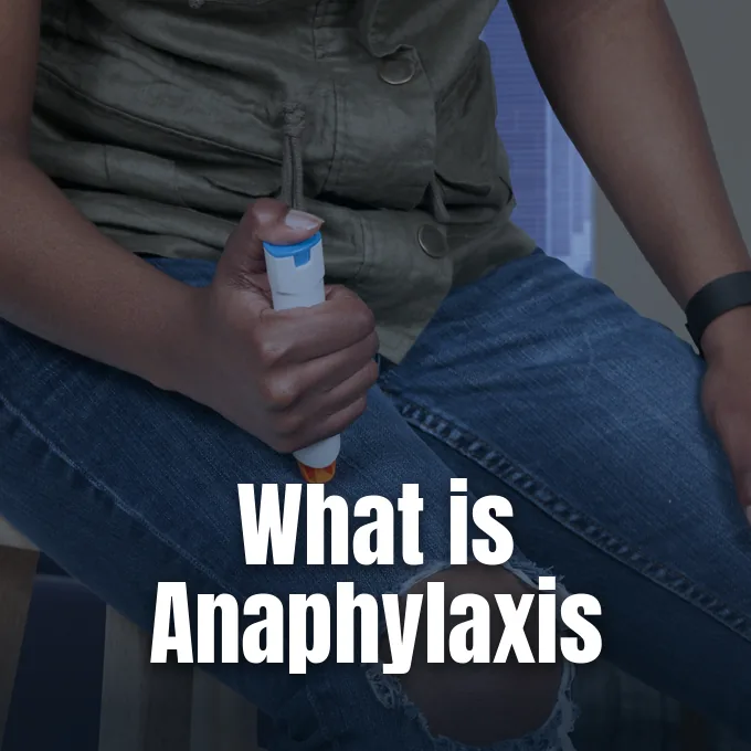
Histamine also relaxes vascular smooth muscle, causing peripheral arterioles to dilate and lowering systemic blood pressure. When histamine binds to endothelial receptors, it spurs the enzymatic release of nitric oxide and endothelium-derived hyperpolarizing factors, which diffuse into nearby muscle cells to prompt relaxation. In mild localized reactions, this vasodilation produces cutaneous flushing and warmth. Intravenous injections of histamine have been known for more than a century to induce a prompt drop in arterial tension. During severe systemic reactions, broad vasodilation paired with fast plasma loss into interstitial spaces precipitates low blood pressure and circulatory collapse [10]. These vascular pathways form the biological basis of anaphylaxis and severe allergic reactions.
In local sensory nerves, histamine stimulates unmyelinated C-fibers to carry itch and pain cues to the spinal cord [10, 12]. Stimulated sensory endings release neuropeptides, such as substance P and calcitonin gene-related peptide, directly into nearby tissue. These neuropeptides act back on resident mast cells to stimulate additional mediator discharge, creating a two-way loop that fuels neurogenic inflammation and local discomfort. In airways, ongoing receptor binding causes lasting muscle spasms and airway constriction [10].
Histamine Function in Stomach Acid Secretion
In the digestive tract, tracing gastric histamine function explains why food intake spurs prompt hydrochloric acid release from parietal cells [10, 12]. When food enters the stomach, hormonal gastrin and vagal acetylcholine spur mucosal enterochromaffin-like cells. These cells discharge histamine directly into the mucosal lamina propria, where it diffuses across short distances to engage H2 receptors on neighboring acid-secreting parietal cells [10, 12]. Acid generation begins promptly [10].
Histamine binding to H2 receptors turns on Gs proteins, activating adenylate cyclase in the parietal cell membrane [10, 12]. Adenylate cyclase converts ATP into cyclic AMP to activate protein kinase A, which prompts tubulovesicles carrying hydrogen-potassium ATPase proton pumps to fuse with the apical membrane surface. Pumps move ions fast. By exchanging potassium for hydronium ions, these membrane pumps pour hydrochloric acid into the stomach lumen, dropping local pH toward 1.5 to break down food proteins and kill swallowed microbes [10].
This discovery led to the creation of histamine H2-receptor antagonists, such as cimetidine and famotidine, which revolutionized peptic ulcer treatment. By blocking H2 receptors on parietal cells, these drugs curb resting and meal-driven acid output. Doctors give these blockers to treat peptic ulcer disease and manage reflux, giving a trusted option alongside proton pump inhibitors like omeprazole [3, 12]. Clinical tests also use histamine to check stomach acid output [3, 6].
Brain Neurotransmission Governing Daily Wakefulness
Within the mammalian brain, histaminergic cell bodies sit only in the tuberomammillary nucleus of the posterior hypothalamus. Although this nucleus contains a small group of neurons, its axons branch across the cortex, striatum, thalamus, and spinal cord. Histamine release displays a circadian rhythm, reaching its highest output during active wakefulness and dropping to minimal levels during slow-wave and rapid eye movement sleep. In the striatum, histaminergic inputs tune basal ganglia loops that help regulate motor control and motivated actions. By depolarizing cortical neurons and waking subcortical centers through H1 receptors, histaminergic transmission promotes waking drive, curiosity, and sharp focus throughout daytime hours [10, 12].
Because central histamine promotes waking, older allergy pills cause strong daytime drowsiness. Compounds such as diphenhydramine cross the blood-brain barrier and block neuronal H1 receptors, calming the brain. Newer second-generation antihistamines do not easily penetrate brain tissue, avoiding sleepiness during allergy care [10, 12]. Tracking these brain paths clarifies how human immune signaling and sleep share chemical messengers across distinct organ systems [10].
Histamine also modulates protein function directly through a chemical process termed histaminylation. First described in 2012, this reaction involves the covalent attachment of histamine to glutamine residues on target proteins through transamidation. Laboratory investigations have identified histaminylated sites on both histone and non-histone proteins in hypothalamic neurons and nearby tissues. Histone histaminylation appears to guide key gene readouts of the circadian clock, revealing an epigenetic facet of central histamine function that operates beyond cell surface receptors [10].
Scombroid Toxicity in Dietary Fish
Bacterial species multiply whenever protein-rich foods are stored under warm conditions, converting free histidine into active amines during food storage [4, 8]. Fermented items naturally accumulate measurable histamine as aging microbes grow. Aged cheeses (gouda, parmesan, cheddar), dry salami, sauerkraut, and soy sauce often hold high levels, while wine contains 2 to 10 mg/L and Japanese sake carries 20 to 40 mg/L. These fermented goods can trigger headaches in sensitive people [4, 10].
Scombroid fish poisoning is an acute illness caused by eating spoiled marine fish. Named after the Scombridae family of tuna and mackerel, it also affects sardines and anchovies when surface bacteria turn abundant free histidine into heat-stable histamine that cooking, baking, and canning cannot destroy. Victims soon develop facial flushing, sweating, throbbing headaches, dizziness, mouth burning, gut cramps, and diarrhea [4, 8, 11].

Food safety agencies enforce limits to protect consumers. Bronte Boyadzhyan at United Food Labs and James M. Hungerford in a Zenodo report showed that poor cooling triggers bacterial amine growth, leading the FDA to set an action limit of 50 ppm in commercial fish [4, 8]. Labs test seafood with HPLC and ELISA assays. Rapid kits verify dockside fish [4, 8].
Enzymatic Clearance of Ingested Amine
Two primary enzymes break down histamine in humans: diamine oxidase and histamine N-methyltransferase. Diamine oxidase, made from the AOC1 gene in small bowel enterocytes, clears dietary amine before it reaches the bloodstream. Inside cells, the HNMT gene product methylates histamine in the brain, airway tissues, and kidneys to keep overall histamine function in balance [10, 11].
When clearing enzymes falter, circulating amine pools rise [10, 11]. Variants in the AOC1 gene, such as rs2052129, rs2268999, rs10156191, and rs1049742, reduce diamine oxidase output and trigger gut distress, while HNMT variants like HNMT-464 TT and HNMT-1639 TT appear in children with airway sensitivity. People with low enzyme levels often suffer headaches and flushing after eating aged foods, disrupting normal histamine activity [10, 11].
Histamine intolerance is not recognized in the International Classification of Diseases (ICD-11) [9, 11]. In a trial by Bent and colleagues from the American Academy of Allergy, Asthma & Immunology (AAAAI) testing 59 patients, placebo-controlled food challenges ruled out histamine intolerance in 85% of cases. Nearly two-thirds of patients had symptoms after placebos. Conditioned worry often mimics true food intolerance, and low enzyme levels appear widely in healthy people. Testing alone cannot prove disease [9].
- WEBSITE National Institute of Standards and Technology. (2026). Histamine. NIST Chemistry WebBook, NIST Standard Reference Database Number 69. [Article Link]
- WEBSITE European Bioinformatics Institute. (2017). Histamine (CHEBI:18295). Chemical Entities of Biological Interest. [Article Link]
- DICTIONARY ENTRY Dictionary.com. (2023). Histamine. In Dictionary.com Unabridged. Random House. [Article Link]
- WEBSITE Boyadzhyan, B. (2025). Histamine Testing in Food. United Food Labs. [Article Link]
- WEBSITE Royal Society of Chemistry. (2026). Histamine (ChemSpider ID 753). ChemSpider. [Article Link]
- WEBSITE Kyoto Encyclopedia of Genes and Genomes. (n.d.). Histamine (KEGG DRUG D08040). [Article Link]
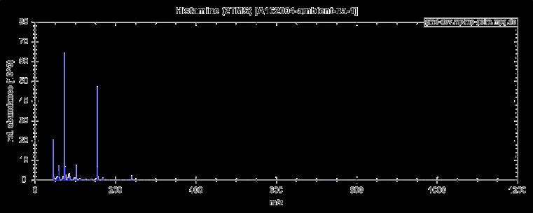
- WEBSITE Boelling, C., Liebig, F., Erban, A., & Kopka, J. (2026). Histamine (2TMS). Golm Metabolome Database. [Article Link]
- ACADEMIC JOURNAL Hungerford, J. M. (2010). Scombroid Poisoning: A Review. Zenodo. [Article Link]
- WEBSITE American Academy of Allergy, Asthma & Immunology. (2023). Histamine intolerance: fact or fiction? [Article Link]
- WEBSITE Wikipedia contributors. (n.d.). Histamine. In Wikipedia, The Free Encyclopedia. [Article Link]
- WEBSITE Wikipedia contributors. (n.d.). Histamine intolerance. In Wikipedia, The Free Encyclopedia. [Article Link]
- WEBSITE Wikipedia contributors. (n.d.). Histamine receptor. In Wikipedia, The Free Encyclopedia. [Article Link]
Cover image: Courtesy of NIAID Ryan Kissinger / Wikimedia Commons, Public domain


