





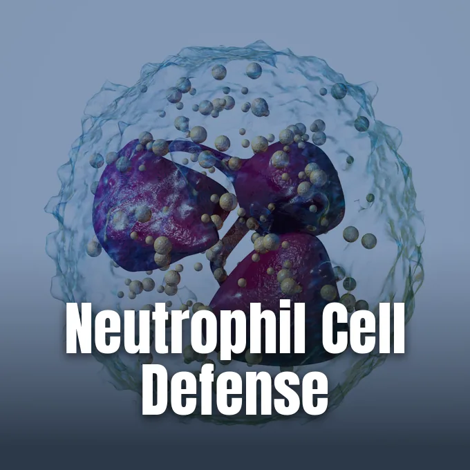
What are neutrophils and how do they defend our body against dangerous germs? Neutrophils form the largest white cell group in human blood, making up 40% to 70% of all traveling leukocytes as the first line of innate immunity [10]. When bacteria or fungal spores pierce skin barriers, these agile defenders swim through blood vessels to trap invaders before colonies spread into deeper organs [1]. Adaptive antibodies take days to form, but these rapid white cells enter wounded tissue within minutes to contain acute contamination. Marrow daily output reaches 1011 cells [10].
- What Are Neutrophils?
- Bone Marrow Production and Lifespan
- Structural Hallmarks Under the Microscope
- Chemical Guidance Toward Infected Tissues
- Defensive Roles of Neutrophil Granulocytes
- Selective Recognition of Microbial Sugars
- Clinical Value of Polymorphonuclear Leukocytes
- Why Extracellular Traps Provoke Inflammation
What Are Neutrophils?
Neutrophils form the largest group of granulocytes, a white cell class that also includes eosinophils and basophils within the polymorphonuclear leukocyte family. Early cytologists named these cells from their neutral pink tint under hematoxylin and eosin stains, contrasting with the red hue of eosinophils and dark blue spots of basophils. In routine clinical care, doctors test red cell surface markers to classify ABO blood types for safe transfusions, but they check neutrophil tallies to gauge immediate protection against foreign bugs. Because these white cells flow day and night through blood vessels, they meet invading bugs much faster than slower monocyte or macrophage cells [10]. In medical reviews by Mayadas, Culleré, and Lowell at the Annual Review of Pathology, pathologists noted that initial cellular waves alert nearby tissues to dangerous bacterial threats [1].
Circulating cells exist mainly as mature segmented forms with multilobed nuclei, though immature band forms also enter blood pools during heavy infection. Healthy segmented cells carry nuclei divided into two to five distinct lobes linked by thin chromatin filaments. When tissues suffer physical trauma or acute bacterial invasion, these cells cluster in dense waves to wall off injured zones. Rapid cell death creates the whitish-yellow substance commonly known as pus, showing an active local immune fight. Bands enter blood early [10].
Beyond killing bugs, neutrophils actively drive acute pain perception at injury sites. They release soluble mediators that sensitize peripheral nociceptors, alerting the central nervous system to localized physical wounds [10]. Mutual signaling helps innate containment transition into broader adaptive immunity [2].
Bone Marrow Production and Lifespan
The human body generates neutrophils on a massive scale inside spongy bone marrow spaces. Stem cells produce vast numbers daily, keeping a deep store to replace short-lived cells lost during routine defense. Anatomical studies show that neutrophils spread through three major body pools in a fixed ratio of 28:1:25, with twenty-eight parts resting in bone marrow reserves, one part circulating in peripheral blood, and twenty-five parts stationed across connective tissues [10]. As Amulic and Zychlinsky described in the Annual Review of Immunology, marrow keeps an immediate supply ready for rapid deployment [2].
In circulating blood, inactive neutrophils have a brief lifespan ranging between 5 and 135 hours. After moving across vessel walls into inflamed connective tissues, their survival typically lasts only one to two days [10].
Immunologists suggest that the brief lifespan of neutrophils serves as an evolutionary adaptation to preserve host health. Many intracellular parasites, such as Leishmania, survive by invading host phagocytes, so rapid cell death deprives these microbes of safe shelter. Furthermore, because digestive enzymes and reactive chemicals can inflict random harm on healthy flesh, a short lifespan curbs harmful collateral swelling during acute responses. When these white cells exhaust their functional span, resident tissue macrophages clear them away using cell surface recognition molecules like PECAM-1 and phosphatidylserine [10]. In research reviewed by Mayadas, Culleré, and Lowell, splenic reserves release additional white cells to protect vital organs during acute coronary distress [1].
Structural Hallmarks Under the Microscope
Under light microscopy, neutrophils reveal clear physical traits that enable cytologists to separate them from other leukocytes. When suspended freely in fluid, human neutrophils maintain a spherical shape with an average diameter of 8.85 μm, but flatten to 12 to 15 micrometers on glass slides. Their hallmark nucleus contains three to five connected lobes packed with dense chromatin, reflecting an advanced stage of cell growth. As these cells mature in the marrow, their nucleoli fade away, an uncommon biological event among nucleated human cells. Clinical hematologist David Dale noted that automated cell counters and manual slide reviews rely on these distinct nuclear lobes for diagnostic accuracy [10].
Nuclear shape also displays sex variance, as up to 17% of female neutrophil nuclei feature a visible drumstick-shaped appendage containing the inactivated X chromosome. Within the cytoplasm, cellular shapes show clear signs of functional focus. The Golgi apparatus remains small, mitochondria and ribosomes appear sparse, and the rough endoplasmic reticulum is entirely absent. Instead of maintaining complex protein-synthesis machinery, the cytosol carries roughly 200 defense granules, about one third of which represent azurophilic granules loaded with potent digestive enzymes [10].
Deviations in nuclear lobulation provide valuable diagnostic clues during routine manual blood film reviews. Doctors define hypersegmentation as the presence of neutrophils containing six or more nuclear lobes, or a blood sample where more than 3% of neutrophils carry five lobes. Diet gaps in vitamin B12 or folic acid cause megaloblastic anemia, which triggers this pronounced nuclear shift. Cytotoxic drugs like Hydroxyurea, Hydroxycarbamide, Methotrexate, and 5-fluorouracil also induce hypersegmented nuclei during cancer therapy. A 1996 United States clinical study noted that baseline nuclear segmentation averages slightly higher among individuals of Black ancestry [12].
Chemical Guidance Toward Infected Tissues
In healthy blood vessels, resting neutrophils travel as passive spheres, but upon detecting chemical alarms they morph into irregular amoeboid hunters. They extend dynamic pseudopods and crawl along vascular surfaces toward chemical signals emitted by damaged endothelium, mast cells, and resident macrophages. Key chemical signals guiding this path include interleukin-8 (IL-8), complement components C3a and C5a, leukotriene B4, formyl peptides such as fMLP, and hydrogen peroxide. Specific surface receptors detect tiny traces of these chemicals, directing cells along chemical trails toward the core of infection [10].
Leaving the bloodstream requires a coordinated multistep process known as leukocyte extravasation. Circulating neutrophils marginate along vessel walls, where endothelial selectins capture them and initiate loose rolling adhesion. Through this transendothelial migration, granulocytes quickly reach foreign invaders [10].
Directional steering inside migrating neutrophils relies on complex internal signaling chains. Phosphoinositide 3-kinases (PI3Ks) generate specialized lipid messengers at the leading edge of the cell, functioning as internal compasses that establish structural polarity. Such lipid signals activate regulatory GTPases, including Rac1, hematopoietic Rac2, and RhoG, which rebuild the actin cytoskeleton to propel the leading membrane forward [10]. As Mayadas and colleagues detailed in mouse models, collective swarming clusters neutrophils around localized infections to build dense barriers against microbial escape [1],. Integrins anchor the cells firmly before tissue penetration begins [10].


Defensive Roles of Neutrophil Granulocytes
To destroy foreign invaders, neutrophil granulocytes employ three main defensive mechanisms: phagocytosis, degranulation, and extracellular trap release. Phagocytosis begins when microbial surfaces become coated with host antibodies or complement opsonins, enabling neutrophil surface Fc and complement receptors to engulf the pathogen into an internal phagosome. Inside this pocket, the cell triggers a respiratory burst driven by the enzyme NADPH oxidase, which rapidly generates vast amounts of superoxide radicals. Superoxide dismutase enzymes, including Cu/ZnSOD and MnSOD, convert this radical into hydrogen peroxide, which the green enzyme myeloperoxidase then transforms into hypochlorous acid (HClO) to kill trapped bacteria [10].
Degranulation provides a parallel chemical defense by releasing antimicrobial proteins into surrounding tissues. Granules discharge their contents in a hierarchical order triggered by rising intracellular calcium concentrations, releasing secretory vesicles first, followed by tertiary granules, specific granules, and dense azurophilic granules. Intracellular transport complexes containing SNARE proteins, RAC2, and RAB27 coordinate this exocytic machinery, ensuring controlled enzyme delivery. In microscopic urinalysis, swollen polymorphonuclear neutrophils displaying active Brownian motion among their cytoplasmic granules are historically called glitter cells [10].
Although neutrophils possess formidable antimicrobial firepower, several adapted microbes have evolved defensive countermeasures to avoid destruction. Intracellular pathogens including Chlamydia pneumoniae and Neisseria gonorrhoeae can delay spontaneous neutrophil apoptosis, extending host cell survival to secure an intracellular breeding ground. In contrast, virulent bacteria such as Streptococcus pyogenes promote rapid cell lysis and accelerated necrosis, killing the neutrophil before it can digest engulfed bacteria [10]. Work by Mayadas, Culleré, and Lowell confirms that these microbial countermoves drive persistent tissue injury during chronic infections [1].
Selective Recognition of Microbial Sugars
For decades, standard biology textbooks described neutrophils as crude, blind killers that devoured any foreign particle crossing their path. However, research published in Cell Host & Microbe by scientists at the Whitehead Institute of Biomedical Research changed this view. Led by postdoctoral researcher Ifat Rubin-Bejerano and MIT biology professor Gerald Fink in Cambridge, Massachusetts, the team discovered that neutrophils distinguish subtle differences among cell wall carbohydrates. As Fink said about the findings, “Ifat’s work has shown that the cells aren’t little Pac-Men, but can discriminate one pathogen from another”. Whitehead researchers proved this in 2007 [7].
Rubin-Bejerano discovered that human neutrophils respond selectively to beta-1,6-glucan, a scarce sugar found on fungal surfaces, while largely ignoring the far more abundant beta-1,3-glucan. When microscopic beads were coated with beta-1,6-glucan, neutrophils rapidly engulfed them, but they rarely ingested beads coated with beta-1,3-glucan. Biochemical tracking revealed that specific blood serum proteins bind beta-1,6-glucan and then connect to receptor molecules on the neutrophil membrane. Testing this mechanism on Candida albicans, the leading cause of fungal bloodstream infections, researchers digested surface beta-1,6-glucan with enzymes and observed a 50 percent reduction in neutrophil engulfment [7].
Metabolic tests in human volunteers further confirm leukocyte sensitivity to dietary carbohydrates. In clinical nutrition trials, ingestion of simple sugars including glucose, fructose, sucrose, honey, and orange juice greatly reduced the ability of white cells to engulf bacteria, whereas fasting strengthened their bacterial uptake without altering total cell numbers [10]. To explore medical treatments based on these fungal sugar responses, Rubin-Bejerano co-founded a biotechnology company named ImmuneXcite with support from the National Institutes of Health. Modulating these carbohydrate pathways may eventually help clinicians stimulate immune defenses in vulnerable patients [7].
Clinical Value of Polymorphonuclear Leukocytes
In clinical medicine, laboratory doctors check the absolute neutrophil count (ANC) to judge how well a patient can resist microbial infection [8]. Calculated as the total white blood cell count multiplied by the combined percentage of segmented neutrophils and bands, a normal adult ANC ranges from 1,500 to 8,000 cells/μL, corresponding to 2.5–7.5 × 10⁹/L [9], [10]. While blood banks track Rh factor antigens to verify blood transfusion compatibility, oncologists rely on ANC numbers to monitor bone marrow suppression in chemotherapy patients. When counts drop below 1,500 cells/μL, clinicians diagnose neutropenia [9]. Tools like MDCalc and Cliniculator provide standard clinical calculators [8],. Human adult counts span 1,500 to 8,000 [9].
Guidelines from the National Comprehensive Cancer Network classify neutropenia into mild (1,000–1,499 cells/μL), moderate (500–999 cells/μL), and severe (<500 cells/μL) tiers [9]. Severe cases create urgent risks when mucosal barriers break down [8]. Chemotherapy nadir hits 7 to 14 days after treatment [9]. Doctors diagnose neutropenic fever if an oral thermometer reads 38.3°C (101°F) once, or holds over 38.0°C (100.4°F) for more than an hour. Guidelines by Freifeld and the Infectious Diseases Society of America advise fast broad-spectrum antibiotics. Oral fevers above 38.3°C demand care [8].
To manage prolonged marrow suppression, physicians may administer Filgrastim, marketed as Neupogen, a recombinant G-CSF medication that stimulates marrow stem cells to produce neutrophils. Clinical scoring systems like the MASCC index and the CISNE calculator help identify stable cancer patients suitable for outpatient observation [8]. When counts exceed 8,000 cells/μL, clinicians identify neutrophilia caused by infection, inflammation, or metabolic stress [9]. Hematologist David Dale described how a left shift of immature band forms signals accelerated bone marrow output [11].
Why Extracellular Traps Provoke Inflammation
In 2004, Volker Brinkmann and colleagues discovered that dying neutrophils can expel web-like structures of decondensed chromatin decorated with antimicrobial granule proteins [3],. Granulocytes expel web-like structures termed neutrophil extracellular traps (NETs), which ensnare and kill bacteria outside the cell during an active death pathway called NETosis [10]. Sørensen and Borregaard confirmed in the Journal of Clinical Investigation that the enzyme PAD4 drives this response in vivo by citrullinating histones to loosen nuclear architecture. While this process neutralizes extracellular microbes, it also exposes intracellular components that can trigger harmful autoantibody production [3]. Brinkmann first named NETs in 2004 [10].
The damaging power of excessive trap release becomes evident during severe sepsis. As Zhang, Wang, Qu, Li, and Miao reported in Clinical and Translational Medicine, runaway neutrophil activation floods blood vessels with circulating NET webs and inflammatory cytokines. Circulating DNA fibers degrade the delicate endothelial glycocalyx, greatly increasing blood vessel permeability. Consequently, vascular breakdown impairs microcirculatory blood flow, driving poor blood flow and multiorgan failure. Defensive traps turn into drivers of systemic vascular injury [4].
Beyond acute sepsis, faulty neutrophil activity drives chronic inflammation in multiple organ systems [5]. In skin conditions like psoriasis, Chiang, Cheng, Kořínek, Lin, and Hwang showed in Frontiers in Immunology that dense neutrophil infiltrates release granule enzymes and NETs that feed persistent cutaneous lesions [6]. In broader systemic pathology, Herrero-Cervera, Soehnlein, and Kenne demonstrated in Cellular and Molecular Immunology that sustained neutrophil activation promotes harmful blood clots and tissue injury in atherosclerosis, diabetes mellitus, and nonalcoholic fatty liver disease. Controlling these swelling cascades without impairing basic microbial defense remains an active focus of current immunological research [5]. Psoriasis affects 2% to 3% globally [6].
- ACADEMIC JOURNAL Mayadas, T. N., Culleré, X., & Lowell, C. A. (2013). The Multifaceted Functions of Neutrophils. Annual Review of Pathology: Mechanisms of Disease, 9(1), 181-218. [Article Link]
- ACADEMIC JOURNAL Amulic, B., Cazalet, C., Hayes, G. L., Metzler, K. D., & Zychlinsky, A. (2012). Neutrophil Function: From Mechanisms to Disease. Annual Review of Immunology, 30(1), 459-489. [Article Link]
- ACADEMIC JOURNAL Sørensen, O. E., & Borregaard, N. (2016). Neutrophil extracellular traps — the dark side of neutrophils. Journal of Clinical Investigation, 126(5), 1612-1620. [Article Link]
- ACADEMIC JOURNAL Zhang, H., Wang, Y., Qu, M., Li, W., Wu, D., Cata, J. P., & Miao, C. (2023). Neutrophil, neutrophil extracellular traps and endothelial cell dysfunction in sepsis. Clinical and Translational Medicine, 13(1), e1170. [Article Link]
- ACADEMIC JOURNAL Herrero-Cervera, A., Soehnlein, O., & Kenne, E. M. (2022). Neutrophils in chronic inflammatory diseases. Cellular and Molecular Immunology, 19(2), 177-191. [Article Link]
- ACADEMIC JOURNAL Chiang, C., Cheng, W., Kořínek, M., Lin, C., & Hwang, T. (2019). Neutrophils in Psoriasis. Frontiers in Immunology, 10, 2376. [Article Link]
- PRESS RELEASE Kneller, A. (2007). White blood cells are picky about sugar. Whitehead Institute of Biomedical Research. [Article Link]
- WEBSITE MDCalc. (2026). Absolute Neutrophil Count (ANC) Calculator. [Article Link]
- WEBSITE Cliniculator. (2025). Absolute Neutrophil Count (ANC) Calculator. [Article Link]
- WEBSITE Wikipedia. (n.d.). Neutrophil. [Article Link]
- WEBSITE Wikipedia. (n.d.). Absolute neutrophil count. [Article Link]
- WEBSITE Wikipedia. (n.d.). Hypersegmented neutrophil. [Article Link]
Cover image: Blausen Medical / Wikimedia Commons, CC BY-SA 4.0


