




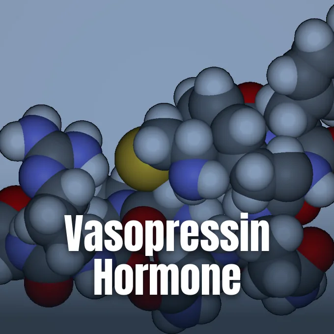
What is vasopressin? It is a peptide hormone made in the brain and stored in the Posterior Pituitary to manage water retention and vascular tone [7]. Known as Antidiuretic Hormone or AVP, the molecule acts on kidney tubules to return solute-free water into the bloodstream when body fluid tonicity climbs [11]. In clinical care across the United States and Europe, medical teams give Vasostrict, Pitressin, or Pressyn to treat vasodilatory shock and endocrine failure [10]. The hormone keeps fluid levels steady while supporting arterial pressure [11].
- What Is Vasopressin and Where Is It Produced?
- Synthesis Pathways in Hypothalamic Nuclei
- Vasopressin Signaling in the Collecting Duct
- Receptor Distribution Across Human Tissues
- Why Pressor Therapy Supports Septic Shock
- Clinical Evaluation of Arginine Vasopressin Deficiency
- Structural Comparisons With Oxytocin
- Plasma Detection Limits and Copeptin Assays
What Is Vasopressin and Where Is It Produced?
Vasopressin synthesis begins deep in the Hypothalamus. Large magnocellular neurons in the Paraventricular Nucleus and Supraoptic Nucleus transcribe the AVP gene into a large pre-prohormone precursor [11]. The cell packages this protein into secretory vesicles that travel along thin axons through the Pituitary Stalk down to the Posterior Pituitary lobe [8]. As these vesicles move toward axonal terminals, enzymes cleave the precursor molecule into arginine vasopressin, neurophysin II, and copeptin, allowing the finished nine-amino-acid peptide to collect inside axonal dilations termed Herring bodies until release into nearby capillary beds [1, 11]. The human body depends on this neurohypophyseal tract for fluid regulation [8].
Basal peptide levels circulate in picomolar ranges that often fall below the detection limits of common clinical assays [1, 6]. The hormone breaks down rapidly in human blood, showing a short half-life of 16 to 24 minutes [11]. Sensory osmoreceptors in the anterior Hypothalamus track solute changes of less than one percent, while vascular baroreceptors track blood volume loss and arterial wall stretch [8]. If arterial blood volume drops, sensor unloading overrides osmotic signals, triggering hormone release despite low salt levels [11].
Parvocellular neurosecretory cells in the Paraventricular Nucleus also synthesize the peptide. These cells release the hormone at the Median Eminence into the hypophyseal portal system feeding the Anterior Pituitary gland, where it acts with Corticotropin-Releasing Hormone to promote ACTH release during physiological stress [3, 11]. Sparapani and colleagues noted in Biomedicines that this hypothalamic wiring links osmotic control directly to systemic stress pathways [3].
Synthesis Pathways in Hypothalamic Nuclei
The chemical structure of arginine vasopressin, registered under ChemSpider identifier 559126 and Chemical Abstracts Service registry 113-79-1, comprises nine amino acid residues. Its peptide sequence, Cys-Tyr-Phe-Gln-Asn-Cys-Pro-Arg-Gly-NH2, possesses a disulfide bridge between cysteine residues at positions 1 and 6, forming a cyclic six-residue ring linked to a tripeptide tail that ends with a primary carboxamide group [9]. Cataloged under molecular formula C46H65N15O12S2 with an exact mass of 1083.4379 daltons in the ChEBI database of the European Bioinformatics Institute, the molecule acts as a Bronsted base in neutral fluids [7, 10]. A stable cyclic shape allows the peptide to bind target G-Protein Coupled Receptors on cell membranes [3, 12].
Gene transcription in the brain follows a diurnal rhythm set by the Suprachiasmatic Nucleus. In this circadian circuit, known as the transcription-translation feedback loop (TTFL), Per2 protein builds up and is phosphorylated by CK1E. The accumulated Per2 then represses the transcription factors Clock and BMAL1 to reduce its own levels in the cell. This inhibitory feedback loop also shuts down transcription factors for the AVP gene, modulating baseline hormone output throughout day and night [11].
Angiotensin II acts as a potent secretagogue that spurs peptide release when renal blood flow drops. Atrial Natriuretic Peptide from cardiac atria inhibits hormone output, while Cortisol also exerts negative feedback on hypothalamic cells. Ethanol intake suppresses peptide discharge by blocking voltage-gated calcium channels in neurohypophyseal terminals, leading to prompt diuresis and fluid loss. Alcohol stops fluid conservation in the kidneys [11].
Vasopressin Signaling in the Collecting Duct
The chief bodily role of vasopressin involves water saving within the renal collecting system [8]. Circulating hormone binds to V2 receptors on the basolateral membrane of principal cells in cortical and medullary collecting ducts, coupling to Gs alpha subunits to activate adenylate cyclase. Adenylate cyclase generates cyclic adenosine monophosphate (cAMP), which prompts protein kinase A to phosphorylate storage vesicles carrying Aquaporin-2 (AQP2) water channels [11, 12]. Phosphorylated vesicles fuse with the apical plasma membrane, inserting functional water pores that permit water to leave tubular filtrate and re-enter hypertonic renal tissue down an osmotic gradient. Solute-free water returns to the blood, yielding concentrated urine with low volume [11].
Cellular activation does not cease when the receptor undergoes internal uptake. Internalized V2 receptors form stable mega-complexes with Beta-Arrestin and heterotrimeric Gs proteins, sustaining cAMP generation from endosomal spaces to maintain prolonged stimulation of the Aquaporin-2 gene [11, 12]. Episodic calcium release from internal cell stores further coordinates vesicle movement to the cell surface [11]. Without active V2 signaling, collecting duct cells remain sealed to water [8].
The hormone also optimizes medullary solute gradients by regulating urea and sodium transporters. In inner medullary collecting ducts, the hormone promotes cell surface expression of urea transporters, allowing urea to move down concentration gradients into the interstitium. In the thick ascending limb of the Loop of Henle, the hormone stimulates the NKCC2 cotransporter. Phosphorylation of specific serine and threonine sites on the cytoplasmic tail of the transporter increases its presence on the cell membrane. Higher NKCC2 activity enhances sodium chloride uptake into medullary tissue, multiplying the osmotic driving force that pulls water through aquaporin channels [11].
Receptor Distribution Across Human Tissues
Vasopressin acts through three separate G-Protein Coupled Receptors: V1A, V1B, and V2 [12]. The AVPR1A gene, with a complementary DNA sequence of 1472 base pairs, encodes a 418-amino-acid polypeptide featuring seven transmembrane alpha-helices. Binding studies confirm that glutamate at residue 54 and arginine at residue 46 form key docking points for the peptide ligand, which can be blocked by Relcovaptan (SR-49059). When stimulated, V1A couples to Gaq/11 proteins to activate phospholipase C, producing inositol trisphosphate and diacylglycerol, which mobilize calcium from cellular stores to contract vascular myocytes and hepatocytes [1, 12, 13]. Platelets also express V1A docking targets, where hormone binding elevates intracellular calcium to facilitate platelet clumping and thrombosis [12].
V1A docking targets regulate microvascular tone within the kidneys. Located in high density on medullary interstitial cells and vasa recta, they constrict efferent arterioles while sparing afferent vessels, reducing inner medullary blood flow without diminishing outer medullary blood supply. Selective vascular constriction shields the deep renal medulla against solute wash-out [12].
The V1B receptor, also designated the V3 receptor, comprises 424 amino acids. It shares 45% sequence homology with V1A and Oxytocin Receptors, but only 39% homology with the V2 subtype. Expressed in Anterior Pituitary corticotropes and hippocampal CA2 pyramidal cells, it coordinates neuroendocrine responses to emotional and social stress [5, 12]. In contrast, V2 docking sites localize predominantly to renal collecting duct cells and hepatocytes, where receptor stimulation triggers the release of factor VIII and von Willebrand factor into systemic blood, targets frequently blocked by Vaptans like Conivaptan and Tolvaptan [7, 12]. Loss-of-function mutations in the AVPR2 gene abolish antidiuretic responses, causing hereditary Nephrogenic Diabetes Insipidus [8, 12].



Why Pressor Therapy Supports Septic Shock
In vasodilatory shock, systemic arterial resistance plummets because vascular smooth muscle loses resting tone. First-line clinical fluid therapy relies on intravenous fluids and Norepinephrine infusions, but patients often require escalating catecholamine doses that trigger arrhythmias, myocardial ischemia, and peripheral tissue injury. Intensive care teams add vasopressin infusions to raise mean arterial blood pressure while sparing catecholamine exposure [4, 11]. The hormone stimulates vascular V1A targets through intracellular calcium pathways that bypass desensitized adrenergic signaling in acidotic tissues [11, 12]. Restored vascular tone preserves organ perfusion during shock [11].
Early pressor efficacy was examined in a clinical trial published in JAMA by Gordon, Mason, Perkins, Cecconi, and colleagues. Conducted in 18 adult intensive care units in the United Kingdom between 2013 and 2015, the randomized trial evaluated early vasopressin titrated up to 0.06 units per minute against Norepinephrine. Patients were randomly assigned to vasopressin and hydrocortisone, vasopressin and placebo, norepinephrine and hydrocortisone, or norepinephrine and placebo. The primary outcome measured kidney failure-free days over 28 days [4].
Intravenous vasopressin infusions have also been used since 1970 to treat bleeding esophageal varices. By constricting mesenteric arterioles, the medication reduces blood inflow into the portal venous circulation, lowering variceal pressures and helping stabilize acute hemorrhage. Synthetic analogs like Terlipressin provide longer systemic vasoconstriction in portal hypertension. However, strong vasoconstriction carries clinical risks, including myocardial infarction, angina, abdominal cramping, nausea, sweating, pale skin, and intestinal hypoperfusion. Monitoring cardiac rhythm is mandatory during active infusion [11].
Clinical Evaluation of Arginine Vasopressin Deficiency
When lesions damage the hypothalamic-pituitary axis, hormone synthesis drops, producing arginine vasopressin deficiency, previously called central diabetes insipidus or Wolfram syndrome in rare genetic presentations. As Carmichael outlines in the Merck Manual Professional Edition, approximately 10% of hypothalamic neurosecretory neurons must survive to prevent symptomatic disease. Acquired causes include basal skull fractures, neurosurgical trauma, suprasellar tumors, Langerhans Cell Histiocytosis, sarcoidosis granulomas, and intracranial infections like encephalitis or meningitis. Without antidiuretic hormone, collecting ducts can’t reabsorb water, forcing patients to excrete between 3 and 30 liters of dilute urine daily with a specific gravity below 1.005 and osmolality under 200 mOsm/kg. Nocturia is almost always present, waking patients multiple times each night [8].
Differentiating central deficiency from nephrogenic resistance and primary polydipsia needs a standardized water deprivation test. Supervised patients stop fluid intake while clinicians monitor hourly body weight and urine osmolality until orthostatic hypotension appears, body weight drops by 5%, or sequential urine osmolality fails to rise by more than 30 mOsm/kg. In healthy individuals, urine osmolality exceeds 700 to 800 mOsm/kg after fluid restriction, whereas patients with complete central deficiency produce dilute urine below plasma osmolality [8].
Injecting five units of aqueous vasopressin or ten micrograms of Desmopressin confirms the diagnosis by causing urine osmolality to surge by 50% to over 100% in complete central deficiency. Partial deficiency yields a 15% to 50% increase. Nephrogenic resistance shows minimal change under 50 mOsm/kg, while primary polydipsia shows no change. Maintenance therapy relies on Desmopressin, an analog that provides 12 to 24 hours of selective antidiuresis with minimal pressor effects [8].
Structural Comparisons With Oxytocin
Vasopressin shares an evolutionary kinship with oxytocin, another nonapeptide produced in adjacent hypothalamic magnocellular neurons [5, 11]. In mammals, genes for both hormones sit on the same chromosome separated by fewer than 15,000 base pairs, arising from an ancestral gene duplication. Both molecules contain nine amino acids linked by a disulfide bridge between sites 1 and 6. Their chemical structures differ at only two sites: the pressor peptide carries phenylalanine at position 3 and arginine at position 8, whereas oxytocin carries isoleucine at position 3 and leucine at position 8. In swine, lysine replaces arginine at position 8, producing lysine vasopressin or Lypressin. Because of their matching nonapeptide structures, high concentrations of AVP can stimulate uterine oxytocin receptors, while oxytocin retains modest antidiuretic activity at renal V2 receptors [11].
In the central nervous system, Benarroch reports in Neurology that both peptides act as neuromodulators governing social cognition, emotional processing, and hippocampal synaptic plasticity. However, oxytocin displays anxiolytic and calming actions, whereas central vasopressin release promotes anxiety, autonomic arousal, and vigilance during environmental stress [5].
Neurobiological studies in rodents show how receptor patterns influence social organization. Monogamous Prairie Voles express dense AVPR1A docking sites in the ventral forebrain and ventral pallidum, coordinating reward circuits that consolidate partner bonds, whereas promiscuous Montane Voles show sparse binding in these reward nuclei despite sharing 99% coding sequence identity [11, 13]. Male mice lacking the AVPR1A gene exhibit impaired social recognition and reduced anxiety on maze tests, while Sprague-Dawley Rats and Brattleboro Rats show diurnal variations in receptor expression [13].
Plasma Detection Limits and Copeptin Assays
Direct measurement of circulating vasopressin poses persistent analytical challenges [1, 6]. Leng and Sabatier reported in the Journal of Neuroendocrinology that clinical bioassays and immunoassays yield plasma concentrations differing by two orders of magnitude in identical patient samples. The nonapeptide circulates at picomolar levels, exhibits an in vivo half-life under 24 minutes, and binds extensively to platelets and plasma proteins [1, 6]. High-molecular-weight plasma components interfere with antibody binding, undermining assay accuracy [6].
To overcome analytical hurdles, diagnostic medicine relies on copeptin as a surrogate biomarker for hormone release. Formed as the C-terminal segment of pre-provasopressin, copeptin is secreted into circulation in an exact 1:1 molar ratio with arginine vasopressin, remaining stable in room-temperature plasma for several days without adhering to blood platelets [1, 2]. Clinical assays quantify copeptin readily using automated sandwich immunoluminometric tools. Bankir, Bichet, and Morgenthaler showed in the Journal of Internal Medicine that circulating copeptin concentrations parallel plasma osmolality during water deprivation testing, providing robust diagnostic split between central deficiency, nephrogenic resistance, and primary polydipsia [1, 8].
Maternal copeptin also provides diagnostic value in pregnancy disorders. In a prospective investigation published in Hypertension, Santillan, Grobe, Scroggins, Min, Sandgren, Pearson, Leslie, Hunter, Zamba, and Gibson-Corley measured maternal plasma copeptin throughout pregnancy. Plasma copeptin was significantly higher in women who developed preeclampsia than in normotensive controls, maintaining statistical significance after adjusting for body mass index, maternal age, and preexisting hypertension. Abnormal hormone secretion plays an early pathogenic role in preeclampsia [2].
- ACADEMIC JOURNAL Bankir, L., Bichet, D. G., & Morgenthaler, N. G. (2017). Vasopressin: physiology, assessment and osmosensation. Journal of Internal Medicine, 282(4), 284-297. [Article Link]
- ACADEMIC JOURNAL Santillan, M. K. O., Santillan, D. A., Scroggins, S. M., Min, J. Y., Sandgren, J. A., Pearson, N. A., Leslie, K. K., Hunter, S. K., Zamba, G. K. D., Gibson-Corley, K. N., & Grobe, J. L. (2014). Vasopressin in Preeclampsia. Hypertension, 64(4), 852-859. [Article Link]
- ACADEMIC JOURNAL Sparapani, S., Millet-Boureima, C., Oliver, J., Mu, K., Hadavi, P., Kalostian, T., Ali, N., Avelar, C. M., Bardies, M., Barrow, B., Benedikt, M., Biancardi, G., Bindra, R., Bui, L., Chihab, Z., Cossitt, A., Costa, J., Daigneault, T., Dault, J.,. Forget, A. (2021). The Biology of Vasopressin. Biomedicines, 9(1), 89. [Article Link]
- ACADEMIC JOURNAL Gordon, A. C., Mason, A. J., Thirunavukkarasu, N., Perkins, G. D., Cecconi, M., Cepkova, M., Pogson, D. G., Aya, H. D., Anjum, A., Frazier, G. J., Santhakumaran, S., Ashby, D., Brett, S. J., & Investigators, f. t. V. (2016). Effect of Early Vasopressin vs Norepinephrine on Kidney Failure in Patients With Septic Shock. JAMA, 316(5), 509-509. [Article Link]
- ACADEMIC JOURNAL Benarroch, E. E. (2013). Oxytocin and vasopressin. Neurology, 80(16), 1521-1528. [Article Link]
- ACADEMIC JOURNAL Leng, G., & Sabatier, N. (2016). Measuring Oxytocin and Vasopressin: Bioassays, Immunoassays and Random Numbers. Journal of Neuroendocrinology, 28(10). [Article Link]
- WEBSITE European Bioinformatics Institute. (2015). Vasopressin (CHEBI:9937). ChEBI. [Article Link]
- WEBSITE Carmichael, J. (2025). Arginine Vasopressin Deficiency (Central Diabetes Insipidus). Merck Manual Professional Edition. [Article Link]
- WEBSITE ChemSpider. (2026). Vasopressin (ChemSpider ID 559126). Royal Society of Chemistry. [Article Link]
- WEBSITE Kyoto Encyclopedia of Genes and Genomes. (n.d.). Vasopressin (KEGG DRUG D00101). [Article Link]
- WEBSITE Wikipedia contributors. (n.d.). Vasopressin. Wikipedia, The Free Encyclopedia. [Article Link]
- WEBSITE Wikipedia contributors. (n.d.). Vasopressin receptor. Wikipedia, The Free Encyclopedia. [Article Link]
- WEBSITE Wikipedia contributors. (n.d.). Vasopressin receptor 1A. Wikipedia, The Free Encyclopedia. [Article Link]
Cover image: Fvasconcellos / Wikimedia Commons, Public domain


