





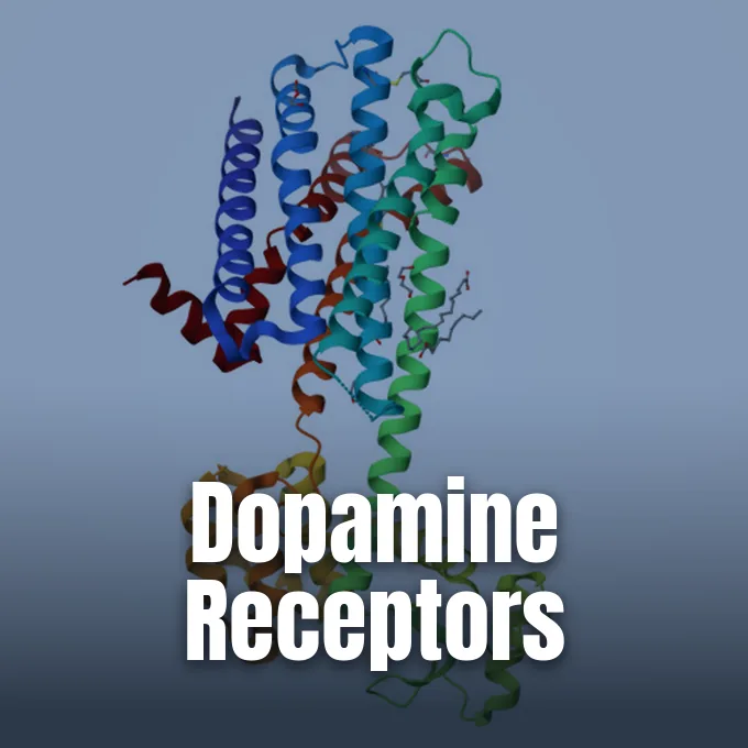
Dopamine receptors translate chemical signals from dopamine into specific actions in the brain and body tissues. First proposed as separate entities in 1976, these membrane proteins guide movement, motivation, memory, and hormone release [1]. When dopamine binds, it triggers internal pathways that either raise or lower second messengers. Brain cells rely on this balance to function normally. Signal faults contribute to mental illness, shaping how doctors at clinical centers evaluate brain disorders [12].
What Are Dopamine Receptors?
How do brain cells read chemical cues in the synapse? Dopamine receptors belong to the large G protein-coupled receptor superfamily, grouping into five distinct subtypes labeled D1 through D5 that divide into two major families based on their structural homology and pharmacological binding profiles. They span the cell membrane. Pharmacologists once thought D6 or D7 subtypes existed, but biochemical tests found none [1].
Subtype concentrations vary across brain structures in humans and mice. Overall density follows a rank order where D1 is the most abundant, followed in order by D2, D3, D5, and D4 [1]. Levels of D1 and D2 run 10 to 100 times higher than levels of D3, D4, or D5. The dorsal striatum, which includes the caudate and putamen, joins the ventral striatum in the nucleus accumbens as the primary zone for D1 and D2 messenger RNA, while smaller amounts occupy cortical layers, the amygdala, and the hypothalamus [2]. Solomon H. Snyder and Philip Seeman tracked these binding sites in early radioligand assays to map basal ganglia distribution [3].
Receptor density shapes how neural circuits react to dopamine. When dopamine levels drop, patients experience deep motor and mood changes, which doctors recognize as common symptoms of dopamine deficiency in clinical neurology. Membrane proteins also bind interacting partners like NCS-1 in the dentate gyrus to guide memory formation and exploratory behavior [3]. Cellular couplings ensure that neurons adjust their response to shifting motor demands [12].
The D1 and D2 Families
The D1-like family consists of D1 and D5 proteins, encoded respectively by the DRD1 and DRD5 genes [1]. Both subtypes couple to the Gsα subunit, while D1 can also recruit Golf within the olfactory tubercle and striatum [2]. They activate adenylyl cyclase. Enzyme activity drives up cyclic adenosine monophosphate (cAMP) levels, activating protein kinase A to phosphorylate DARPP-32, which inhibits protein phosphatase 1 and amplifies excitatory signals in receiving neurons [1].
In contrast, D2-like members include D2, D3, and D4, which couple to Giα to block adenylyl cyclase and cut cAMP production [1]. Beyond this basic pathway, D2 activates an Akt, beta-arrestin 2, and PP2A complex that inhibits Akt, thereby freeing glycogen synthase kinase 3 to alter cell survival and movement [12]. Cyclin-dependent kinase 5 keeps D1 activity balanced, preventing excess excitation in striatal networks. Signaling paths also regulate inward-rectifying potassium channels and modify NMDA currents during repetitive firing [1]. Cells maintain balance through these opposing actions. Paul Greengard first outlined these kinase cascades when studying how psychiatric drugs alter post-synaptic striatal targets in medicine [3].
Splicing of the DRD2 gene produces two distinct protein isoforms called D2Sh and D2Lh [3]. The short D2Sh isoform functions presynaptically as an autoreceptor, adjusting transmitter synthesis and release to prevent synaptic flooding [1]. The long D2Lh variant acts as a classic post-synaptic receptor. A third variant replaces valine at position 270 with VVQ [3].
Molecular Architecture of Binding Pockets
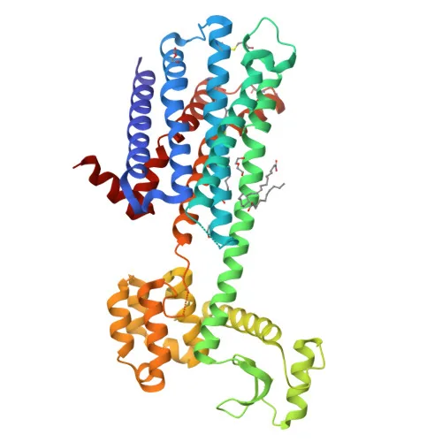
High-resolution crystallography has clarified how dopamine receptors distinguish between therapeutic drugs. In 2018, S. Wang, B. Roth, and fellow pharmacologists at the University of North Carolina at Chapel Hill resolved the crystal structure of the D2 receptor bound to the atypical antipsychotic drug risperidone. Their model, deposited in the RCSB Protein Data Bank as 6CM4 and reported in Nature, revealed an orthosteric binding site next to a second pocket where engagement with allosteric modulators like SB269652 alters receptor function without displacing orthosteric ligands. Binding kinetics differ markedly. Atomic determinants explain why risperidone binds tightly to D2 while showing distinct clinical actions from older typical neuroleptics [4].
Receptor activation depends on transitions between the active D2HighR state and the inactive D2LowR state. Agonist binding shifts transmembrane helices 5 and 6 outward, creating an opening for G proteins at the cytoplasmic surface [3]. Antagonists lock the protein in its inactive state. Because antipsychotic drugs bind this inactive shape, mapping contact residues gives medicinal chemists a reliable template to design drugs that avoid off-target receptors and reduce side effects like sedation or tremors [4].
Structural work on D1 reveals contrasting features that support Gs coupling. Agonist binding moves transmembrane helix 6 outward and extends helix 5 by two helical turns, expanding the intracellular interface to 1,520 square angstroms. Serine residues S198, S199, and S202 act as microswitches that trigger receptor activation. In transmembrane helix 7, residues V317 and W321 provide hydrophobic contacts that help the binding cavity accommodate non-catechol agonists like SKF-38393 [2].

How Dopamine Receptors Direct Learning
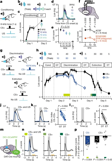
How do neural circuits convert unexpected events into learned habits? In a 2020 study published in Nature, Yusuke Iino, Haruo Kasai, and Sho Yagishita showed that reward learning requires balanced actions between D1 and D2 subtypes. In head-restrained mice, tone conditioning caused broad stimulus generalization that depended on D1 receptors in the nucleus accumbens. Discrimination learning then refined this behavior when mice detected a brief pause in dopamine release, lasting just 0.4 seconds, known as a dopamine dip [5].
When this dip occurs, D2 receptors (D2R) disinhibit adenosine A2A receptor signaling in spiny projection neurons, prompting enlargement of dendritic spines via CaMKII. Dendritic enlargement allows mice to distinguish reward tones from unrewarded tones. Extinction learning does not use dopamine dips or D2 spiny projection neurons. When methamphetamine disrupted dopamine transmission, mice lost discrimination; applying a D2 receptor antagonist reversed the defect and restored spine enlargement [5].
Synaptic learning also operates within defined timing rules. In an unreviewed preprint on bioRxiv, Kenji Yamaguchi and colleagues measured how quickly mice link conditioned auditory tones to sucrose rewards in the nucleus accumbens. Reinforcement occurs only when reward delivery arrives within 1 second after cue presentation. Conditioning failed without functional D1 dopamine receptors and CaMKII enzymes. Replacing sensory tones with optogenetic stimulation of synaptic inputs did not shorten this 1-second limit, confirming that dopamine timing in the ventral striatum sets defined biochemical bounds for behavioral reinforcement in the mammalian brain [6].

Addiction Circuits in the Nucleus Accumbens
Drugs of abuse hijack reinforcement circuits in the mesolimbic dopamine pathway. Dopamine drives incentive salience—the motivational urge of wanting—rather than sensory pleasure or liking [1]. Stimulants like cocaine block the dopamine transporter, leaving excess dopamine in the synapse. Over time, repeated drug exposure downregulates post-synaptic D2 receptors, dulling normal reward responses and prompting animals to seek more drug to overcome reduced signaling capacity [7].
Restoring receptor counts in the brain can reverse drug intake. At Brookhaven National Laboratory, Panayotis Thanos and researchers supported by the NIAAA and Department of Energy injected an adeno-associated virus carrying the D2 gene into the nucleus accumbens of rats. The treatment produced a 75 percent drop in cocaine self-administration in trained rats, lasting six days before baseline consumption returned. Gene therapy proved that boosting D2 receptor levels blunts drug-taking behavior in rodents [7].
Receptor subtypes also influence psychostimulant toxicity. S. Ares-Santos and R. Moratalla found that genetic deletion of either D1 or D2 dopamine receptors protects striatal neurons against methamphetamine damage. In D1 knockout mice, protection comes from reduced dopamine turnover and hypothermia blockade, while D2 deletion cuts transporter activity [8]. Popular interest in receptor downregulation has popularized lifestyle trends like dopamine detox methods that aim to recalibrate everyday motivation. Behavioral strategies reflect growing awareness of how neural receptors adapt to chronic stimulation [12].
Genetic Variations in DRD4 Alleles
Human cognitive traits vary with receptor genetics. The dopamine D4 receptor, encoded by the DRD4 gene on chromosome 11p15.5, displays polymorphic diversity within its third exon, where a variable number tandem repeat alters an intracellular loop [1]. Geneticists focus on the 7-repeat allele. The 7-repeat allele occurs frequently in children diagnosed with attention-deficit hyperactivity disorder [9].
In a clinical trial update, M. Gornick, A. Addington, and J. Rapoport analyzed 166 pediatric ADHD cases and 282 controls. The 7-repeat allele appeared significantly more often in ADHD cases than controls, yielding an odds ratio of 1.2 with P = 0.028. Following 107 children from a baseline mean age of 9 to a follow-up mean age of 15 revealed unexpected cognitive trends, where ADHD children carrying the 7-repeat allele scored higher on the WISC-III cognitive test (P = 0.013–0.07) and had better long-term clinical outcomes. Long-term data suggest the allele confers a more benign form of the syndrome [9].
Biochemically, the 7-repeat allele shows reduced gene expression compared to shorter variants. It blunts dopamine signaling in the prefrontal cortex without causing major cognitive deficits. Medications like methylphenidate and amphetamine compensate for this muted signaling by blocking the dopamine transporter, raising extracellular dopamine to sharpen attention in school-age children [1].
Dopamine Receptors in Neurodegenerative Illness
Neurodegenerative disorders dismantle dopaminergic circuits, altering receptor density in the basal ganglia. In Parkinson’s disease (PD), loss of dopamine neurons in the substantia nigra causes transmitter depletion in the striatum. While levodopa remains the standard replacement therapy, long-term use triggers dyskinesias. To delay levodopa therapy, neurologists prescribe dopamine receptor agonists like rotigotine, pramipexole, apomorphine, or pergolide, though these drugs carry risks of heart valvulopathy and dopamine dysregulation syndrome [10].
Dopaminergic deficits also appear in Alzheimer’s disease. A systematic review and network meta-analysis by Xi Pan and coworkers, published in Frontiers in Aging Neuroscience, examined dopaminergic markers in 512 patients and 500 healthy controls. The analysis found significantly lower dopamine levels in Alzheimer’s patients, with a standardized mean difference of -1.56, while receptor levels dropped substantially to a standardized mean difference of -5.05 for D1 and -1.13 for D2. Meta-analytic findings confirm that dopamine receptors decline in addition to cholinergic loss during dementia progression [11].
Similar changes occur in Huntington’s disease and multiple sclerosis. C. Rangel-Barajas and colleagues noted that dopamine receptors reorganize as a compensatory mechanism before contributing to neuronal death [12]. Understanding these shifts helps scientists track disease stages and design neuroprotective agents [10].
Signaling Roles Outside the Brain
Dopamine receptors also operate throughout peripheral organs. In the cardio-pulmonary system, arterial smooth muscle expresses D1, D2, D4, and D5, mediating vasodilation [1]. Clinicians use the selective D1 partial agonist fenoldopam to treat acute hypertension because it lowers blood pressure without crossing the blood-brain barrier [2]. In the heart, receptors on the myocardium increase contractile force and cardiac output without raising heart rate [1].
Metabolic control in the kidneys and pancreas also requires dopaminergic signaling. Renal proximal tubule cells express high receptor densities, where dopamine regulates sodium excretion and fluid balance. In the endocrine pancreas, insulin-producing beta cells in the Islets of Langerhans express D2 receptors and co-secrete dopamine with insulin. Bound D2 acts as a negative regulator, inhibiting insulin secretion. Because antipsychotic medications block D2 receptors, they disrupt this pancreatic brake, causing weight gain and glycemic dysregulation as metabolic side effects. Adipose tissue also expresses D1, D2, D4, and D5 to regulate lipid breakdown and thermogenesis [1].
Receptors form heteromers with other G protein-coupled receptor (GPCR) partners, including adenosine A2A, ghrelin, and alpha-1B adrenoceptors [1]. Heteromeric pairs alter ligand affinity and downstream signaling. Through these multifaceted connections, dopamine pathways maintain homeostasis across both brain and body tissues [12].
- WEBSITE Wikipedia contributors. (2026). Dopamine receptor. Wikipedia, The Free Encyclopedia. [Article Link]
- WEBSITE Wikipedia contributors. (2026). Dopamine receptor D1. Wikipedia, The Free Encyclopedia. [Article Link]
- WEBSITE Wikipedia contributors. (2026). Dopamine receptor D2. Wikipedia, The Free Encyclopedia. [Article Link]
- ACADEMIC JOURNAL Wang, S., Che, T., Levit, A., Shoichet, B. K., Wacker, D., & Roth, B. L. (2018). Structure of the D2 dopamine receptor bound to the atypical antipsychotic drug risperidone. Nature, 555(7695), 269–273. [Article Link]
- ACADEMIC JOURNAL Iino, Y., Sawada, T., Yamaguchi, K., Tajiri, M., Ishii, S., Kasai, H., & Yagishita, S. (2020). Dopamine D2 receptors in discrimination learning and spine enlargement. Nature, 579(7800), 555–560. [Article Link]
- PREPRINT Yamaguchi, K., Maeda, Y., Sawada, T., Iino, Y., Tajiri, M., Nakazato, R., Kasai, H., & Yagishita, S. (2019). The minimal behavioral time window for reward conditioning in the nucleus accumbens of mice. bioRxiv. [Article Link]
- ONLINE NEWS Brookhaven National Laboratory. (2008). Gene therapy for addiction: Flooding brain with ‘pleasure chemical’ receptors works on cocaine, as on alcohol. ScienceDaily. [Article Link]
- ACADEMIC JOURNAL Ares-Santos, S., Granado, N., & Moratalla, R. (2013). The role of dopamine receptors in the neurotoxicity of methamphetamine. Journal of Internal Medicine, 273(5), 437–453. [Article Link]
- ACADEMIC JOURNAL Gornick, M., Addington, A., Shaw, P., Bobb, A., Sharp, W., Greenstein, D., Arepalli, S., Castellanos, F., & Rapoport, J. (2007). Association of the dopamine receptor D4 (DRD4) gene 7-repeat allele with children with attention-deficit/hyperactivity disorder (ADHD): An update. American Journal of Medical Genetics Part B: Neuropsychiatric Genetics, 144B(3), 379–382. [Article Link]
- ACADEMIC JOURNAL Hisahara, S., & Shimohama, S. (2011). Dopamine receptors and Parkinson’s disease. International Journal of Medicinal Chemistry, 2011, 1–16. [Article Link]
- ACADEMIC JOURNAL Pan, X., Kaminga, A. C., Wen, S. W., Wu, X., Acheampong, K., & Liu, A. (2019). Dopamine and dopamine receptors in Alzheimer’s disease: A systematic review and network meta-analysis. Frontiers in Aging Neuroscience, 11, 175. [Article Link]
- ACADEMIC JOURNAL Beaulieu, J., Espinoza, S., & Gainetdinov, R. R. (2014). Dopamine receptors – IUPHAR Review 13. British Journal of Pharmacology, 172(1), 1–23. [Article Link]


