





Research into how tetanus works begins with Clostridium tetani, a spore-forming anaerobic bacterium first described in medical literature by Greek physician Hippocrates in the fifth century BCE. When dormant endospores enter deep beneath the skin into an oxygen-deprived wound, they turn into rod-shaped vegetative cells and release tetanospasmin. This potent neurotoxin enters peripheral motor nerves, journeys backward along axonal pathways into the spinal cord, and stops the essential release of inhibitory neurotransmitters from central Renshaw cells. Deprived of normal calming signals, alpha motor neurons fire continuously, locking major muscle groups into painful, uncontrolled contractions [1].
- How Tetanus Works at Wound Sites
- Clostridium Tetani Spores in the Soil
- Molecular Steps: How Tetanus Works in Nerve Cells
- Why Muscle Spasms Trigger Lockjaw and Opisthotonos
- Four Distinct Clinical Forms of Tetanus
- Emergency Care with Antitoxin Neutralization
- Immunity Gaps: How Tetanus Works Without Antibodies
- Global Vaccination Schedules and Prevention
How Tetanus Works at Wound Sites
Skin breaks let spores in. Popular belief links tetanus to rusty iron, but rust does not make bacteria. Instead, rough oxidized nails lying outdoors in soil or manure provide porous surfaces where resilient bacterial spores collect over time before penetrating deep human tissue [7].
Once under the skin, endospores need an anaerobic environment with low oxidation-reduction potential to wake up. Healthy human tissue has rich arterial blood flow that delivers oxygen, which stops Clostridium tetani from multiplying. But crush wounds, dead tissue, burns, and coinfections consume local oxygen. In those devitalized, oxygen-starved tissue pockets, local oxidation-reduction potential drops, prompting dormant spores to shed their protective coats and germinate into active vegetative bacilli [5].
As vegetative cells multiply, they release two exotoxins: tetanolysin and tetanospasmin. Tetanolysin acts as a hemolysin that breaks red blood cells, but it has no known role in spastic symptoms. Tetanospasmin is the true cause of disease. A detailed analysis of how tetanus works in human tissue reveals that this neurotoxin is among the deadliest poisons known, with an estimated human lethal threshold of just 2.5 nanograms per kilogram of human body mass. For a 70-kilogram person (about 154 pounds), a tiny dose of 175 nanograms can kill. The Centers for Disease Control and Prevention notes that prior to vaccination, tetanus was fatal in most clinical cases [1].
Clostridium Tetani Spores in the Soil
Microbial spores resist decay. The bacterium Clostridium tetani is a slender, motile, gram-positive rod that lives without oxygen. When environmental moisture or essential nutrients run out, each bacillus forms a round spore at one end, giving the cell a characteristic drumstick shape under the microscope. Microscopic records and crystallographic data at the RCSB Protein Data Bank document this structural form. These tough endospores live in garden soil, farm manure, street dust, and the digestive tracts of sheep, cattle, horses, dogs, cats, rats, and domestic chickens worldwide [1].
Endospores endure boiling water, resist common chemical disinfectants like phenol, and easily withstand autoclaving at 249.8 degrees Fahrenheit (121 degrees Celsius) for 10 to 15 minutes. Chemical disinfectants cannot destroy them. In dry soil, spores stay infectious for more than 40 years [3].

A comprehensive understanding of how tetanus works across various injury types helps doctors identify non-obvious risks. Surveillance records from the United States show that puncture wounds cause half of all acute cases, while lacerations account for a third and minor scrapes contribute about 9%. Accidental punctures from stepping on an outdoor nail caused nearly a third of all puncture wounds. Other risk points include chronic diabetic foot ulcers, gangrene, frostbite, animal bites, burns, and subcutaneous drug injection using contaminated heroin [2].
Molecular Steps: How Tetanus Works in Nerve Cells
Modern investigations into how tetanus works at the synaptic level show that tetanospasmin has a modular structure tailored for nerve invasion. The tetX gene on the PE88 plasmid codes for a 150-kilodalton protein precursor. Bacterial proteases split this 150-kilodalton precursor into a 100-kilodalton heavy chain and a 50-kilodalton light chain joined by an essential disulfide bridge to enable cell entry [8].
After binding to the motor nerve terminal, the toxin enters the cell through endocytosis. While related Clostridium botulinum neurotoxins stay at the peripheral neuromuscular junction to block acetylcholine release, tetanus toxin journeys backward up motor axons toward the spinal cord. It rides inside non-acidified vesicles using dynein molecular motors along axonal microtubules at steady speeds. This retrograde axonal transport takes between 2 and 14 days, depending on the distance between the injured limb and the spinal gray matter. When the transport vesicle reaches motor neuron dendrites in the spinal gray matter, the toxin exits into the synaptic cleft by transcytosis to target nearby inhibitory interneurons [10].
The toxin binds to presynaptic receptors on inhibitory interneurons, known as Renshaw cells, and enters acidic endosomes. Research shows this entry depends on synaptic vesicle protein 2 (SV2) recycling. In the acidic intravesicular environment, the heavy chain forms a channel that helps the light chain cross into the cytosol to reach its intracellular target. Host thioredoxin reductase enzymes reduce the disulfide bond, freeing the light chain. As an active zinc endopeptidase, this light chain cleaves synaptobrevin II at the Gln76-Phe77 peptide link [11].
Why Muscle Spasms Trigger Lockjaw and Opisthotonos
Synaptobrevin, also called vesicle-associated membrane protein (VAMP), forms the SNARE core that lets vesicles fuse with the cell membrane. In a healthy nervous system, inhibitory interneurons release gamma-aminobutyric acid (GABA) and glycine to stop motor neurons from firing continuously. When tetanospasmin cuts synaptobrevin, vesicle fusion stops. Deprived of GABA and glycine, motor neurons fire out of control. This molecular shutdown explains how tetanus works to paralyse normal muscle coordination, making opposing flexor and extensor muscle groups contract simultaneously without relief [12].
Cranial pathways are short. Because shorter cranial nerves deliver tetanospasmin more rapidly than long peripheral leg nerves, facial and jaw muscles succumb first, leading over 50% of patients to present with masseter trismus, commonly termed lockjaw. In addition, spasms across facial and throat muscles produce risus sardonicus, a painful fixed smile, with dysphagia, drooling, fever, and severe difficulty speaking [5].
As disinhibition spreads along the spine, muscle rigidity grips the neck, back, chest, and limbs. Because powerful dorsal back muscles easily overpower weaker abdominal muscles, continuous tonic contractions bend the spine backward into a rigid arch called opisthotonos. Reflex spasms occur without warning, triggered by minor noises, bright lights, drafts, or touch. Contractions can break bones in the spine or tear tendons. When spasms seize throat muscles, laryngospasm blocks the airway, and aspiration pneumonia develops in 50% to 70% of fatal cases examined at autopsy [1]. Because the toxin leaves the cerebral cortex unharmed, patients stay fully conscious throughout these agonizing convulsions.
Four Distinct Clinical Forms of Tetanus
Doctors group tetanus into four clinical forms based on age and symptom spread. Clinical tracking of how tetanus works as symptoms progress highlights the classic downward spread of muscular rigidity. Generalized tetanus is the most common form, accounting for more than 80% of all cases. In severe cases, autonomic nerve instability causes swinging blood pressure, high heart rate, fever, and profuse sweating, requiring weeks of intensive medical support. Muscle spasms continue for three to four weeks before easing, and full recovery takes months in intensive care. Modern surveillance shows that even with intensive hospital therapy, tetanus is fatal in approximately 11% of reported cases, mainly among unvaccinated individuals over age 60 [1].
Other forms are rare. In localized tetanus, muscle stiffness stays limited to the injured body part, carrying a low 1% fatality rate although it can occasionally evolve into generalized illness. Cephalic tetanus makes up under 3% of cases following head injuries or middle ear infections, presenting with distinct cranial nerve palsies that can advance to generalized illness [6].
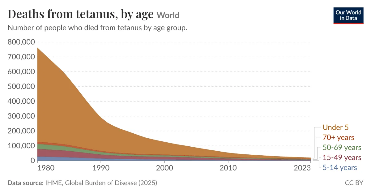
Neonatal tetanus, also called trismus nascentium, strikes newborn infants born to mothers who lack protective antibodies. Neonatal infection occurs when people use unsterile instruments to cut the umbilical cord or cover the healing stump with contaminated soil or animal dung. Symptoms show up four to 14 days after birth, averaging seven days, as infants stop nursing and suffer rigid spasms. In 1998, neonatal tetanus killed about 215,000 newborns worldwide before global vaccination initiatives curtailed transmission [4].
The World Health Organization notes that unclean delivery practices and lack of maternal immunization remain the driving factors behind neonatal infections.

Emergency Care with Antitoxin Neutralization
Diagnosis rests on clinical signs because lab blood tests cannot measure circulating toxin. Because of how tetanus works deep inside central nerve synapses, standard blood tests cannot detect circulating toxin. Wound cultures isolate C. tetani in only 30% of patients. The bedside spatula test helps confirm the diagnosis: touching the posterior pharyngeal wall causes patients to bite down involuntarily in 94% of cases, while healthy people gag [6].
Medical teams act quickly to neutralize free toxin before it enters nerve endings. Clinicians administer human tetanus immune globulin (TIG), usually prescribing a single intramuscular dose of 500 units for children and adults to bind circulating toxin. If TIG is unavailable, doctors administer intravenous immune globulin (IVIG), which also contains protective antitoxin antibodies. Part of the dose is injected around the wound edges. Doctors clean the wound thoroughly, cutting away dead tissue and removing foreign debris to destroy the low-oxygen environment that bacteria need to multiply [1]. Intravenous metronidazole is given for 10 days to eradicate remaining vegetative bacteria at the site.
Bound toxin stays fixed. Because antitoxin cannot dislodge tetanospasmin once it binds nerve cells, biological healing requires weeks while damaged motor terminals sprout entirely new synaptic connections. Inside a quiet, darkened intensive care unit designed to avoid sensory triggers, continuous diazepam infusions and intravenous magnesium relax rigid muscles while stabilizing erratic autonomic blood pressure swings. When respiratory muscles spasm, endotracheal intubation or early tracheotomy with mechanical ventilation protects the patient’s airway until endogenous nerve function returns [6].
Immunity Gaps: How Tetanus Works Without Antibodies
Does surviving severe tetanus protect a person from catching it again? The answer is no, showing another unique aspect of how tetanus works in humans. Tetanospasmin is so biologically potent that an amount capable of causing death is far too small to trigger a protective antibody response from the immune system. Patients who recover develop no natural immunity. As a result, anyone who recovers from clinical tetanus remains fully susceptible and needs a complete course of vaccine [4].
Vaccine development took decades of research across Europe and Asia. In 1884, Italian researchers Antonio Carle and Giorgio Rattone at the University of Turin proved tetanus was transmissible by injecting pus from a fatal human case into animals. In 1889, Japanese physician Kitasato Shibasaburo isolated Clostridium tetani, showed it caused disease in animals, and discovered that antibodies neutralized the toxin. Edmond Nocard demonstrated passive protection using animal antitoxin in 1897. In 1924, scientists discovered that treating the toxin with formaldehyde yielded an inactivated tetanus toxoid that created safe immunity, leading to widespread military use during World War II [1].

Antibodies fade over time. National health surveys conducted in the United States show that while over 80% of young people carry protective antibodies, that number drops to 27.8% among adults aged 70 and older. Older adults rarely receive ten-year boosters, leaving them at greatest risk [13].
Global Vaccination Schedules and Prevention
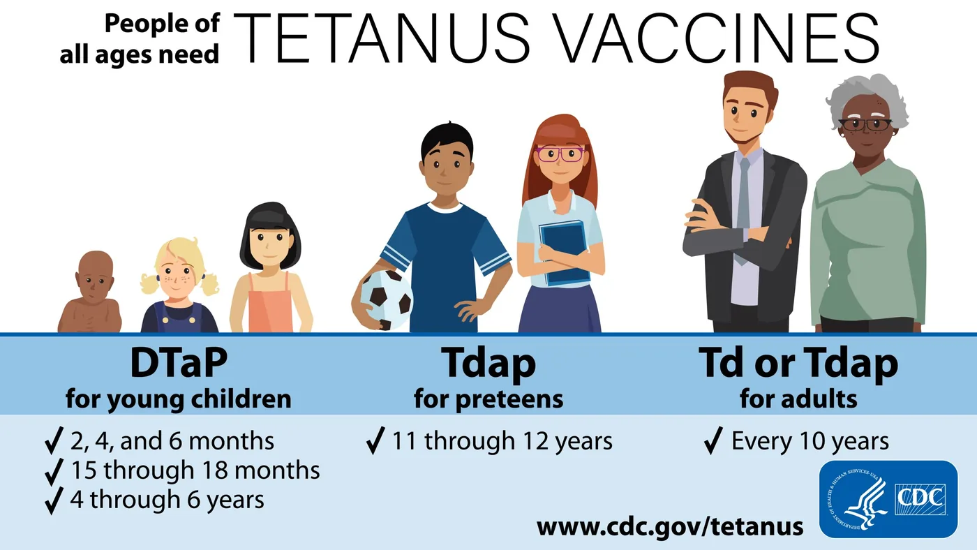
Vaccines protect children early. The World Health Organization recommends six childhood doses starting at six weeks of age. Young infants receive DTaP, which protects against diphtheria, tetanus, and acellular pertussis. Older children and adults receive Tdap or Td boosters every 10 years, with Tdap recommended once before age 65. By 2025, about 85% of infants globally had received three initial doses [9].
Maternal immunization coupled with clean delivery practices protects newborn babies. A maternal dose of Tdap given between weeks 27 and 36 of pregnancy transfers protective antibodies across the placenta to the baby. The Maternal and Neonatal Tetanus Elimination initiative, launched in 1999 by UNICEF, WHO, and the United Nations Population Fund (UNFPA), achieved an 84% reduction in newborn deaths between 2000 and 2021. In Uganda, after a major public health campaign, tetanus was eliminated in 2011. As of July 2026, only eight countries worldwide have yet to eliminate maternal and neonatal tetanus [4].
Wound care follows clear rules. People with contaminated injuries who have had fewer than three lifetime doses, or an unknown vaccine history, receive both tetanus toxoid and human tetanus immune globulin. If an injured person has had three or more doses, a booster is given only if five or more years have passed since their last shot. Prior to vaccines, the United States saw between 500 and 600 cases per year (about 0.4 cases per 100,000 population). Today, clean wound care and high vaccine coverage keep annual cases below 40 [1].
- REPORT Centers for Disease Control and Prevention. (2021). Chapter 21: Tetanus. In Epidemiology and Prevention of Vaccine-Preventable Diseases. [Article Link]
- WEBSITE Centers for Disease Control and Prevention. (2024). About Tetanus. [Article Link]
- WEBSITE MedlinePlus. (2025). Tetanus: MedlinePlus Medical Encyclopedia. [Article Link]
- REPORT World Health Organization. (2024). Tetanus Fact Sheet. [Article Link]
- WEBSITE Medscape. (2024). Tetanus: Background, Pathophysiology, Etiology. [Article Link]
- WEBSITE CheckOrphan. (2014). Tetanus Overview and Treatment. [Article Link]
- WEBSITE Wikipedia contributors. (2024). Tetanus. [Article Link]
- WEBSITE Wikipedia contributors. (2024). Tetanus toxin. [Article Link]
- WEBSITE Wikipedia contributors. (2024). Tetanus vaccine. [Article Link]
- ACADEMIC JOURNAL Dong, M., Masuyer, G., & Stenmark, P. (2018). Botulinum and Tetanus Neurotoxins. Annual Review of Biochemistry, 88(1), 811-837. [Article Link]
- ACADEMIC JOURNAL Yeh, F. L., Dong, M., Yao, J., Tepp, W. H., Lin, G., Johnson, E. A., & Chapman, E. R. (2010). SV2 Mediates Entry of Tetanus Neurotoxin into Central Neurons. PLoS Pathogens, 6(11), e1001207. [Article Link]
- ACADEMIC JOURNAL Hassel, B. (2013). Tetanus: Pathophysiology, Treatment, and the Possibility of Using Botulinum Toxin against Tetanus-Induced Rigidity and Spasms. Toxins, 5(1), 73-83. [Article Link]
- ACADEMIC JOURNAL Gergen, P. J., McQuillan, G. M., Kiely, M., Ezzati-Rice, T. M., Sutter, R. W., & Virella, G. (1995). A Population-Based Serologic Survey of Immunity to Tetanus in the United States. New England Journal of Medicine, 332(12), 761-767. [Article Link]
- WEBSITE Knapp, M., Segelke, B., & Rupp, B. (1998). The 1.61 Angstrom Structure of the Tetanus Toxin Ganglioside Binding Region. RCSB Protein Data Bank. [Article Link]


