






The myelin sheath function speeds nerve signaling while shielding delicate axons in the vertebrate nervous system, allowing complex neural circuits to communicate with fine temporal precision [1]. By wrapping nerve fibres in concentric lamellae, this cellular wrap coordinates nerve conduction and axon protection. Nerve cells depend on these layered coats to send action pulses over long body distances without losing electrical charge or signal strength [6]. Glial cells manufacture these fatty layers, leaving exposed gaps that let impulses leap forward instead of creeping slowly along the fiber, which preserves sensory and motor timing across central pathways [1, 7].
What Is the Myelin Sheath?
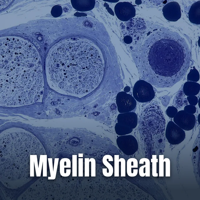
Myelin is a fatty coat produced by glial cells that encircles the long axons of nerve cells in most vertebrates to speed up signal flow. Flemish anatomist Andreas Vesalius first described these white tissue tracts during the sixteenth century while dissecting human brain anatomy. German pathologist Rudolf Virchow named the substance in 1854, borrowing the Greek word myelos because its soft texture reminded him of bone marrow. While an insulated electrical cable offers a familiar physical comparison, living nerve wraps behave differently because they do not form an unbroken jacket along the axon, but instead feature small exposed gaps that interrupt fatty segments at set distances [1].
Nerve bundles wrapped in fat give white matter its pale tone in the brain and spinal cord. Thousands of parallel axons travel side by side through major neural pathways like the optic nerve and the corticospinal tract. In the human corpus callosum, more than 200 million axons run together to link the left and right cerebral hemispheres. Capillaries bring oxygen and glucose to these packed fiber tracts, feeding nearby astrocytes and microglia. Without these fatty wraps, communication between distant brain regions and peripheral limbs breaks down [1].
Jawless fish lack myelin, but nearly all other vertebrates evolved these lipid coats to manage larger bodies with fast reflex responses. Uncoated fibers leak charge, forcing action waves to move slowly and consume heavy metabolic fuel to restore resting electrical gradients after each signal [1, 6].
Membrane Lipid Architecture
Fresh myelin tissue holds roughly 40% water by weight, while the remaining dry material contains about 60 to 75 percent lipids and 15 to 25 percent proteins [1]. Research by A. E. Kister and I. Kister in Frontiers in Chemistry shows that this rich fat balance makes myelin unique among all animal cell boundaries [6]. The main glycolipid in the wrap is galactocerebroside, whose long lipid chains interlock to steady the stacked membranes. Sphingomyelin adds extra firmness to the multilayered sheath. Biochemical studies confirm that cholesterol forms an essential structural anchor without which compact myelin sheaths fail to form properly [1].
Proteins make up the rest of the dry mass, acting like molecular pins that hold the wrapped membranes together in dense layers. In the central nervous system, proteolipid protein (PLP) is the most common structural protein, binding outer cell surfaces to keep the layers tight. Myelin basic protein (MBP) condenses the inner surfaces into solid coats. Myelin-associated glycoprotein (MAG) anchors the wrap directly to the axon wall [1]. Myelin oligodendrocyte glycoprotein (MOG) develops late on glia, exposing an immunoglobulin domain that was mapped at 1.45 Angstroms resolution in Norway rat studies [5].
In outer body nerves, myelin protein zero (MPZ) replaces PLP, making up over 50% of local sheath proteins. The MPZ gene sits on human chromosome 1 near the Duffy locus, spanning six exons. MPZ dimers and tetramers lock adjacent bilayers together through homotypic adhesion [4].
Saltatory Conduction and Signal Velocity
How does a fatty wrap speed up neural signaling while saving space and cellular energy? By boosting charge resistance and lowering membrane capacitance across the axolemma, the myelin coat stops ionic current from leaking into the wet fluid outside the nerve. Signals do not move as unbroken waves along coated fibers. Instead, impulses travel by saltatory conduction, a term rooted in the Latin verb saltare, which means to leap or hop from one open node to the next. Current entered at one node flows swiftly through the inner core to depolarize the next available gap [1].
Saltatory jumping lets impulses travel up to one hundred times faster than in uninsulated fibers of the same width. Uncoated nerves need wide diameters to gain even modest speed. For instance, squid giant axons reach high velocity only by swelling to nearly one millimeter across. Vertebrates avoid thick nerves because myelin allows a tiny axon of two micrometers to fire with high speed, which permits millions of compact insulated neural pathways to fit inside the human skull [1].
Myelin also feeds the axon with vital energy. A 2012 study proved that glia act like fueling pumps, delivering lactate directly to active axons [1]. Writing in Physiological Reviews, Stadelmann and Simons showed that glia regulate water and ion balance, shielding working axons from metabolic exhaustion during prolonged bursts of electrical firing [7].
Glial Cells Producing the Sheath
Two distinct classes of glial cells manufacture myelin, dividing labor between the central brain and outer nerves. In the brain and spinal cord, oligodendrocytes build every internodal wrap. A single oligodendrocyte sends out dozens of thin cellular arms, wrapping coats around up to fifty separate axons at the same time to link local networks [1, 8]. Studies by S. Kuhn and Y. Dombrowski in the journal Cells show that adult brains keep a pool of oligodendrocyte progenitor cells. These backup progenitor cells can grow into fresh myelinating glia to replace damaged or lost coats throughout adult life [8].
Outside the brain, Schwann cells build myelin for motor and sensory nerves. Each myelinating Schwann cell devotes its entire body to a single internode on one axon. Non-myelinating Schwann cells, termed Remak cells, gather thin fibers into protective Remak bundles [1]. Schwann cells also produce peripheral myelin protein 22 to reinforce the sheath alongside MPZ. Signals from the axon instruct Schwann cells when to start wrapping and how many membrane layers to build [1, 4].
Nerves smaller than one micrometer in diameter stay unmyelinated. When an axon grows wider, it signals nearby glia to begin spiral wrapping, winding around the axon repeatedly to squeeze out inner cytoplasm until dozens of tight membrane sheets encircle the core [1].

Why Myelin Sheath Function Needs Nodes
Although fatty wraps insulate internodes, proper myelin sheath function needs regular uninsulated breaks along the axon to keep signals from dying out. These tiny gaps, discovered by French anatomist Louis-Antoine Ranvier, are called nodes of Ranvier and span only one micrometre each. At every node, the bare axonal membrane, or axolemma, touches outer fluid. Voltage-gated sodium channels pack into these narrow gaps in dense clusters, while covered segments beneath the myelin lack sodium gates almost entirely, which lets action pulses recharge at regular intervals without decaying across long anatomical distances [1].
When a wave reaches a node of Ranvier, voltage-gated sodium gates open swiftly to admit positive ions. Inflowing sodium pushes the local membrane potential up to about +35 millivolts. Potassium gates then open to let potassium ions escape, rapidly returning the nerve wall to its resting negative charge. Sodium-potassium pumps then burn fuel to eject sodium and take back potassium. Because ions cross only at nodes, nerves burn far less fuel than uncoated fibers [1].
Internodal length describes the physical span between neighboring nodes, which thick axons expand to take larger leaps. If nodes sit too far apart, current fades before reaching the next gate. If gaps sit too close, signals slow down, forcing the nervous system to balance internodal length and node spacing to achieve optimum signal velocity [1].
Brain Myelination Timeline
Myelin growth follows a steady schedule that begins during pregnancy and continues for decades as neural networks mature [3]. Human myelination starts during the third trimester, near the twenty-sixth week of fetal life [1]. Pediatric data from the University of Utah shows that a newborn brain weighs just 400 grams at birth. By twelve months, infant brain weight reaches 1,000 grams, and by age two the organ attains 80 percent of adult size. Brain weight reaches its mature mark of 1400 grams by eighteen years of age, with axonal growth and progressive myelin wrapping driving nearly all of this weight gain [3].
Brain regions do not gain myelin at the same time. Pathways for reflexes and survival, including cranial nerves and brainstem tracts, myelinate first. Insulation along the corticospinal pathway commences around the thirty-sixth gestational week, reaching full cover by two years of age. Arm nerves myelinate before leg nerves. Magnetic resonance research by A. L. MacKay and C. Laule in Brain Plasticity confirms that myelin water imaging can track myelination in living people well into their twenties and thirties [3, 9].
Rear brain lobes myelinate before front zones. Vision and touch areas in the parietal and occipital lobes gain fat coats before frontal regions mature. Frontal networks that guide deep thought and impulse control finish last, though adults can still add myelin to active grey matter circuits throughout life [1, 3].
How Illness Impairs Myelin Sheath Function
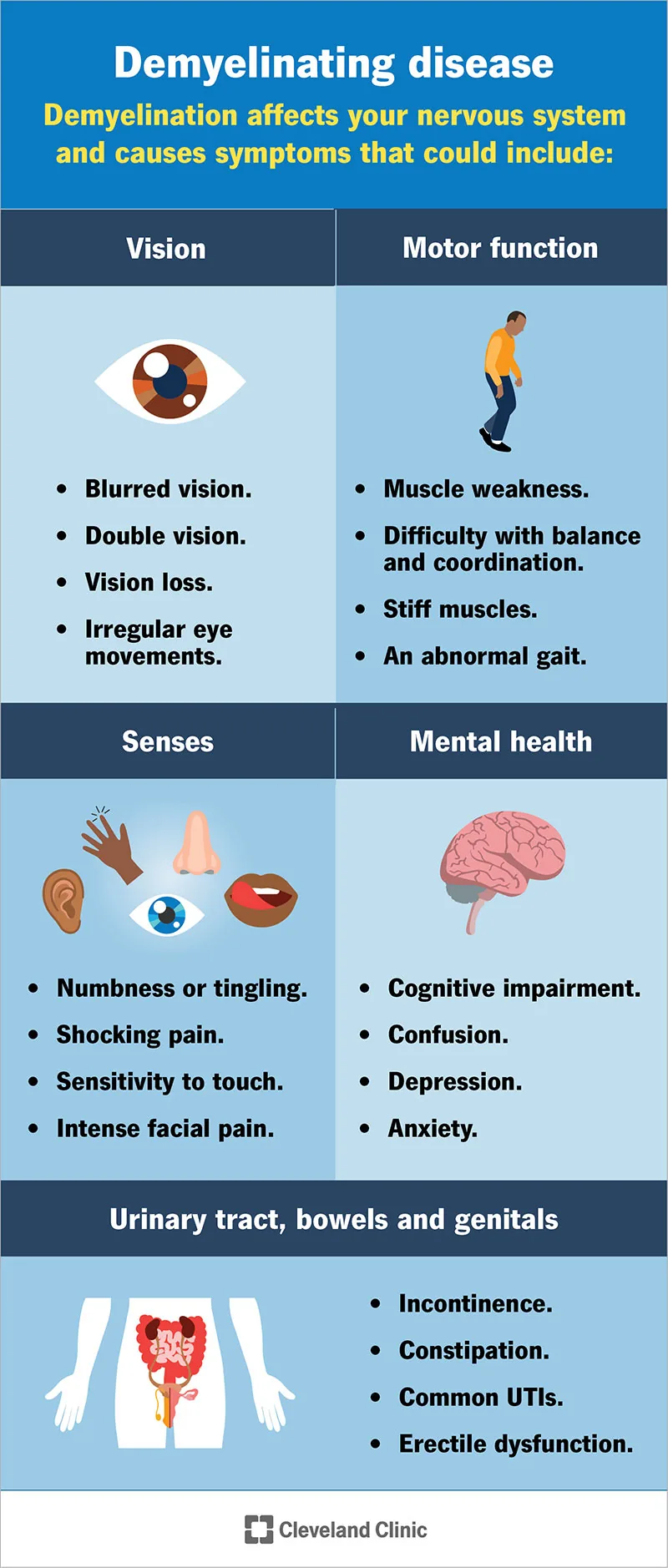
When illness injures nerve insulation, healthy myelin sheath function breaks down and electrical impulses fail to cross bare segments. Demyelinating conditions emerge when immune cells mistake myelin for foreign invaders. In North America, the most widespread central demyelinating condition is multiple sclerosis, with clinical estimates from the Cleveland Clinic reporting that roughly 1 million people in the United States live with multiple sclerosis. Magnetic resonance imaging detects demyelinated spots as bright white patches in the brain and spinal cord, where damaged coats slow or halt nerve flow until uncovered axons wither from lack of support [1, 2].
Myelin loss causes varied symptoms depending on which nerve tracts suffer harm. People face muscle weakness, balance trouble, numbness, and vision loss from optic neuritis [2, 5]. In the journal Brain, G. D. Ponath and D. Pitt showed that astrocytes swallow myelin debris through receptor endocytosis, which triggers NF-κB activation and chemokine secretion to worsen local tissue inflammation [11]. Chronic demyelination leaves bare axons vulnerable to permanent damage [2, 7].
Outer nerves face genetic faults in MPZ on chromosome 1 near the Duffy locus, sparking Charcot–Marie–Tooth disease and Dejerine–Sottas disease during early childhood. Guillain–Barré syndrome attacks outer sheaths after infections, leading to muscle weakness and lost sensory reflexes [1, 4].
Nerve Regeneration and Myelin Sheath Function
When nerve fibers suffer physical cuts, their ability to heal depends on whether they run through outer body nerves or the central nervous system. In peripheral nerves, severed axons can regrow because Schwann cells clear dead myelin debris and lay down chemical paths that guide new growth cones toward target muscles. The basal lamina and surviving glia protect this regrowth. Still, repair is rarely flawless. Regenerating fibers sometimes miss their original muscle connections, and injured motor neurons may perish before reinnervation finishes, leaving patients with partial functional loss [1].
Central nervous system axons face much harsher limits after injury. In adult mammalian brains, severed fibers cannot regrow across lesion gaps [1]. Surviving oligodendrocyte progenitor cells travel into hurt zones to attempt remyelination, but chemical blockers in myelin debris stall their growth into mature cells [8]. In Alzheimer’s disease, S. E. Nasrabady and A. M. Brickman reported in Acta Neuropathologica Communications that white matter loss, iron buildup, and oxidative stress damage myelin, while amyloid-beta poisons oligodendrocytes [10].
Safeguarding myelin sheath function remains a top goal in modern neurology and neurobiology. Scientists test therapies to awaken quiet progenitor cells, restore leaping conduction, and protect bare axons from decay, while researchers like Stadelmann and Kuhn continue mapping how glia build and fuel myelin to protect lifelong brain health [6, 7, 8].
- WEBSITE Wikipedia. Myelin. [Article Link]
- WEBSITE Cleveland Clinic. (2023). What Is a Demyelinating Disease? [Article Link]
- WEBSITE University of Utah. (2020). Pediatric Neurologic Examination Videos & Descriptions: Developmental Anatomy. [Article Link]
- WEBSITE Wikipedia. Myelin protein zero. [Article Link]
- WEBSITE Wikipedia. Myelin oligodendrocyte glycoprotein. [Article Link]
- ACADEMIC JOURNAL Kister, A. E., & Kister, I. (2023). Overview of myelin, major myelin lipids, and myelin-associated proteins. Frontiers in Chemistry, 10, 1041961. [Article Link]
- ACADEMIC JOURNAL Stadelmann, C., Timmler, S., Barrantes‐Freer, A., & Simons, M. (2019). Myelin in the Central Nervous System: Structure, Function, and Pathology. Physiological Reviews, 99(3), 1381-1431. [Article Link]
- ACADEMIC JOURNAL Kuhn, S., Gritti, L., Crooks, D. A., & Dombrowski, Y. (2019). Oligodendrocytes in Development, Myelin Generation and Beyond. Cells, 8(11), 1424. [Article Link]
- ACADEMIC JOURNAL MacKay, A. L., & Laule, C. (2016). Magnetic Resonance of Myelin Water: An in vivo Marker for Myelin. Brain Plasticity, 2(1), 71-91. [Article Link]
- ACADEMIC JOURNAL Nasrabady, S. E., Rizvi, B. M., Goldman, J. E., & Brickman, A. M. (2018). White matter changes in Alzheimer’s disease: a focus on myelin and oligodendrocytes. Acta Neuropathologica Communications, 6(1), 22. [Article Link]
- ACADEMIC JOURNAL Ponath, G. D., Ramanan, S., Mubarak, M., Housley, W., Lee, S., Sahinkaya, F. R., Vortmeyer, A. O., Raine, C. S., & Pitt, D. (2016). Myelin phagocytosis by astrocytes after myelin damage promotes lesion pathology. Brain, 140(2), 399-413. [Article Link]
Cover image: NIH History Office from Bethesda / Wikimedia Commons, Public domain


