





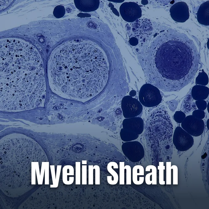
Nerve cells rely on a myelin sheath to insulate axons and accelerate electrical signal transmission across the body. In both the brain and peripheral pathways, specialized glial cells generate this multilayered coat in regular segments separated by narrow uninsulated gaps [1]. Bare axons conduct action potentials at much slower speeds, and persistently exposed nerve fibers deteriorate over time [9]. Early anatomists mapped these pale glistening tracks by hand, and modern clinical imaging now tracks sheath integrity to evaluate neurological health [7].
What Is the Myelin Sheath?
Anatomists first described white matter tracks in the sixteenth century, when Andreas Vesalius recorded pale pathways in deep human brain tissue. Rudolf Virchow named the substance in 1854. Borrowing the Greek root myelos for bone marrow because of the shared ivory hue and soft texture, Virchow classified the material as a distinct nerve element. Twentieth-century electron microscopy showed that living glial cell membranes wrap tightly around the axon in dense concentric layers [9]. In Frontiers in Chemistry, Kister and Kister explained that myelin functions as an insulated biological sheath that speeds nerve impulses while offering trophic support to distant parts of the axon [1].
With the sole exception of primitive jawless fish, all living vertebrate groups wrap their nerve fibers in myelin coats. The appearance of this insulating layer enabled vertebrates to develop complex brains and rapid motor coordination without needing massive axonal diameters. Invertebrate animals must rely on huge axon thickness, such as the giant nerve fiber of Loligo squids, to conduct electrical impulses quickly [9].
A myelinated nerve fiber functions similarly to an insulated copper wire, but living nervous tissue exhibits far more biological complexity than plastic cable. Instead of forming a single continuous pipe, the coat is divided into separate internodal segments punctuated by nodes of Ranvier measuring roughly one micrometer in length. Each insulated segment provides electrical shielding that keeps ionic currents from leaking outward across the axolemma [9]. By preventing signal dissipation along the fiber, myelin sheath assembly organizes ion channels, stabilizes the axonal cytoskeleton, and supplies needed nutrients to distant neural outposts [1].
Lipid and Protein Composition
Fresh myelin tissue holds roughly 40% water. Dried biochemical samples yield between 60% and 75% lipids and 15% to 25% proteins, a composition that distinguishes myelin from ordinary plasma membranes containing equal shares of fat and protein [9].
The primary glycolipid component is galactocerebroside, whose galactose headgroups pack together tightly to stabilize multilayered myelin sheath architecture. Glial cells need cholesterol to build myelin, failing completely to synthesize the coat if cholesterol production stops. Intertwined chains of sphingomyelin add toughness to the layers. Proteins make up the remaining dry mass, with proteolipid protein (PLP) serving as the main component in brain myelin and myelin basic protein (MBP) adhering opposing inner membrane leaflets to create compact myelin [1, 9]. Myelin oligodendrocyte glycoprotein coats the outer surface, but myelin-associated glycoprotein binds axonal proteins to anchor the insulating layers [9].
In peripheral body nerves, the dominant protein is myelin protein zero (MPZ or P0), encoded by a 7,000-base gene containing six exons located near the Duffy locus on human chromosome 1. Theodor Schwann first identified these peripheral wraps. The 219-amino-acid P0 chain carries an outer immunoglobulin fold, a single transmembrane region, and a basic inner tail. P0 makes up over 50% of peripheral myelin protein, forming homotypic dimers and tetramers that bind adjacent membrane layers together like glue [10].
Rapid Impulse Conduction
Bare fibers conduct impulses slowly. Action potentials propagate as an unbroken wave of charge down the naked membrane, burning large reserves of cellular fuel. Myelinated axons bypass that biological limitation through saltatory conduction, where impulses leap between nodes of Ranvier because concentric glial membrane wraps raise resistance across the cell wall and lower membrane capacitance. Signals reach distant targets in milliseconds. Conduction speeds along an intact myelin sheath easily outpace uninsulated fibers of equal width, helping large vertebrates coordinate swift motor reflexes and sensory processing [9].
Bare axons leak electrical current into surrounding fluids. By widening the physical distance between intracellular axoplasm and extracellular fluid, myelin insulation prevents opposing electrical charges from accumulating across the axolemma. Internodal stretches completely lack sodium channels, and exposed nodal gaps leave bare axonal membrane accessible to surrounding ions [9].
At each one-micrometer node of Ranvier, voltage-gated sodium channels sit in dense clusters, opening swiftly so positive sodium ions rush inward to depolarize the axolemma to roughly +35 mV. The incoming positive charge then diffuses electrotonically down the axoplasm to the next node, tripping adjacent voltage-gated channels in a rapid cascade. Because cytoplasmic diffusion is decremental by nature, signal amplitude declines over distance, requiring nodes of Ranvier to sit at regular intervals to sustain impulse propagation. Voltage-gated potassium channels quickly restore the resting membrane potential, and ATP-dependent sodium-potassium pumps work continuously along internodes to restore baseline ionic gradients [9].
Myelin Sheath Formation by Glial Cells
Two distinct glial cell families construct myelin based on body anatomy [1]. In the brain and spinal cord, oligodendrocytes build layered myelin sheath segments, extending cytoplasmic foot processes to insulate separate internodes on dozens of neighboring axons. Schwann cells wrap single peripheral axons [9]. Both glial types spiral around the axon core and squeeze out cytoplasm to produce compact concentric membrane wraps [1]. Axon caliber regulates this program, as fibers exceeding one to two micrometers stimulate wrapping, with internodal length correlating directly with axon thickness [9].
In the central nervous system, mature oligodendrocytes derive from migratory oligodendrocyte progenitor cells (OPCs) that remain widely distributed in adult brain tissue. Writing in Cells, Kuhn and Dombrowski reported that high metabolic turnover leaves oligodendrocytes vulnerable to toxic stress, but resident adult progenitor cells can divide and differentiate to regenerate lost myelin coats following tissue injury [3].
Peripheral nerve fibers display small pockets of residual cytoplasm trapped between membrane layers during Schwann cell wrapping. These funnel openings form Schmidt-Lanterman clefts. Lanterman clefts appear ab initio across the radial thickness of the sheath, providing open cytoplasmic communication pathways through compact myelin [11]. Pioneer histologist Robert Remak showed that non-myelinating Schwann cells ensheath bundles of small-diameter unmyelinated axons within protective Remak bundles, and astrocytes support unmyelinated fibers in central tracts [9].



Timeline of Nervous System Development
Early embryonic milestones prepare the brain for myelination, beginning when neural tube closure finishes by 28 days of gestation. Prosencephalization then shapes the cerebral hemispheres and diencephalon, with neuronal proliferation peaking between two and four months before neuroblasts migrate radially from three to five months. Major brain fissures form during the fifth month, secondary sulci by seven months, and tertiary sulci by nine months gestation. By 36 weeks gestation, proliferation is largely complete, and individual neuroblasts expand from 5 microns to 50 microns in diameter, marking a 1,000-fold jump in cellular volume. Brain weight tracks this rapid anatomical expansion: the newborn brain weighs roughly 400 grams, reaches 1,000 grams at one year, attains 80 percent of adult size by age two, and reaches its final adult weight of 1400 grams by eighteen years of age [7].
Myelin begins forming early in the third trimester near gestational week 26 and advances swiftest during the first two years of childhood [7, 9]. Sensory roots and brainstem tracts myelinate first to support neonatal reflexes. University of Utah clinical records document that the corticospinal tract begins myelinating at 36 weeks gestation and completes the process by 24 months of age [7].
Upper limb and trunk axons gain myelin before leg fibers, matching the progressive head-to-toe acquisition of early infant motor skills. Posterior lobes finish first. In the cerebrum, rear frontal, parietal, and occipital lobes finish before anterior frontal and temporal zones, so by age two primary cerebral tracts are largely covered to support crawling, walking, and speech acquisition [7, 9]. However, interconnections within the association cortex continue gaining myelin into the second and third decades of life [7].
Diseases That Damage the Myelin Sheath
Severe neurological dysfunction follows when inflammatory processes strip away insulating coats from healthy nerve fibers. In demyelinating conditions, the immune system mistakes myelin proteins for foreign antigens and mounts an attack that causes conduction slowing or complete signal block. Cleveland Clinic diagnostic records identify multiple sclerosis (MS) as the most prevalent demyelinating illness in North America, with a 2019 epidemiological study estimating that nearly one million individuals live with MS in the United States. Lesions can strike the brain, optic nerves, and spinal cord [8].
Peripheral nerves also suffer from genetic mutations in structural proteins. Charcot-Marie-Tooth disease and infant Dejerine-Sottas disease frequently arise from point mutations in the MPZ gene encoding myelin protein zero. When mutated amino acid substitutions impair P0 homotypic adhesion, peripheral fibers fail to compact properly or degenerate prematurely [10]. Acquired autoimmune neuropathies like Guillain-Barré syndrome and chronic inflammatory demyelinating polyneuropathy similarly attack limb nerves [8, 9]. Denuded fibers wither and die, making early care at centers like the Cleveland Clinic important for managing symptoms before nerve fibers degenerate beyond repair [8]. Genetic leukodystrophies and Canavan disease cause broad myelin decay throughout the central nervous system [9].
In Brain, Ponath, Ramanan, Mubarak, and Pitt showed that astrocytes ingest myelin debris through receptor-mediated endocytosis during acute lesion breakdown. Ingesting cellular debris triggers NF-κB transcription factor activation inside astrocytes, stimulating the secretion of inflammatory chemokines that aggravate local tissue injury [5]. In metabolic disorders, untreated pernicious anaemia causes subacute combined degeneration of the spinal cord, impairing speech, balance, and cognitive awareness [9].
Imaging Methods and Biomarkers
Physicians rely on magnetic resonance imaging (MRI) to identify demyelinating lesions and track therapeutic response over time. Clinical MRI scanners use pulsed radio waves and magnetic fields to visualize focal inflammatory plaques in the brain and spinal cord [8]. Scans readily pinpoint damage across major white matter tracts such as the corpus callosum, which contains over 200 million axons interconnecting the cerebral hemispheres [9].
Advanced quantitative neuroimaging techniques now measure tissue myelin content directly rather than merely displaying focal white matter scars. In an extensive methodological review published in Brain Plasticity, MacKay and Laule evaluated myelin water imaging, an MRI technique that isolates water trapped between concentric lipid bilayers as an in vivo biomarker for myelination. Multi-echo T2 relaxation scans now take under 10 minutes for full-brain runs. Standard data analysis employs non-negative least squares algorithms to calculate the myelin water fraction, but multi-component gradient-echo sequences like mcDESPOT provide complementary measurements. Histological validation in post-mortem tissue confirms high specificity for intact myelin sheath segments, although myelin debris can confound measurements during acute inflammation [2].
Quantitative neuroimaging shows that white matter pathology extends into neurodegenerative conditions beyond classic demyelinating diseases. In Acta Neuropathologica Communications, Nasrabady, Rizvi, Goldman, and Brickman reviewed clinical evidence showing that white matter abnormalities and oligodendrocyte vulnerability represent significant early pathological features of Alzheimer’s disease. Grey matter plaques draw attention, but white matter decay begins early in disease progression. Oxygen loss, iron overload, and excitotoxicity all compromise oligodendrocytes, suggesting that therapies protecting myelin could serve as valuable treatment targets across diverse neurodegenerative conditions [6].
Axonal Nourishment Beyond Insulation
For decades, neurobiology textbooks treated myelin solely as a passive electrical insulator, but recent discoveries have transformed that classical perspective. In 2012, experimental research showed that myelinating glial cells actively feed underlying axons, operating as localized fueling stations along the nerve fiber length [9]. Stadelmann, Timmler, Barrantes-Freer, and Simons detailed in Physiological Reviews that oligodendrocytes are embedded in an active network of interconnected glial and neuronal cells. These glial partners provide energy substrates, regulate local ion and water balance, and adjust support in response to activity-dependent neuronal signaling, keeping axons alive through heavy impulse firing [4].
Myelinating cells sculpt axon anatomy. Signaling from a mature myelin sheath promotes the phosphorylation of neurofilaments, which expands axonal diameter and thickens the fiber along internodal segments. Glial interactions also guide the clustering of sodium and potassium channels at the nodes of Ranvier, and modulates the transport of mitochondria along the nerve fiber [9].
Central nerve fibers in the mammalian brain fail to regenerate, but peripheral Schwann cells provide a guiding physical track along which damaged axons can regrow toward target muscle tissues. Regrowth is imperfect. Some regenerating axons fail to reach correct muscle targets, and severely damaged motor neurons may die before completing repair [9]. Current neurobiological research focuses on neutralizing inhibitory central nervous system molecules and stimulating adult oligodendrocyte progenitor cells to restore damaged neural circuits [1, 3].
- ACADEMIC JOURNAL Kister, A. E., & Kister, I. (2023). Overview of myelin, major myelin lipids, and myelin-associated proteins. Frontiers in Chemistry, 10, 1041961. [Article Link]
- ACADEMIC JOURNAL MacKay, A. L., & Laule, C. (2016). Magnetic Resonance of Myelin Water: An in vivo Marker for Myelin. Brain Plasticity, 2(1), 71-91. [Article Link]
- ACADEMIC JOURNAL Kuhn, S., Gritti, L., Crooks, D. A., & Dombrowski, Y. (2019). Oligodendrocytes in Development, Myelin Generation and Beyond. Cells, 8(11), 1424. [Article Link]
- ACADEMIC JOURNAL Stadelmann, C., Timmler, S., Barrantes-Freer, A., & Simons, M. (2019). Myelin in the Central Nervous System: Structure, Function, and Pathology. Physiological Reviews, 99(3), 1381-1431. [Article Link]
- ACADEMIC JOURNAL Ponath, G. D., Ramanan, S., Mubarak, M., Housley, W., Lee, S., Sahinkaya, F. R., Vortmeyer, A. O., Raine, C. S., & Pitt, D. (2016). Myelin phagocytosis by astrocytes after myelin damage promotes lesion pathology. Brain, 140(2), 399-413. [Article Link]
- ACADEMIC JOURNAL Nasrabady, S. E., Rizvi, B. M., Goldman, J. E., & Brickman, A. M. (2018). White matter changes in Alzheimer’s disease: a focus on myelin and oligodendrocytes. Acta Neuropathologica Communications, 6(1), 22. [Article Link]
- WEBSITE University of Utah. (2020). Pediatric Neurologic Examination Videos & Descriptions: Developmental Anatomy. NeuroLogic Examination. [Article Link]
- WEBSITE Cleveland Clinic. (2023). What Is a Demyelinating Disease? [Article Link]
- WEBSITE Wikipedia contributors. (2026). Myelin. Wikipedia, The Free Encyclopedia. [Article Link]
- WEBSITE Wikipedia contributors. (2026). Myelin protein zero. Wikipedia, The Free Encyclopedia. [Article Link]
- WEBSITE Wikipedia contributors. (2026). Myelin incisure. Wikipedia, The Free Encyclopedia. [Article Link]
Cover image: Nephron / Wikimedia Commons, CC BY-SA 3.0


