






What is astigmatism, and why does it blur human vision? In simple terms, astigmatism is a common optical eye flaw where the front window of the eye or the crystalline lens curves unevenly. Instead of being round like a ping-pong ball, an astigmatic eye curves like the back of a spoon. Light rays passing through cannot focus on a single spot on the retina, making images look blurry or stretched at both near and far distances. The condition affects between 30% and 60% of adults in Europe and Asia [6]. Corrective eyeglasses, soft toric contacts, and modern laser surgery help bend these mismatched light paths back into shape so people can enjoy clear sight [1].
- What Is Astigmatism and Why It Occurs?
- Light Refraction on Toric Surfaces
- Comparing Lenticular and Corneal Astigmatism
- How Keratoconus Distorts Corneal Shape
- Diagnostic Testing with Keratometry and Tomography
- Why Uncorrected Cylinder Degrades Visual Clarity
- Toric Lenses for Astigmatism Correction
- Corneal Laser Surgery Options
What Is Astigmatism and Why It Occurs?
The word astigmatism comes from Greek roots, joining a- meaning without and stigma meaning a spot [7]. English polymath Thomas Young first described the eye flaw in 1801 [6]. In 1837, English astronomer Sir George Biddle Airy made a special cylinder glass to fix the uneven focus in his own eye [3].
Astigmatism is common in all human groups across Europe and Asia. In a broad systematic review synthesizing data from MEDLINE, EMBASE, and Cochrane Library records, Zhang and Bullimore observed that population rates ranged from 8% to 62%, with numbers climbing highest among adults aged 70 or older [13]. Twin studies in 2007 suggested that inherited genes play a modest role in corneal shape. Later genome-wide scans uncovered specific chromosomal loci. Researchers identified variants near the PDGFRA gene on chromosome 4q12 in Asian groups, an association replicated in Northern European cohorts alongside candidate markers including CLDN7, ACP2, and TNFAIP8L3 [6].
Most boys and girls are born with mild corneal asymmetry that stays steady through adult life. In other people, blur starts after direct eye injury, severe corneal infection, or surgical cuts from cataract removal [1, 5]. Dr. Audrey Tai at Athena Eye Care in Mission Viejo, California, notes that scar tissue pulls the cornea out of round. Healed incisions flatten tissue along one meridian, changing how light enters the pupil and creating new visual blur [1, 5].
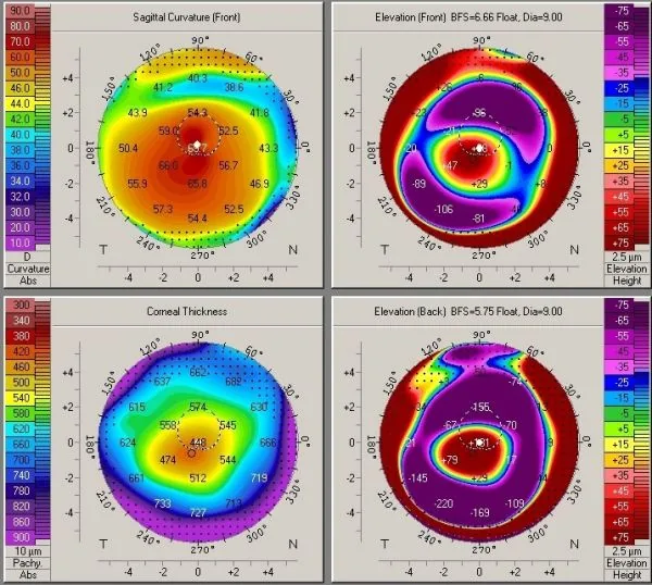
Light Refraction on Toric Surfaces
In a normal eye, the cornea and crystalline lens form smooth spherical surfaces that curve equally in all directions. Light rays bend evenly, meeting cleanly at one sharp point on the retina. In an astigmatic eye, the front surface curves like an American football (lying on its side) or a spoon [5, 6]. Because perpendicular optical meridians possess distinctly different surface curvatures, incoming light rays bend with unequal power across the vertical and horizontal planes. The steep curve bends light fast, while the flat curve focuses light farther back [7].
Optical analysts at Telescope Optics explain that astigmatism acts as an off-axis wavefront aberration where surface radii differ across planes, following Seidel aberration rules and Zernike polynomials. Light splits into two perpendicular line foci. Tangential rays meet first in a horizontal line, while sagittal rays travel deeper before meeting in a vertical line. Midway between these two separate focal planes lies the circle of least confusion, an intermediate circular cross-section where the converging light bundle achieves its sharpest overall focus before corrective cylindrical lenses are introduced [4, 7]. This middle plane on the Petzval surface gives the best visual compromise for an untreated eye [4].
The 3D bundle of light between these two lines is called the conoid of Sturm, described by mathematician Jacques Charles François Sturm. Correcting vision means collapsing this cone until both lines meet on the retina. When the circle of least confusion rests right on the retinal plane, the eye reaches meridional balance, letting eye doctors test cylinder power with high precision [3].
Comparing Lenticular and Corneal Astigmatism
Dr. Andrew A. Dahl at MedicineNet notes that most astigmatic blur comes from the cornea, which provides two-thirds of the focusing power of the eye [5]. Doctors test this front surface with manual keratometers or video tools to check corneal steepness [1, 2]. Regular corneal astigmatism occurs when the steepest and flattest curves sit 90 degrees apart. When these axes wander away from right angles, irregular astigmatism creates wavy distortions that simple eyeglasses cannot fix [5].
Total ocular refraction depends on internal eye anatomy as well as the front corneal surface. At the Cullen Eye Institute at Baylor College of Medicine in Houston, Texas, Koch, Weikert, and Wang analyzed 715 corneas across 435 consecutive patients using a dual-Scheimpflug analyzer. They found that posterior corneal astigmatism averaged -0.30 diopter. In 86.6% of eyes, the steep posterior curve ran vertically (within 60 to 120 degrees). While front curves often tilt from vertical to horizontal as people age, posterior alignment stays vertically stable [8]. In Bologna, Italy, Savini and Versaci confirmed that posterior curvature averaged 0.54 diopter with the Sirius topographer, demonstrating that relying solely on anterior measurements overestimates with-the-rule astigmatism by 0.22 diopter [11].
Internal eye parts also create cylinder blur. Inside the eye, the crystalline lens can develop uneven curves, called lenticular astigmatism [5]. In a pediatric group of 165 children, Lin evaluated ocular residual astigmatism using Thibos vector analysis and Spearman rank correlations in Scientific Reports, discovering a median residual value of 0.57 diopter. In 84.8% of these young patients, this internal flaw counteracted front corneal steepness, working naturally to reduce the total blur of the eye [12].
How Keratoconus Distorts Corneal Shape
When corneal steepening and tissue thinning progress out of control, the condition may indicate an ectatic corneal disorder rather than ordinary benign refractive error [1, 2]. Keratoconus is an asymmetric corneal disease where the central cornea bulges forward into a cone. German professor Burchard Mauchart first recorded the disorder in 1748, naming it staphyloma diaphanum. British doctor John Nottingham described it as conical cornea in 1854, and Swiss physician John Horner introduced the term keratoconus in 1869 [2].
Keratoconus affects about 1 in 700 people and often starts in puberty across populations in the Middle East, India, China, and Australia. Eye rubbing is the main risk. Clinical surveys on EyeWiki suggest that environmental influences account for 92% of ectatic progression, while direct genetic mutations contribute only 8%. Chronic, vigorous eye rubbing linked to allergic conjunctivitis damages fragile stromal collagen layers. In a pediatric cohort of 305 eyes, a baseline maximum keratometry reading of 55 diopters or higher predicted faster disease worsening, showing why children with Down syndrome or atopy must never rub their eyes [2].
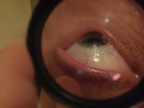
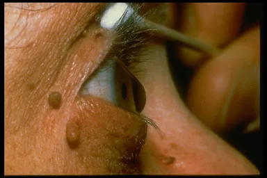
Slit-lamp checks show clear signs of disease. Eye doctors spot fine vertical stress lines in the stroma called Vogt striae, along with brown Fleischer iron rings around the base of the cone. In late stages, the cone pushes out the lower eyelid when looking down, a clinical sign described by Edwin A. Munson. If Descemet membrane breaks, fluid rushes into the stroma, causing sudden pain and cloudiness known as acute hydrops [2].


Diagnostic Testing with Keratometry and Tomography
Eye doctors test and measure cylinder blur through quick, painless exams. Testing starts with reading letter lines on a Snellen chart, followed by automated refraction and slit-lamp biomicroscopy [1, 5]. When evaluating babies or young children who cannot speak, eye doctors use retinoscopy to shine a streak of reflected light across the pupil [1]. An abnormal scissoring motion of the red light reflex warns the doctor of high astigmatism or early keratoconus before test lenses are placed in a trial frame [2].
Refining cylinder power and axis requires the Jackson cross cylinder, an optical tool created in the nineteenth century. In 1849, Sir George Gabriel Stokes built the Stokes lens, using two rotating cylinders to yield powers (from zero to 4.00 diopters) sphere with -8.00 diopters cylinder. Dr. W. S. Dennett updated the lens frame in 1885, and Dr. Edward Jackson showed its diagnostic power in 1887. Dr. W. H. Crisp helped promote the cross cylinder worldwide. When a doctor flips or twirls the modern cross cylinder in front of the patient, the plus and minus axes trade places. By choosing which view looks darker and sharper, the patient helps the doctor set the axis within five degrees [3].
Modern eye clinics map corneal shape with computerized Placido disks and rotating Scheimpflug cameras. Advanced diagnostic systems like the Pentacam, Sirius, and Galilei track thousands of elevation points across front and back surfaces [2, 8, 11]. These 3D maps detect subclinical keratoconus early and help surgeons calculate accurate intraocular lens powers before cataract surgery with instruments like the IOLMaster, Lenstar, and Atlas [8, 10].
Why Uncorrected Cylinder Degrades Visual Clarity
Uncorrected astigmatic blur exhausts the human eye [6]. In an investigational simulation published in the Journal of Cataract & Refractive Surgery, Wolffsohn, Bhogal-Bhamra, and Shah tested how induced cylinder impairs daily visual performance. When presbyopic adult volunteers experienced uncorrected cylinder from zero to -4.00 diopters, distance visual acuity and near reading speed dropped sharply across 95%, 50%, and 10% contrast targets. C-Quant straylight tests showed increased measurement variability, while computer driving simulator tests proved that uncorrected cylinder caused slower braking reaction times. Participants also reported fuzzy text on mobile phones and computers, confirming that uncorrected cylinder disrupts ordinary work [9].
Beyond persistent optical blurring across visual distances, uncorrected cylindrical error places continuous strain on the internal focusing muscles of the human eye. Ciliary muscles make ongoing, reflex adjustments to pull shifting focal lines onto the retina. As this muscular effort continues, patients develop asthenopia marked by aching orbits, burning sensations, and heavy fatigue after reading books or working on digital screens [5, 6]. Clinical surveys show that adults with untreated cylinder report frequent tension headaches and migraine attacks far more often than people with balanced spherical vision [6].
In young kids, untreated cylinder can hurt developing brain pathways. If one eye has high astigmatism while the other eye sees well, the visual cortex ignores the blurry feed. This visual suppression causes amblyopia (commonly known as lazy eye), producing permanent vision loss if doctors do not prescribe glasses before early childhood ends [1, 6].
Toric Lenses for Astigmatism Correction
Eyeglasses offer the easiest, safest way to fix regular corneal or lens asymmetry [1, 6]. Astigmatic spectacle lenses feature cylinder power that curves light along one specific meridian without changing focus in the opposite meridian [3, 5]. Eyeglass prescriptions can use plus or minus format; for example, a script of -0.75 diopter cylinder at 45 degrees is identical in optical effect to +0.75 diopter cylinder at 135 degrees. Minus cylinders run along the steep corneal axis to diverge vertical rays slightly, ensuring that horizontal and vertical light waves meet at the same retinal focal plane [6].
Contact lenses offer a broad field of view without the edge distortions of thick eyeglass frames. Soft toric contact lenses use small prism weights or thin zones to keep the lens from spinning when the wearer blinks [5, 6]. For irregular corneal surfaces, rigid gas-permeable (RGP) contact lenses give sharper sight [2, 6]. The firm plastic vaults over bumps and dips, letting natural tear fluid pool underneath to create a brand-new, smooth spherical surface [2].

Patients with severe keratoconus who cannot wear small corneal lenses often switch to large scleral lenses. These wide lenses rest on the white sclera, arching over the sensitive cornea with a pocket of clean saline fluid. Specialized lens geometries like Rose K lenses, invented by Paul Rose in New Zealand, and custom wavefront lenses restore clear sight for complex eyes [2].
Corneal Laser Surgery Options
Laser surgery offers a lasting way to fix vision by reshaping the clear stroma of the cornea. In surface treatments like photorefractive keratectomy, eye surgeons gently remove the outer epithelial cell layer and apply a cool ultraviolet excimer laser beam to vaporize microscopic layers of stromal tissue. In LASEK and Epi-LASIK, the epithelial layer is loosened with mild alcohol or a blunt mechanical blade, moved aside for the laser beam, and smoothed back into place [1, 5]. As the surface heals over several days, the newly flattened corneal profile allows light to focus cleanly without cylindrical glasses [1].
Laser-assisted in situ keratomileusis remains the most common laser vision surgery in the world. The surgeon cuts a thin corneal flap with a microkeratome or femtosecond laser, lifts the flap, sculpts the exposed bed with an excimer beam, and lays the flap back without stitches. Another method, small incision lenticule extraction, uses a femtosecond laser to cut a thin disc inside the stroma, which the doctor pulls out through a tiny keyhole slit to flatten the steep meridian [5].
Older adults having cataract surgery can fix pre-existing astigmatism by choosing toric intraocular lenses (IOLs) [6, 10]. Skilled eye surgeons place the toric implant inside the capsular bag to cancel out corneal cylinder. Koch and colleagues proved that Holladay 2 formula calculations must include posterior corneal curvature to avoid systematic prediction errors [8, 10]. Eye surgeons can also make small limbal relaxing cuts along the steep axis to ease tissue tension and smooth out the front surface of the eye [6].
- WEBSITE MedlinePlus Medical Encyclopedia. (2024). Astigmatism. [Article Link]
- WEBSITE American Academy of Ophthalmology EyeWiki. (2026). Keratoconus. [Article Link]
- WEBSITE Wunsh, S. E. (2016). The cross cylinder. Ento Key. [Article Link]
- WEBSITE Sacek, V. (2000). Telescope astigmatism. Telescope Optics. [Article Link]
- WEBSITE Dahl, A. A. (2023). Astigmatism overview: Glasses, lens, vision test, causes, treatment. MedicineNet. [Article Link]
- WEBSITE Wikipedia. (2026). Astigmatism. [Article Link]
- WEBSITE Wikipedia. (2026). Astigmatism (optical systems). [Article Link]
- ACADEMIC JOURNAL Koch, D. D., Ali, S. F., Weikert, M. P., Shirayama, M., Jenkins, R. B., & Wang, L. (2012). Contribution of posterior corneal astigmatism to total corneal astigmatism. Journal of Cataract & Refractive Surgery, 38(12), 2080-2087. [Article Link]
- ACADEMIC JOURNAL Wolffsohn, J. S., Bhogal-Bhamra, G. K., & Shah, S. (2011). Effect of uncorrected astigmatism on vision. Journal of Cataract & Refractive Surgery, 37(3), 454-460. [Article Link]
- ACADEMIC JOURNAL Koch, D. D., Jenkins, R. B., Weikert, M. P., Yeu, E., & Wang, L. (2013). Correcting astigmatism with toric intraocular lenses: Effect of posterior corneal astigmatism. Journal of Cataract & Refractive Surgery, 39(12), 1803-1809. [Article Link]
- ACADEMIC JOURNAL Savini, G., Versaci, F., Vestri, G., Ducoli, P., & Næser, K. (2014). Influence of posterior corneal astigmatism on total corneal astigmatism in eyes with moderate to high astigmatism. Journal of Cataract & Refractive Surgery, 40(10), 1645-1653. [Article Link]
- ACADEMIC JOURNAL Lin, J. (2021). The contribution of ocular residual astigmatism to anterior corneal astigmatism in refractive astigmatism eyes. Scientific Reports, 11(1), 1018. [Article Link]
- ACADEMIC JOURNAL Zhang, J., Wu, Y., Sharma, B., Gupta, R., Jawla, S., & Bullimore, M. A. (2023). Epidemiology and burden of astigmatism: A systematic literature review. Optometry and Vision Science, 100(3), 218-231. [Article Link]


