





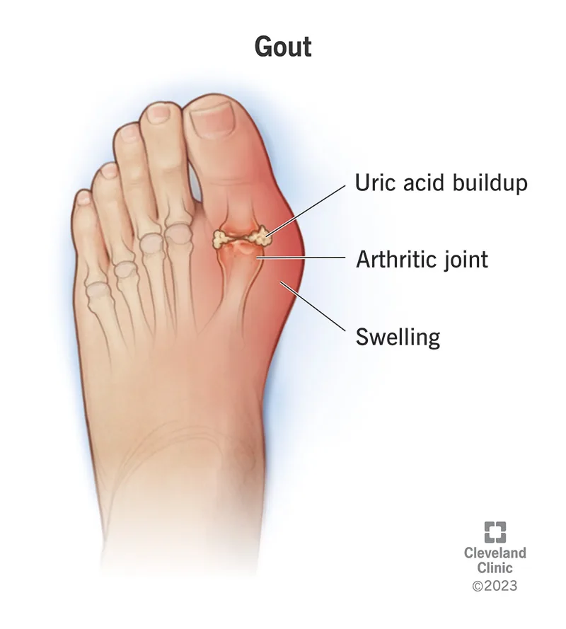
What causes gout is the crystallization of monosodium urate in joint tissue following a persistent build-up of uric acid in the bloodstream. When blood urate levels rise past the chemical saturation point of roughly 6.8 milligrams per deciliter (mg/dL), needle-shaped crystals precipitate into the fluid surrounding vulnerable joints. The local immune system encounters these sharp mineral deposits and mounts an acute inflammatory reaction marked by redness, swelling, warmth, and throbbing pain that wakes people from sleep. Although high urate provides the necessary biological foundation for crystal formation, many people carry elevated levels for decades without ever suffering an acute flare. Gout strikes men far more often than premenopausal women, and unmanaged disease can progress from isolated nocturnal attacks to chronic joint destruction [1].
- How Urate Deposition Causes Gout in Joints
- How Uric Acid Forms from Purines
- Why the Kidneys Fail to Excrete Urate
- Genetic Factors in Gouty Arthritis
- Dietary Triggers and Alcohol Consumption
- Underlying Health Conditions and Medications
- What Causes Gout to Flare Up Suddenly?
- Medical Strategies to Lower Uric Acid
How Urate Deposition Causes Gout in Joints
Gout begins at the microscopic level when monosodium urate precipitates out of supersaturated synovial fluid and lodges within a peripheral joint cavity. Understanding what causes gout in foot joints explains why cooling plays a decisive role. Roughly half of all first attacks strike the big toe at the first metatarsophalangeal junction, a classic medical presentation called podagra. Lower extremities prove uniquely susceptible because distal foot joints register cooler baseline temperatures than the torso, which lowers the solubility of urate salts and accelerates local crystal precipitation during nighttime rest [13].
Crystal accumulation develops through multiple silent phases before patients experience their first painful flare. During the prolonged phase of asymptomatic hyperuricemia, microscopic needle-like crystals settle onto articular cartilage and synovial membranes without producing visible swelling or physical distress. When minor trauma, cold exposure, or rapid shifts in blood chemistry dislodge these deposits into the joint fluid, circulating white blood cells rush to engulf them. Pain peaks within 24 hours. The inflamed skin turns shiny red and becomes so tender that even a bedsheet triggers agony [1].
Repeated bouts of unmanaged joint inflammation cause cumulative damage to cartilage and subchondral bone. When high blood urate persists for years, crystal deposits aggregate into chalky white nodules known as tophi beneath the skin around elbows, knees, fingers, and ear cartilage. Tophi cause bone erosion and permanent stiffness [5].

How Uric Acid Forms from Purines
Uric acid is not an alien toxin; it is the natural end product of purine breakdown in human biochemistry. Purines are nitrogenous bases that form the fundamental building blocks of cellular DNA and RNA, serving basic roles in energy transfer and protein synthesis in every tissue. The body breaks down millions of old cells every day, releasing endogenous purines into the bloodstream for recycling or disposal. A smaller share comes from digesting purine-rich foods [3].
Purine breakdown occurs in the liver. During this sequence, cellular enzymes convert adenine and guanine into intermediate compounds called hypoxanthine and xanthine before final oxidation. The enzyme xanthine oxidase then oxidizes hypoxanthine into xanthine, and later converts xanthine into uric acid. In most non-primate mammals, a functional hepatic enzyme called urate oxidase, or uricase, further degrades uric acid into allantoin, a harmless and highly soluble compound that the kidneys excrete effortlessly into urine [13].
Millions of years ago, ancestral hominids experienced mutational losses that permanently disabled the gene encoding uricase. Consequently, humans maintain baseline blood urate levels up to ten times higher than other mammals. Medical anthropologists argue that this trait provided an evolutionary survival advantage by sustaining arterial blood pressure during dietary salt deprivation and offering neural antioxidant defense [6].
Why the Kidneys Fail to Excrete Urate
Hyperuricemia stems from two distinct physiological imbalances: metabolic overproduction of urate or diminished renal clearance. While overproduction can happen, poor kidney excretion is the main defect that causes gout in most patients. Nephrology research establishes that kidney underexcretion causes approximately 90% of all hyperuricemia cases, while endogenous overproduction accounts for less than 10% [13]. The glomeruli filter large quantities of urate continuously, but proximal renal tubules promptly reabsorb roughly 90% of that filtered load back into the blood, trapping the mineral in circulation [5].
Renal handling of urate depends on transport proteins embedded within the brush-border membranes of proximal tubules. The major reabsorptive carrier, URAT1 (encoded by the SLC22A12 gene), exchanges intracellular anions for luminal urate, pulling the molecule back into tubular cells. Another basolateral transporter, GLUT9 (encoded by the SLC2A9 gene), shuttles urate into peritubular capillaries to complete reabsorption, while uricosuric drug molecules block these pathways to promote urinary elimination [8].

Clinical guidelines define hyperuricemia when serum urate exceeds 7.0 mg/dL in men and 6.0 mg/dL in women. Between 7.0 and 8.9 mg/dL, annual gout incidence remains modest at roughly 0.5%, but once levels exceed 9.0 mg/dL (535 micromoles per liter), annual risk jumps to 4.5%. Yet roughly 90% of hyperuricemic individuals never develop gout, showing that crystallization needs extra triggers [13].
Genetic Factors in Gouty Arthritis
Genetic inheritance plays a major role in predisposing individuals to gouty arthritis, explaining roughly 60% of the variance in population urate concentrations. Polymorphisms in SLC2A9 and ABCG2 weaken urate secretion, approximately doubling risk. When inherited mutations impair excretion, this genetic defect causes gout to emerge early in life [13].
Rare inherited enzyme defects cause severe juvenile hyperuricemia and early joint destruction independent of diet or lifestyle habits. Lesch-Nyhan syndrome, an X-linked recessive disorder arising from a deficiency of hypoxanthine-guanine phosphoribosyltransferase (HPRT), unleashes uncontrolled purine synthesis and massive urate overproduction that damages both the nervous system and kidneys [5]. Kelley-Seegmiller syndrome features a partial deficiency of the same enzyme, presenting with adolescent gout and recurrent uric acid kidney stones [13].
Epidemiological research reveals significant ethnic differences in gout susceptibility worldwide, driven by divergent frequencies of urate-handling gene alleles across human populations. Studies reviewed by Singh and Gaffo show that New Zealand Māori, Han Chinese, and racial minority groups in the United States face markedly higher gout rates than people of European descent. In addition, gout displays a striking sex disparity, and men older than 65 years face particularly high vulnerability, while female incidence climbs after menopause [7]. Estrogen enhances renal urate excretion by modulating tubular transport proteins, which shields women throughout their reproductive years, though female incidence climbs after menopause when circulating estrogen drops sharply and kidney clearance decreases [1].


Dietary Triggers and Alcohol Consumption
While genetics creates the underlying vulnerability, excess intake of purine-rich meat and beer often causes gout attacks in susceptible individuals by flooding the digestive tract with nucleic acids that break down directly into uric acid [4]. Offal cuts such as liver and kidneys, as well as red meats including beef, lamb, and pork, carry heavy purine loads [12]. Certain seafoods, particularly anchovies, sardines, herring, mussels, and scallops, also push blood urate upward [2]. Conversely, clinical trials show that purine-rich vegetables like spinach, peas, beans, and lentils do not heighten gout risk [13].
Fructose represents another major dietary trigger, despite containing no purines. When people drink sugar-sweetened sodas or beverages packed with high-fructose corn syrup, hepatic fructokinase rapidly consumes intracellular adenosine triphosphate (ATP). This rapid reaction depletes cellular phosphate, accumulating adenosine monophosphate (AMP) that accelerates purine breakdown and drives prompt uric acid synthesis [18].
Alcohol consumption disrupts urate balance through a dual physiological mechanism that both stimulates production and impairs excretion. Ethanol metabolism generates lactic acid, which competes directly with urate for tubular secretion in the kidneys, suppressing urate clearance and raising blood concentrations [13]. Brewer’s yeast in beer contains dense concentrations of guanosine, delivering a heavy purine load in addition to ethanol. A prospective study of nearly 45,000 men found that those adhering to a standard Western diet had a 42% greater chance of developing gout than men consuming a DASH-style diet or a Mediterranean diet rich in low-fat dairy and whole grains. The Arthritis Foundation highlights that gradual weight loss and low-fat dairy help lower blood urate [12].
Underlying Health Conditions and Medications
Gout commonly coexists with systemic cardiovascular and metabolic conditions rather than appearing in isolation. Nearly 75% of patients with gout also suffer from metabolic syndrome, a clinical cluster featuring central obesity, hypertension, insulin resistance, and atherogenic dyslipidemia. Clinical studies show that a body mass index of 35 or higher triples a man’s risk of gout compared to normal-weight peers. Visceral adiposity spurs endogenous urate synthesis, while renal damage from long-term hypertension impairs urate removal and causes gout in older adults [13].
Chronic kidney disease is both a major driver and a frequent consequence of sustained hyperuricemia. When progressive glomerulosclerosis destroys functional nephrons, renal clearance declines, causing urate to accumulate in circulation and precipitate in peripheral joints and renal parenchyma [5]. Furthermore, high cell turnover conditions like severe psoriasis, hemolytic anemia, and hematologic malignancies such as leukemia flood the bloodstream with breakdown products from dying cell nuclei [2]. Cytotoxic cancer chemotherapy can also trigger tumor lysis syndrome, releasing sudden waves of intracellular nucleic acids that overwhelm renal excretion. In addition, chronic environmental exposure to lead injures proximal renal tubules, creating a secondary form of arthritis called saturnine gout [13].
A wide range of routine pharmaceutical therapies can inadvertently spark hyperuricemia by altering renal excretion. Thiazide and loop diuretics prescribed for hypertension and congestive heart failure stimulate proximal tubular urate reabsorption while reducing secretion, markedly increasing serum urate levels [5]. Low-dose aspirin (acetylsalicylic acid) suppresses urate excretion, whereas higher anti-inflammatory doses enhance it [4]. The immunosuppressants cyclosporine and tacrolimus, prescribed following organ transplantation, impair tubular transport and sharply increase gout incidence [13].
What Causes Gout to Flare Up Suddenly?
An acute flare occurs when naked monosodium urate crystals interact directly with resident macrophages in the synovial membrane. In stable joints, microscopic crystal surfaces are coated by shielding serum proteins such as apolipoprotein E, which prevents contact with immune cell receptors. When joint trauma, cold, or sudden urate fluctuations strip this protective protein coat away, exposed crystal facets bind to toll-like receptors on synovial macrophages, triggering rapid intracellular assembly of the NLRP3 inflammasome [13].
Assembly of the NLRP3 complex recruits and activates caspase-1, an enzyme that cleaves dormant precursor pro-interleukin-1β into active interleukin-1β (IL-1β). As a potent master cytokine, IL-1β triggers profound vasodilatation and induces endothelial cells to release chemotactic factors that draw thousands of neutrophils into the joint fluid [9]. Infiltrating neutrophils ingest the sharp crystals, releasing cytotoxic enzymes, reactive oxygen species, and prostaglandins that produce excruciating joint pain within 12 to 24 hours [12].

Sudden shifts in blood urate levels in either direction can trigger acute crystal destabilization and ignite a flare. A rich purine meal, an alcoholic binge, or severe dehydration causes a rapid urate spike that precipitates fresh microcrystals. Conversely, initiating urate-lowering drugs like allopurinol rapidly lowers serum urate, causing existing articular deposits to partially dissolve and shed bare crystal fragments into the synovial cavity [4]. To prevent these paradoxical flares, guidelines mandate co-prescribing low-dose colchicine or an anti-inflammatory drug during the first six months of urate-lowering therapy [8].
Medical Strategies to Lower Uric Acid
Clinical management of gout encompasses two distinct phases: suppressing acute inflammatory flares and establishing long-term urate-lowering therapy. First-line treatments for acute flares include oral nonsteroidal anti-inflammatory drugs (NSAIDs) such as ibuprofen (Advil, Motrin) or naproxen (Aleve, Naprosyn), low-dose colchicine (Colcrys), or corticosteroids. Each NSAID relieves joint inflammation by inhibiting cyclooxygenase pathways [8]. Colchicine halts neutrophil chemotaxis and blocks inflammasome activation [9]. Therapy works best when initiated within 24 hours of flare onset [4].
Long-term disease control relies on urate-lowering therapy to deplete the body’s total crystal reservoir and halt the process that causes gout flares. Reviewer Robert H. Shmerling, MD, Senior Faculty Editor at Harvard Health Publishing, notes that lifestyle changes and medication combine to control the disease [3]. The 2020 American College of Rheumatology guideline recommends allopurinol (Zyloprim, Aloprim) as the preferred first-line agent for all patients, starting at a conservative dosage of 100 mg daily or lower in chronic kidney disease. Febuxostat (Uloric) serves as an alternative xanthine oxidase inhibitor for patients intolerant of allopurinol [8].
When xanthine oxidase inhibitors prove inadequate, uricosuric agents like probenecid (Benemid) enhance renal clearance by blocking tubular URAT1 reabsorption. Pharmacokinetic investigations show that combining allopurinol with probenecid achieves greater hypouricemic efficacy than monotherapy, successfully lowering average plasma urate from 0.30 mmol/L down to 0.09 mmol/L [10]. The established treat-to-target standard aims for serum urate below 6.0 mg/dL, or below 5.0 mg/dL in tophaceous gout [8]. Maintaining these target levels over months dissolves existing deposits, preventing future attacks and reversing joint damage. In severe refractory cases, intravenous pegloticase (Krystexxa) or interleukin-1 inhibitors like anakinra (Kineret) offer additional therapeutic options [11].
- WEBSITE Cleveland Clinic. (2026). Gout. Cleveland Clinic Health Library. [Article Link]
- WEBSITE Healthdirect Australia. (2025). Gout – symptoms, causes and treatment. Healthdirect. [Article Link]
- WEBSITE Restivo, J., & Shmerling, R. H. (2023). Causes of Gout. Harvard Health Publishing. [Article Link]
- WEBSITE Harvard Health Publishing. (2020). Gout. Harvard Health Publishing. [Article Link]
- REPORT National Institute of Arthritis and Musculoskeletal and Skin Diseases. (2017). Gout. National Institutes of Health. [Article Link]
- ACADEMIC JOURNAL Asghari, K. M., Zahmatyar, M., Seyedi, F., Motamedi, A., Zolfi, M., Alamdary, S. J., Fazlollahi, A., Shamekh, A., Mousavi, S. E., Nejadghaderi, S. A., Mohammadinasab, R., Ghazi-Sha’rbaf, J., Karamzad, N., Sullman, M. J. M., Kolahi, A., & Safiri, S. (2024). Gout: global epidemiology, risk factors, comorbidities and complications: a narrative review. BMC Musculoskeletal Disorders, 25(1). [Article Link]
- ACADEMIC JOURNAL Singh, J. A., & Gaffo, A. (2020). Gout epidemiology and comorbidities. Seminars in Arthritis and Rheumatism, 50(3), S11-S16. [Article Link]
- ACADEMIC JOURNAL FitzGerald, J. D., Dalbeth, N., Mikuls, T. R., Brignardello-Petersen, R., Guyatt, G. H., Abeles, A. M., Gelber, A. C., Harrold, L. R., Khanna, D., King, C., Levy, G. D., Libbey, C. A., Mount, D. B., Pillinger, M. H., Rosenthal, A. K., Singh, J. A., Sims, J. E., Smith, B. J., Wenger, N. S., & Poon, S. H. T. (2020). 2020 American College of Rheumatology Guideline for the Management of Gout. Arthritis Care & Research, 72(6), 744-760. [Article Link]
- ACADEMIC JOURNAL Dalbeth, N., Lauterio, T. J., & Wolfe, H. R. (2014). Mechanism of Action of Colchicine in the Treatment of Gout. Clinical Therapeutics, 36(10), 1465-1479. [Article Link]
- ACADEMIC JOURNAL Stocker, S. L., Williams, K. M., McLachlan, A. J., Graham, G. G., & Day, R. O. (2008). Pharmacokinetic and Pharmacodynamic Interaction between Allopurinol and Probenecid in Healthy Subjects. Clinical Pharmacokinetics, 47(2), 111-118. [Article Link]
- WEBSITE National Library of Medicine. (2025). Gout: MedlinePlus Medical Encyclopedia. MedlinePlus. [Article Link]
- WEBSITE Arthritis Foundation. (2026). Gout. Arthritis Foundation. [Article Link]
- WEBSITE Wikipedia contributors. (2026). Gout. Wikipedia, The Free Encyclopedia. [Article Link]
- WEBSITE Wikipedia contributors. (2026). Gout suppressants. Wikipedia, The Free Encyclopedia. [Article Link]
- REPORT NIHR Evidence. (2024). Gout flares: How common are side-effects of treatment when starting allopurinol?. National Institute for Health and Care Research. [Article Link]
- WEBSITE Drug Office. (2026). Oral Antigout Drugs. Department of Health, The Government of the Hong Kong Special Administrative Region. [Article Link]
- WEBSITE PharmaChoice. (2024). What Causes Gout in Feet – 5 Food & Drinks to Avoid. PharmaChoice. [Article Link]
- WEBSITE Healthline Editorial Team. (2018). What Causes Gout: Uric Acid, Foods, Triggers, and More. Healthline Media. [Article Link]
- WEBSITE McIntosh, J. (2017). Gout: Symptoms, causes, and treatment. Medical News Today. [Article Link]
- ACADEMIC JOURNAL Cipolletta, E., Tata, L. J., Nakafero, G., Avery, A. J., Mamas, M. A., & Abhishek, A. (2022). Association Between Gout Flare and Subsequent Cardiovascular Events Among Patients With Gout. JAMA, 328(5), 440. [Article Link]


