






When an aggressive virus enters a living cell, the infected host cell faces destruction, yet it can send chemical warnings that save surrounding tissues before the invaders multiply and spread. What are interferons, and how do they coordinate this cellular rescue work? Known simply as Interferons (IFNs), these soluble signaling proteins belong to the cytokine family and serve as frontline messengers that sound a chemical alarm throughout the human body. After their release into tissue fluid, they attach to receptors on neighboring cells to trigger hundreds of defensive genes before the pathogen can take hold [8].
The 1957 Discovery by Isaacs and Lindenmann
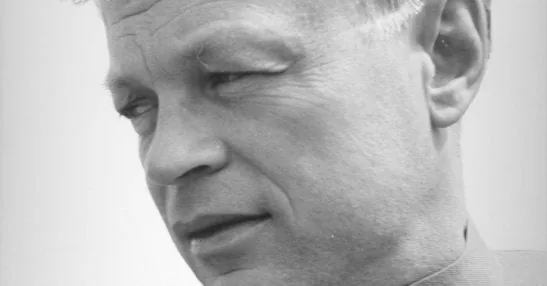
The story of cellular defense began with a lab puzzle in London during 1957. At the National Institute for Medical Research in London, Swiss postdoctoral fellow Dr. Jean Lindenmann worked alongside British virologist Dr. Alick Isaacs to explore how influenza viruses grow in host tissues. Lindenmann had arrived from Switzerland as a visiting researcher eager to learn viral culture methods. They treated influenza virus particles with heat to kill them, and then placed the dead viral material onto healthy animal tissue cultures to study whether cells developed immunity [3].
When the two scientists later exposed those treated cells to live influenza virus strains, the live germs surprisingly failed to take hold and multiply inside the cultured tissue dishes. Isaacs and Lindenmann initially wondered whether the heat-killed germs physically blocked cellular doors, or whether dead viral particles released a secret antibiotic agent into the surrounding fluid. Tests proved them wrong. The living cells had responded to the dead germs by making a protective protein that actively interfered with live viral growth. Because this active factor interfered with live infection, Isaacs and Lindenmann named it interferon in their 1957 report on viral interference, opening an entirely new field of experimental medicine, oncology, and cellular immunology [3].
Lindenmann soon returned to the University of Zurich to study why some mouse strains resisted influenza while other strains died, leaving broader drug development to fellow researchers around the globe. He was 90. He passed away in hospice care in Zurich from prostate cancer complications, his son Christian confirmed. Laboratories worldwide then set out to isolate related interferons across mammalian species [3].
What Are Interferons?
To grasp how human bodies resist germs, one must consider what are interferons at a basic biological level. They are soluble glycoprotein messengers that belong to the cytokine family, alerting host tissues to viruses, microbes, and growing tumor cells. These soluble glycoprotein messengers act in both autocrine and paracrine ways, binding to receptors on the cell that released them as well as on distant cells across the entire body [7]. Most cells make IFNs. Almost every cell in the human body can make IFNs when it senses foreign viral genes. Instead of directly killing invaders by themselves, natural interferons tell nearby healthy cells to build an internal defensive wall that blocks viral growth before the infection can spread further [8].
The warning cycle begins when internal sensors spot viral parts inside an infected cell. Membrane sensors known as toll-like receptor 3 (TLR3) catch double-stranded viral RNA, while cytoplasmic sensors like RIG-I and MDA5 scan the fluid inside the cell for foreign genetic strands. Such binding events quickly activate transcription factors named IRF3 and NF-κB. Both factors move into the cell nucleus to start the production of new interferons [8].
Once made, the proteins pass outside the cell into surrounding fluid to warn nearby tissues. They bind matching receptors sitting on nearby cells [7]. While the damaged cell dies to trap the infection, healthy neighbors change their machinery to block incoming germs. In addition to stopping viruses, IFNs activate killer immune cells and increase antigen presentation. They also help coordinate with other cytokines like Interleukin-10 and Interleukin-12 during complex immune reactions [8].
Three Major Classes of Interferons
Biologists divide interferons into three main groups based on receptor binding and protein folds [8]. In humans, the Type I group forms the largest family and includes thirteen subtypes of interferon-alpha, along with beta, omega, epsilon, and kappa forms. A cluster of functional Type I genes sits on chromosome 9 at band 9p21.3 across 400 kilobases, including the IFNA1 gene and pseudogenes. In mice, matching genes cluster on chromosome 4, where Swiss researcher Shaw and Charles Weissmann mapped them in 1982 [9]. The human interferon-beta crystal structure was deposited in 1997 and released in 1998 in the SCOPe database under PDB entry 1au1 [11]. All Type I forms bind to a shared receptor pairing made of IFNAR1 and IFNAR2 chains. Fibroblasts make interferon-beta, while plasmacytoid dendritic cells make interferon-alpha during sudden viral attacks [9].
Type II interferon contains only one member, called interferon-gamma (IFN-γ), which has distinct immunological roles in host defense. Its single human gene, IFNG, sits on a separate chromosome and codes for an intertwined protein dimer. Full length human IFN-γ is 143 amino acids long, while crystallized structural models cataloged in the SCOPe database under PDB code 1d9c span 126 amino acids in an intertwined dimer. Only one gene exists [10].

The third family consists of Type III interferons, known as interferon-lambda, which bind to the heterodimeric IFNLR receptor complex (Type III). Their receptors sit mainly on epithelial cells that line the lungs and gut. Because these receptors stay on body linings, Type III proteins protect barrier tissues without causing whole-body inflammation [8].
The JAK-STAT Antiviral Signaling Cascade
When an IFN binds to its receptor on the cell surface, it sets off a signal chain that heads straight for the nucleus. This pathway is the Janus kinase-STAT pathway, linking outer receptor binding to the activation of human defense genes. Receptor binding brings together Janus kinases such as JAK1 and TYK2, which add phosphate groups to docking sites on the receptor tail [8].
Docking sites pull in cytoplasmic messenger proteins called signal transducers and activators of transcription, or STATs. In Type I and Type III signaling cascades, receptor kinases rapidly phosphorylate STAT1 and STAT2, which team up with a third protein factor called IRF9 to build a three-part molecular machine named ISGF3. The complex is ISGF3. The ISGF3 machine enters the nucleus and binds tightly to conserved DNA sequences called interferon-stimulated response elements (ISREs) found in front of hundreds of defense genes [8]. That binding event turns on hundreds of downstream defense genes [2].
Type II signaling works in a similar way, using STAT1 pairs that bind gamma-activated site sequences in DNA. Cells also use backup routes, including p38 MAP kinase and PI3K pathways, to adjust their growth and survival. Protein CRKL and Rap1 pathways also help fine-tune cell survival during viral attacks [8]. Through this broad network, a quick touch on the outer cell wall reprograms cellular defenses within hours [2].

How Effector Proteins Block Viral Replication
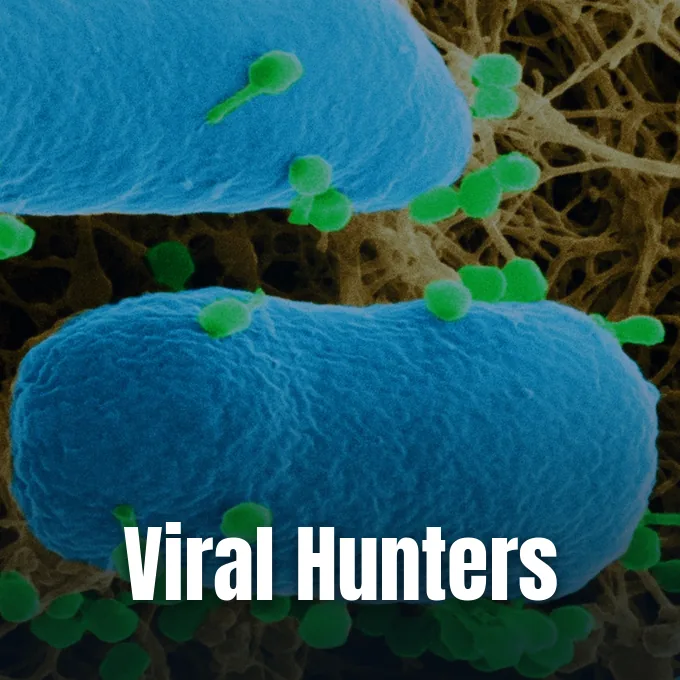
The real defense shield built by interferons comes from hundreds of proteins made by interferon-stimulated genes, or IFN-stimulated genes (ISGs) [2]. A key weapon is the RNA-dependent protein kinase R (PKR), which stays quiet in the cytoplasm until it detects double-stranded viral RNA inside the host cell. When turned on, PKR adds a phosphate to eukaryotic initiation factor 2 (eIF-2), trapping the recycling protein eIF2B and abruptly shutting down protein synthesis inside the cell. The cell stops making proteins. By halting this cellular assembly line, the host cell denies the virus the essential building blocks and structural proteins it needs to produce new infectious particles [8].
Another defense weapon is the 2-5A synthetase family, which spots viral RNA and makes short oligoadenylate chains. These small chains activate a quiet cellular enzyme called RNase L. RNase L cuts quickly. Once awake, RNase L cuts up both viral genetic strands and host cellular RNA, destroying viral messages before they can guide viral production [8].
Host cells also call upon Mx proteins that trap viral capsids so they cannot reach copying machinery inside the cell [2]. Furthermore, IFNs boost p53 activity, pushing dying host cells into planned suicide when an infection cannot be stopped. At the same time, they increase MHC Class I and Class II proteins on the cell surface, letting killer T cells and natural killer cells spot and destroy sick cells quickly. The immunoproteasome helps cut up viral peptides for display [8].
Viral Strategies Against Interferon Defenses
Because IFN defenses threaten viral survival, successful viruses have evolved clever tricks to disarm host interferons before the alarm system can take control. Many germs make specialized proteins that hide viral RNA or block host sensors before the cell can even produce an alert. In influenza A infections, a non-structural viral protein called NS1 binds double-stranded RNA, hiding it from host defense sensors like RIG-I and PKR before an alert can sound. The virus hides its RNA. By masking these foreign signs, influenza delays the host alarm long enough to spread through lung tissue [2].
Other germs cut off communication after host cells have already released their chemical warnings [2]. For example, Japanese encephalitis virus and dengue virus produce viral factors that block STAT1 and STAT2 activation, stopping them from entering the nucleus to start defense transcription. Without STAT proteins inside the nucleus, host cells cannot read their antiviral gene library, which leaves nearby tissues open to infection. Herpesviruses such as HCMV and KSHV also disrupt host defense signals [8].
Some large DNA viruses use decoy tricks to soak up protective proteins outside the cell. Poxviruses, such as the Vaccinia virus, release a soluble decoy protein called B18R that mimics the normal host receptor and binds extracellular IFNs before they touch host cells. B18R acts as a decoy. It catches circulating interferons in tissue fluids and binds them tight, keeping them from ever reaching actual receptors on neighboring cells [8].
Approved Clinical Uses in Medicine
Seeing how well these molecules stop viruses and slow cell growth, doctors turned them into medical treatments. In 1986, health regulators approved the first lab-made recombinant interferon to treat hairy cell leukemia, marking an early success for immunotherapy. Drug makers soon built synthetic interferons to fight long-term infections and several forms of cancer. The development of recombinant DNA methods allowed scientists to produce human proteins in bacteria in large amounts [1].
For years, recombinant interferon-alpha served as the primary treatment for chronic hepatitis B and hepatitis C, helping thousands of patients clear dangerous viral loads from liver tissue [1]. To make treatment easier, drug makers joined the protein to polyethylene glycol (PEG) to create peginterferon drugs. Adding PEG keeps the drug in the blood longer, so patients need fewer injections each week. Newer direct-acting antiviral pills now cure most hepatitis C cases faster and with far fewer side effects, yet manufactured interferons remain essential clinical tools against Kaposi sarcoma, malignant melanoma, and persistent genital warts [6].
Interferon-beta injections serve a different role by treating multiple sclerosis, an autoimmune illness where the body attacks its own nerves. The medicine calms inflammation in the brain and spinal cord, preventing nerve damage and lowering flare-ups [1]. For chronic granulomatous disease and malignant osteopetrosis, doctors use interferon gamma-1b, sold as Actimmune. The drug is Actimmune [6].
Side Effects and Health Risks
While medical interferons help treat dangerous diseases, they can cause heavy side effects that need close medical oversight [5]. The most common problem is a group of flu-like symptoms, including fever, chills, fatigue, head pain, and muscle aches that hit within hours of a subcutaneous injection [1]. Patients may also notice temporary skin redness or mild swelling around the injection site. Many patients also notice hair thinning, stomach pain, loss of appetite, and dizziness as treatment goes on [5]. These issues often improve with rest, fluids, and standard fever drugs [1].
Longer drug courses can trigger mental health issues that require careful attention [5]. Many patients experience depression, severe anxiety, or thoughts of self-harm while taking these drugs [6]. Doctors check patients for mood disorders before starting therapy and can prescribe antidepressant pills or lower the drug dose if depression appears [5].
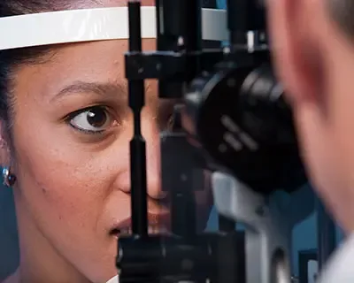
Synthetic interferons can also harm the eyes by causing retinopathy, a condition where retinal blood vessels leak or swell, threatening vision if left untreated. The American Academy of Ophthalmology strongly recommends a detailed eye checkup before starting therapy, along with routine clinic visits using a slit lamp and ophthalmoscope to spot retinal vessel damage early. Such eye damage is reversible. If retinal swelling develops, eye doctors can inject anti-VEGF drugs like Lucentis or Avastin into the eye, or give steroid shots to protect long-term sight [4].
- WEBSITE Cleveland Clinic. (2024). What Are Interferons? [Article Link]
- ACADEMIC JOURNAL Katze, M. G., He, Y., & Gale, M. (2002). Viruses and interferon: a fight for supremacy. Nature Reviews Immunology, 2(9), 675-687. [Article Link]
- ONLINE NEWS Kolata, G. (2015). Jean Lindenmann, Who Made Interferon His Life’s Work, Is Dead at 90. The New York Times. [Article Link]
- WEBSITE Turbert, D. (2026). What Is Interferon? American Academy of Ophthalmology. [Article Link]
- ONLINE NEWS Wessels, D. (2018). Interferon side effects: Signs, causes, and risks. Medical News Today. [Article Link]
- WEBSITE Watson, S. (2024). Your Guide to Interferons. WebMD. [Article Link]
- ONLINE NEWS Dutta, S. S. (2020). Interferons and Interleukins. News-Medical. [Article Link]
- WEBSITE Wikipedia contributors. (2026). Interferon. Wikipedia, The Free Encyclopedia. [Article Link]
- WEBSITE Wikipedia contributors. (2026). Interferon type I. Wikipedia, The Free Encyclopedia. [Article Link]
- WEBSITE Wikipedia contributors. (2026). Interferon gamma. Wikipedia, The Free Encyclopedia. [Article Link]
- WEBSITE SCOPe. (2020). SCOPe 2.08: Structural Classification of Proteins — Extended PDB Entry 1au1. [Article Link]
- WEBSITE SCOPe. (2020). SCOPe 2.08: Domain d1d9ca_: Bovine Interferon-Gamma. [Article Link]


