





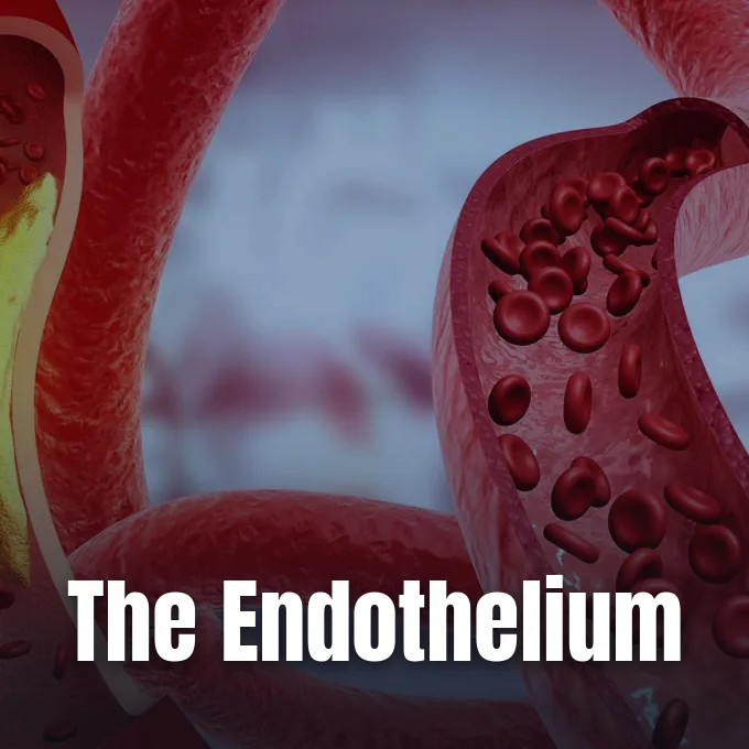
When clinicians look at the vascular system, they often focus on the beating heart and thick muscular arteries. But what is endothelium, and why does this microscopic cell layer guide vascular health throughout human life? The endothelium forms an active single layer of flat cells that lines every blood vessel and lymph conduit in the human body. In an adult, this inner sheet covers about 60,000 miles of blood pathways, according to clinical data published by the Cleveland Clinic. By surface area, the tissue extends across 3,000 to 6,000 square meters, making it one of the largest functional surfaces in human anatomy. It houses over 1 trillion individual cells. These cells constantly exchange biochemical messages with passing blood and lymph fluids [1, 5]. Rather than acting as a simple passive tube, this expansive monolayer functions as a true endocrine organ that regulates vessel diameter, governs fluid passage, and prevents unwanted blood clots during daily activity [1, 15].
What Is Endothelium in Human Vessels?
Swiss anatomist Wilhelm His coined the term endothelium in 1865 to separate this deep vascular lining from outer epithelial coverings. The tissue originates from embryonic mesoderm. Because these cells build their internal skeleton out of vimentin fibers instead of keratin proteins, the Foundational Model of Anatomy classifies them apart from common epithelial tissues, noting that they attach firmly to an underlying basal lamina [5].
Epithelial sheets cover body surfaces that meet the open external environment, including the outer skin and the digestive tract, whereas endothelial cells line fully enclosed fluid circuits [1]. In straight stretches of healthy arteries and veins, these flat squamous cells align along the flow of blood. They adapt their shape to minimize fluid shear forces. Tight junctions link adjacent cell borders together, creating a selective seal that prevents unregulated fluid loss into tissue spaces [5].
Each cell is thin, running only a few hundred nanometers in thickness, as shown in laboratory studies by Harvard Medical School researchers [2]. At its longest span, a single endothelial cell extends about 50 micrometers along the vessel axis, which means it spans roughly half the width of a human hair that measures 100 micrometers across. They are invisible to the naked eye. Medical teams must use electron or fluorescence microscopes to observe how these tiny cell borders manage the passage of water, ions, and small nutrients into tissue spaces [1, 3].
Structural Layers of Vessel Walls
To understand how the inner cell sheet protects blood vessels, vascular biologists examine the three distinct tunics that build arterial and venous walls. The outermost coat is the adventitia, which supplies structural support and anchors vessels to surrounding tissues. Beneath it rests the tunica media, a muscular middle coat composed of smooth muscle cells and elastic fibers that contract or relax to change lumen diameter [3].
The innermost layer is the tunica intima, formed by the endothelial monolayer resting directly upon its basal lamina. Blood cells and platelets glide over this slick floor without sticking during healthy flow, as reported by Medical News Today [1, 3]. Integrin receptors anchor each cell to the underlying matrix. This smooth cellular boundary creates a continuous physical barrier that separates flowing blood from the deeper connective elements of the vessel wall [1, 5].
Facing the bloodstream, the cell surface carries the glycocalyx, a gel-like carbohydrate coat that touches plasma proteins and circulating blood cells. It acts as a protective sieve [3]. In an experimental study published in the American Journal of Physiology, researchers Rowan, Rochfort, and McLoughlin investigated how fluid properties govern microcirculatory balance. Working with isolated murine lung preparations, the team found that perfusion solutions with a viscosity score of 2.5 cut pulmonary edema rates without changing capillary filtration pressure. Blood viscosity works alongside tight cell junctions to preserve fluid balance and prevent tissue swelling in healthy lungs [11].
Three Classes of Vascular Endothelium
Endothelial structures differ across organ systems to meet specific local metabolic demands and transport duties. In 1959, American anatomists Bennett, Luft, and Hampton used electron microscopy to divide endothelia into continuous, fenestrated, and discontinuous types across mammalian organ beds. Continuous endothelium is the most common variety, featuring an intact basal lamina and tightly sealed cell borders. This continuous cell sheet lines blood vessels in the brain, heart, lungs, and skeletal muscle. It shields delicate tissues from fluid shifts [5].
Where tissues require rapid exchange of solutes and fluids, vascular beds exhibit fenestrated or discontinuous architectures. Fenestrated endothelium maintains an intact basal lamina but features circular pores measuring 50 to 60 nanometers across, each bridged by a thin diaphragm. These openings predominate in capillaries of the renal glomerulus, endocrine glands, and intestinal mucosa. They allow water, salts, and peptide hormones to filter into nearby tissues while retaining blood cells and large proteins safely within the lumen [5].

Discontinuous endothelium features wide intercellular gaps of 100 to 200 nanometers and possesses an incomplete or absent basement membrane. Found in liver sinusoids, spleen, and bone marrow, these broad openings allow whole blood cells and large proteins to pass freely between circulation and tissue spaces. The inner lining also protects against clot formation. In 1958, Scottish hematologist A. S. Todd at the University of St Andrews demonstrated that human vascular endothelial linings possess intrinsic fibrinolytic activity, producing plasminogen activators that dissolve microscopic fibrin deposits [5].
Regulation of Vascular Tone
The vascular endothelium actively guides blood flow by adjusting vessel diameter to meet tissue demands. As blood flows over the cellular lining, shear stress activates mechanosensitive ion channels on the luminal membrane. This mechanical force stimulates endothelial nitric oxide synthase, an enzyme known as eNOS. The active enzyme converts L-arginine into nitric oxide, a short-lived gas that diffuses into vascular smooth muscle cells within the tunica media [4, 5]. Natural Heart Doctor founder Jack Wolfson describes nitric oxide as an essential vasodilator that keeps vessel walls relaxed and open to promote healthy blood pressure [4].
Within vascular smooth muscle, nitric oxide stimulates soluble guanylyl cyclase, producing cyclic guanosine monophosphate to relax muscle fibers and lower vascular resistance. The gas acts fast. Endothelial cells also produce prostacyclin, or PGI2, via cyclooxygenase pathways to relax vascular muscle and inhibit platelet clumping [5, 15]. In addition, endothelium-derived hyperpolarizing factor opens potassium channels in resistance vessels to support dilation when nitric oxide levels decline [5].
When bodily conditions require vessel constriction, cells release contracting peptides including endothelin-1 and thromboxane A2 [4, 5]. This narrows the lumen [4]. French cardiovascular researcher Michel Félétou showed that endothelial surface enzymes also direct the production of systemic vasoactive hormones like angiotensin II, allowing the monolayer to coordinate regional blood delivery during exercise, cold exposure, and quiet rest [1, 15].


Cellular Self-Repair After Mechanical Injury
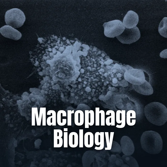
Vascular endothelial cells routinely withstand high fluid pressures, mechanical stretch, and immune cell crossings without losing structural continuity. During acute inflammation, white blood cells breach the endothelial monolayer to reach infected tissues. At Beth Israel Deaconess Medical Center, a vascular biology research team led by Christopher Carman and Roberta Martinelli investigated how cells survive these transcellular breaches. Using high-resolution confocal microscopy, Carman and Martinelli observed seven leukocytes pass directly through a single cell body within ten minutes without tearing the plasma membrane. The cell quickly healed each opening [2].
Martinelli discovered that pore closure begins when a penetrating leukocyte releases resting isometric tension within the cell skeleton. She compared the cell to a circus tent: “If you cut one of the ropes holding up the tent, two things will happen… the part of the tent that was under tension will undergo a recoil… At the same time, the remaining sets of ropes and stakes will have to bear the extra load”. In response to this mechanical signal, the cell extends ventral lamellipodia that migrate across the pore to seal it. This localized self-repair requires small amounts of hydrogen peroxide generated by NADPH oxidases [2].
In a 2018 preprint on arXiv that had not undergone peer review, Jorge Escribano, Roger Kamm, and Fabian Spill showed that gaps form spontaneously at vertices where three cells meet [9]. Metastatic cancer cells exploit these gaps. Publishing in PLoS Computational Biology, Escribano confirmed that circulating tumor cells preferentially cross vessel walls at these three-cell vertices rather than boring new holes through cell bodies [8]. Center for Vascular Biology Research director William Aird said that vascular leakage accelerates when cells lose their intrinsic reserve to heal mechanical micro-wounds [2].

How Endothelial Cells Guide Immunity
The endothelium acts as an active gatekeeper for the immune system, directing defense cells into inflamed tissue beds. When tissue injury triggers inflammation, endothelial cells express cell-surface adhesion molecules on their luminal boundary [3, 5]. Circulating white blood cells bind to E-selectin, intercellular adhesion molecule 1 (ICAM-1), and vascular cell adhesion molecule 1 (VCAM-1) [5]. Anchored monocytes subsequently crawl across the endothelial sheet into extravascular tissue, where they differentiate into macrophages to clear invading pathogens and cellular debris.
Nuclear factor kappa B steers this inflammatory genetic response inside stimulated endothelial cells. To prevent runaway tissue damage, the pathway also activates the deubiquitinase A20, an enzyme encoded by the TNFAIP3 gene that repairs the endothelial barrier. Healthy cell surfaces also keep blood fluid [5, 16]. They express heparan sulfate proteoglycans that activate antithrombin, neutralizing circulating thrombin and activated factor X before obstructive clots can form [5].
When blood vessel walls rupture, endothelial cells expose tissue factor and release stored von Willebrand factor, prompting rapid platelet clumping to seal the breach [5, 16]. This prevents fatal bleeding [16]. Low oxygen levels prompt cells to initiate angiogenesis, where vascular endothelial growth factor (VEGF) and angiopoietin-2 cooperate to degrade basement membranes and guide new capillary sprouts toward oxygen-starved tissue beds [5]. Harvard professor William Aird said that cells across different vascular beds employ distinct strategies to maintain local hemostatic balance throughout the body [16].
Corneal Endothelium Fluid Pump Dynamics
Specialized endothelial layers perform distinct physiological tasks in ocular and lymphatic structures. The posterior surface of the cornea carries the corneal endothelium, cataloged under anatomical identifier THA 6912 (FMA 63882) by the IFAA [18]. Unlike blood vessel linings derived from mesoderm, the corneal endothelium originates embryologically from the neural crest. This cell layer consists of about 300,000 hexagonal cells arranged in a honeycomb mosaic upon Descemet’s membrane, an acellular collagen sheet [6].
In healthy young adults, corneal endothelial density spans 2,400 to 3,200 cells per square millimeter. Because the cornea must remain avascular to transmit clear light, its cells obtain glucose and solutes by diffusion from the aqueous humor. The monolayer preserves corneal deturgescence through the classic pump-leak mechanism. While nutrients leak into the stroma, active Na+/K+ ATPase pumps and carbonic anhydrase enzymes constantly move water back into the anterior chamber to preserve optical transparency [6].
Human corneal cells cannot divide after birth, existing in a permanent non-dividing state. They divide rarely after birth. When cells die from trauma, nearby cells slide over denuded gaps to maintain the pump. If density falls below 500 cells per square millimeter, corneal edema impairs visual acuity [6]. Lymphatic endothelial cells also line blind-ended lymph capillaries to drain excess fluid and immune cells from peripheral tissues. First described by Hippocrates in 400 BC and rediscovered in the 17th century by Italian anatomist Gasparo Aselli, lymphatic vessels expand during inflammation, as demonstrated by British pathologist Howard Florey in 1937. Modern laboratories identify these lymphatic linings using specific molecular markers including LYVE-1 and podoplanin [7].
Mechanisms of Endothelial Dysfunction
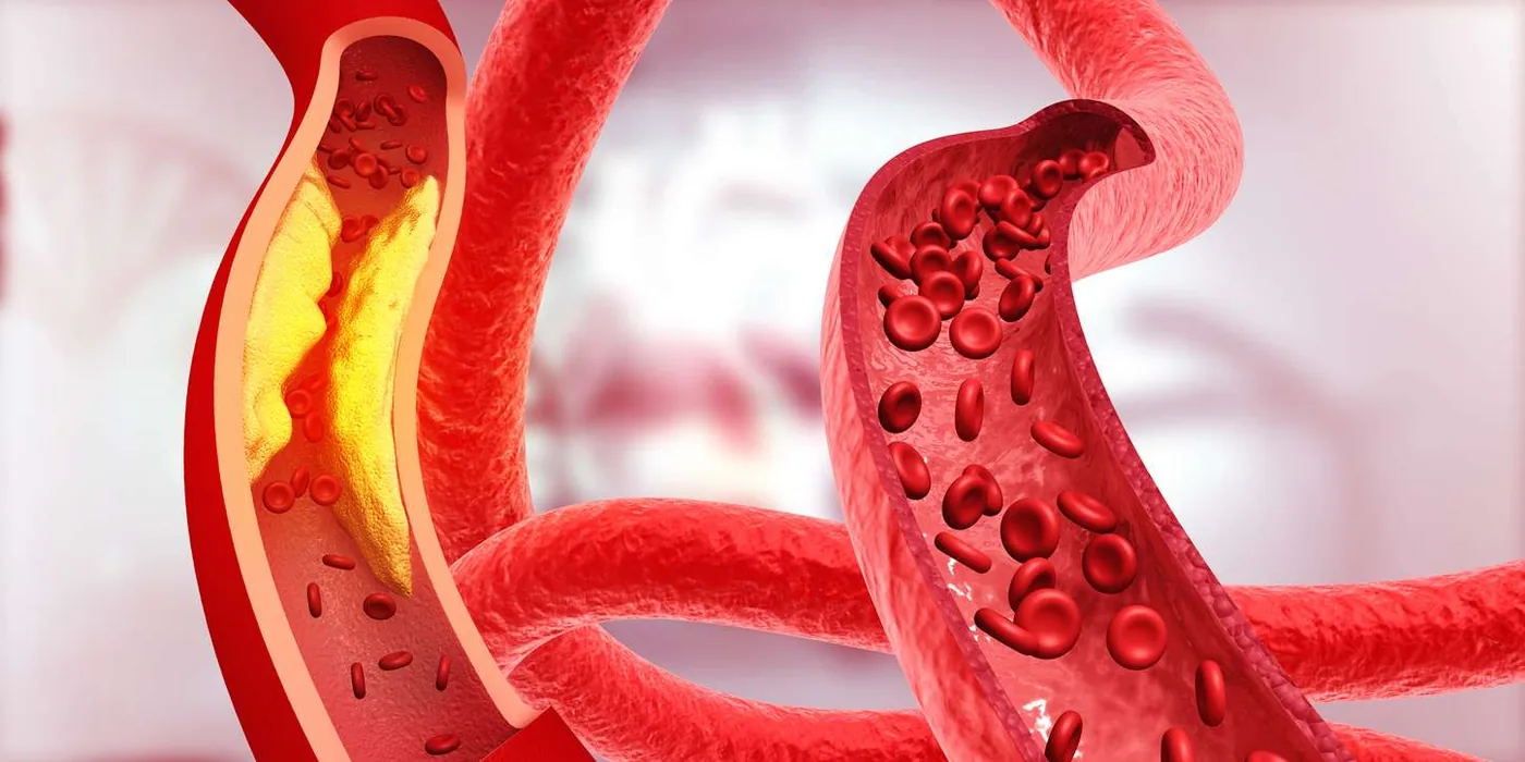
When endothelial cells experience chronic chemical or physical stress, they lose their protective phenotypes in a pathological state termed endothelial dysfunction [1, 3, 10]. Vessel walls lose their flexibility. Persistent high blood pressure, tobacco smoking, sedentary living, and elevated low-density lipoprotein (LDL) particles disrupt regular endothelial signaling pathways [1, 3]. Injured cells produce diminished nitric oxide, largely because high concentrations of asymmetric dimethylarginine competitively inhibit the eNOS pathway, leading to inappropriate arterial narrowing and heightened leukocyte adhesion [1, 5].
Monocytes ingest oxidized LDL particles and transform into foam cells [1, 5]. This builds early fatty streaks. Over decades, growing plaques narrow arterial conduits, markedly increasing the risk of myocardial infarction, peripheral arterial disease, and ischemic stroke, as reviewed by Rajendran and colleagues in the International Journal of Biological Sciences [1, 12]. In severe sepsis, systemic endothelial activation causes glycocalyx shedding, capillary leakage, and microvascular thrombosis [8, 13]. Clinical researchers at an Acute Dialysis Quality Initiative symposium in Bogota, Colombia, reported that widespread microvascular collapse drives multi-organ failure during septic shock [13]. In Cardiovascular Research, Dolmatova and Griendling confirmed that microvascular dysfunction accounts for nearly 700,000 sepsis-related deaths across Europe annually [14].
In cancer care, toxic chemotherapy agents like doxorubicin induce cardiotoxicity by causing endothelial cell death, as demonstrated by Luu and Verma in JACC Basic to Translational Science [17]. In rare instances, endothelial cells turn malignant to form angiosarcoma, a vascular tumor with about 300 cases per year in the United States and a five-year survival rate of 35%. The disease remains difficult to treat. Clinical evidence indicates that regular physical exercise and a Mediterranean diet rich in walnuts improve endothelial nitric oxide production, protecting vascular health over time [1, 5].
- WEBSITE Cleveland Clinic. (2023). What is the Endothelium? [Article Link]
- PRESS RELEASE Prescott, B. (2013). Endothelium, Heal Thyself. Harvard Medical School. [Article Link]
- ONLINE NEWS Jones, R. (2023). What to know about the endothelium. Medical News Today. [Article Link]
- WEBSITE Wolfson, J. (2024). The Endothelium: What You Need to Know. Natural Heart Doctor. [Article Link]
- WEBSITE Wikipedia contributors. (2026). Endothelium. Wikipedia, The Free Encyclopedia. [Article Link]
- WEBSITE Wikipedia contributors. (2026). Corneal endothelium. Wikipedia, The Free Encyclopedia. [Article Link]
- WEBSITE Wikipedia contributors. (2026). Lymphatic endothelium. Wikipedia, The Free Encyclopedia. [Article Link]
- ACADEMIC JOURNAL Escribano, J., Chen, M. B., Moeendarbary, E., Cao, X., Shenoy, V. B., García‐Aznar, J. M., Kamm, R. D., & Spill, F. (2019). Balance of mechanical forces drives endothelial gap formation and may facilitate cancer and immune-cell extravasation. PLoS Computational Biology, 15(5). [Article Link]
- PREPRINT Escribano, J., Chen, M. B., Moeendarbary, E., Cao, X., Shenoy, V., Garcia-Aznar, J. M., Kamm, R. D., & Spill, F. (2018). Balance of Mechanical Forces Drives Endothelial Gap Formation and May Facilitate Cancer and Immune-Cell Extravasation. arXiv. [Article Link]
- ACADEMIC JOURNAL Peng, Z., Shu, B., Zhang, Y., & Wang, M. (2019). Endothelial Response to Pathophysiological Stress. Arteriosclerosis, Thrombosis, and Vascular Biology, 39(11). [Article Link]
- ACADEMIC JOURNAL Rowan, S. C., Rochfort, K. D., Piouceau, L., Cummins, P. M., O’Rourke, M., & McLoughlin, P. (2018). Pulmonary endothelial permeability and tissue fluid balance depend on the viscosity of the perfusion solution. American Journal of Physiology-Lung Cellular and Molecular Physiology, 315(4), L476-L484. [Article Link]
- ACADEMIC JOURNAL Rajendran, P., Rengarajan, T., Thangavel, J., Nishigaki, Y., Sakthisekaran, D., Sethi, G., & Nishigaki, I. (2013). The Vascular Endothelium and Human Diseases. International Journal of Biological Sciences, 9(10), 1057-1069. [Article Link]
- ACADEMIC JOURNAL İnce, C., Mayeux, P. R., Nguyen, T. C., Gómez, H., Kellum, J. A., Ospina-Tascón, G. A., P, G. H., Murray, P. T., & Backer, D. D. (2016). The Endothelium in Sepsis. Shock, 45(3), 259-270. [Article Link]
- ACADEMIC JOURNAL Dolmatova, E. V., Wang, K., Mandavilli, R., & Griendling, K. K. (2020). The effects of sepsis on endothelium and clinical implications. Cardiovascular Research, 117(1), 60-73. [Article Link]
- ACADEMIC JOURNAL Félétou, M. (2011). The Endothelium, Part I: Multiple Functions of the Endothelial Cells — Focus on Endothelium-Derived Vasoactive Mediators. Colloquium Series on Integrated Systems Physiology From Molecule to Function, 3(4), 1-306. [Article Link]
- ACADEMIC JOURNAL Aird, W. C. (2015). Endothelium and haemostasis. Hämostaseologie, 35(01), 11-16. [Article Link]
- ACADEMIC JOURNAL Luu, A. Z., Chowdhury, B., Al-Omran, M. A., Teoh, H., Hess, D. A., & Verma, S. (2018). Role of Endothelium in Doxorubicin-Induced Cardiomyopathy. JACC Basic to Translational Science, 3(6), 861-870. [Article Link]
- WEBSITE Federative International Programme on Anatomical Terminologies. (2013). Corneal endothelium (A15.2.02.022). IFAA. [Article Link]


