





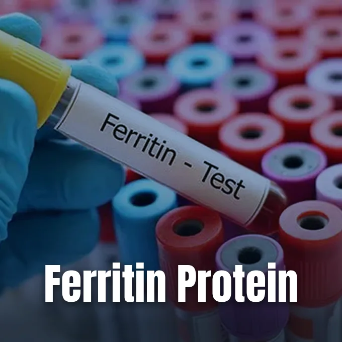
Asking what is ferritin leads directly to the core molecular machinery that manages iron storage inside living human cells [1]. Ferritin is a spherical protein complex that stores iron in a soluble, nontoxic form and releases it when physiological demands arise [13]. Without this protective storage mechanism, free iron ions would react with oxygen compounds to generate cellular damage [9]. The protein serves as the body’s primary iron reservoir, operating in tissues and circulating in small quantities through the bloodstream [4].
What Is Ferritin?
Ferritin functions as a universal iron storage protein found across animals, plants, bacteria, and archaea [13]. Within human cells, it acts as a molecular warehouse, holding iron atoms in reserve until bone marrow needs them to produce fresh red blood cells [1]. Dr. Angela Weyand from the University of Michigan Medical School describes ferritin as an iron savings account that reflects long-term reserves, releasing stored iron whenever dietary intake falls short of daily metabolic requirements [6].
Iron is needed for cellular respiration and DNA synthesis, yet unshielded iron ions present grave biological hazards to living tissue. Under physiological conditions, iron(III) ions have a tiny solubility of roughly 10-18 M, meaning they clump into insoluble rust-like aggregates unless shielded by proteins. The regular breakdown of aged erythrocytes releases roughly 0.54 mmol of iron into circulation every twenty-four hours. Researchers at Duke University and RCSB PDB calculated that a human would have to drink 1013 liters of water every day to prevent rusty kidneys if that mineral fell out of solution. Ferritin stops this crisis by gathering free iron ions, shielding them from reactive ligands, and storing them safely inside a soluble protein shell [9].
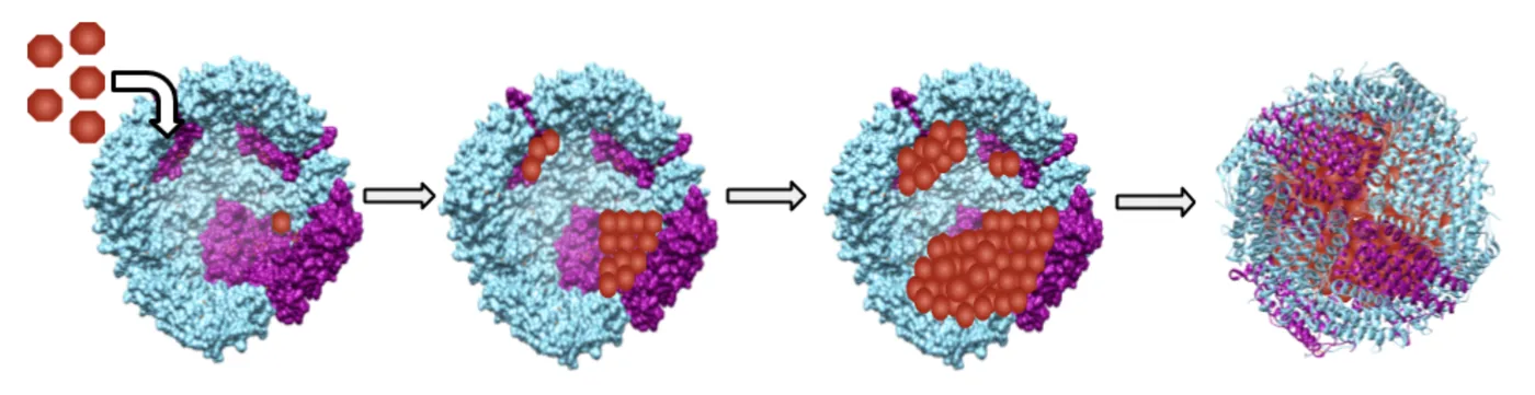
The protein exists in two basic states based on mineral load. When empty of iron, the hollow shell is called apoferritin [13]. Once filled, the central cavity holds up to 4500 iron ions, accounting for up to 24% of the total protein weight [9]. Clumped ferritin in tissue can transform into hemosiderin, an insoluble complex that yields iron much more slowly [13].
Subunit Architecture and Iron Core
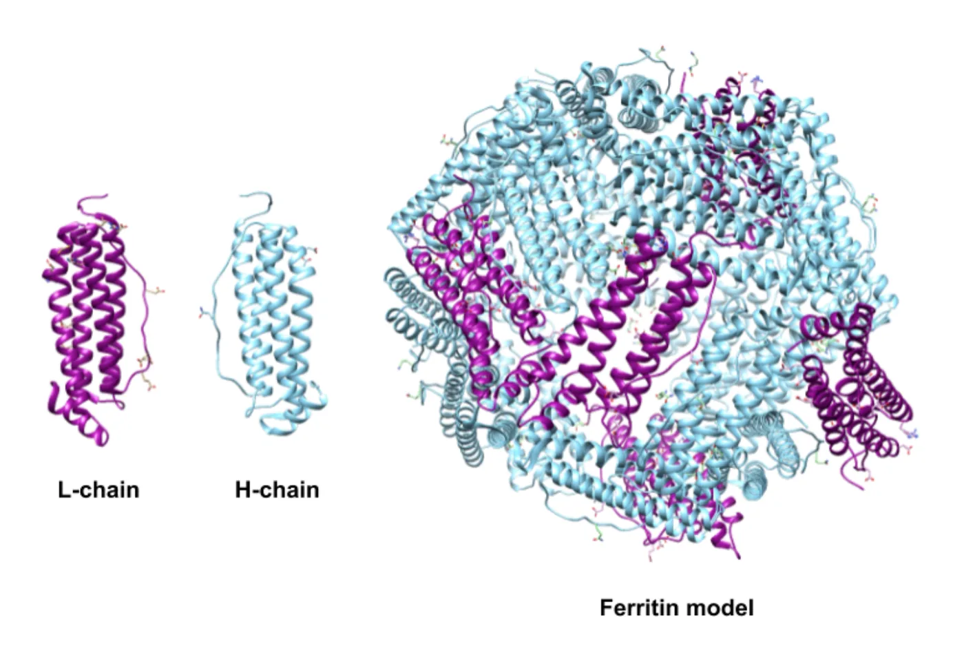
The standard mammalian ferritin molecule is a hollow globular complex with an outer diameter of 12 nanometers and an inner cavity measuring 8 nanometers. It has a total molecular mass of roughly 474 to 480 kilodaltons, built from 24 individual polypeptide subunits [9, 13]. Each subunit folds into four alpha helices, as cataloged in RCSB PDB entry 1fha under SCOPe classification [2]. Together, these 24 subunits assemble in symmetric fashion to construct the spherical shell [9].
In humans, the protein shell combines two subunit types known as heavy (H) and light (L) chains. The H-subunit has 178 amino acids, while the L-subunit contains 171 amino acids. The H-chain oxidizes iron, whereas the L-chain nucleates mineral crystals [9].
The mineral deposited inside the central space matches ferrihydrite, with a chemical composition of 5Fe2O3·9H2O along with phosphate and hydroxide ions [13]. High-resolution cryogenic studies at 1.2 Å resolution by T. Granier, B. Gallois, J.-M. Chevalier, G. Précigoux, and P. Arosio at Université Bordeaux I in Talence, France (postal code 33405) deposited under RCSB PDB code 1LB3 identified that glutamate side chains E57, E60, E61, and E64 direct metal binding at the nucleation site. Lysine residue K140 interacts directly with E61, explaining why mouse L-chain ferritin exhibits different iron incorporation properties than human L-chains [10].
Channel Pathways for Mineral Entry
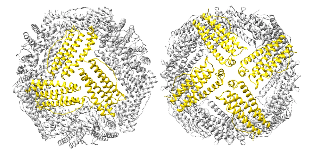
Iron atoms enter the shell through tiny pores where subunits meet. The structure carries eight 3-fold channels and six 4-fold channels. The 3-fold channels have polar aspartate and glutamate side chains that form a water-friendly path [9]. In mouse L-chain crystals, residues H118, D131, and E134 shape this entry gate to grip metal ions [10].
These water-friendly 3-fold channels serve as the main path for ferrous iron (Fe2+) to enter the core, whereas the six 4-fold channels are lined with non-polar leucine residues that create a water-repellent corridor. Structural studies on human ferritin entry pathways in Chemistry LibreTexts indicate these greasy paths let oxygen and hydrogen peroxide diffuse inward while blocking charged metal ions from leaking out. This spatial separation prevents early chemical reactions on the outer protein coat, ensuring that mineral growth stays confined within the inner cavity [9].
When cells need iron for fresh enzymes, they break down stored mineral cores. Lysosomes handle this task by digesting the protein coat through autophagy. Through this breakdown of apoferritin, cells recover iron without spilling dangerous free radicals [13].
How Ferritin Protects Against Chemical Toxicity
Free iron in cells is risky because it sparks free radical creation [4]. When ferrous iron meets hydrogen peroxide, it drives the Fenton reaction, producing ferric iron, hydroxide, and the destructive hydroxyl radical. Hydroxyl radicals tear through cell membranes, alter DNA structures, and break protein bonds, promoting an iron-dependent form of cell death known as ferroptosis. Ferritin prevents this harm by trapping free iron early [9].
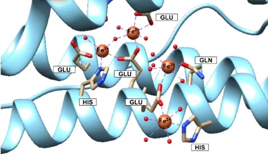
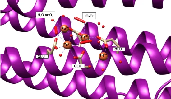
This protective conversion happens at catalytic ferroxidase centers embedded within each H-subunit of the protein shell. In these active sites, amino acid side chains from glutamic acid, glutamine, and histidine bind incoming ferrous ions and coordinate them with molecular oxygen to turn Fe2+ into Fe3+, which then migrates inward to L-chain nucleation sites to crystallize into safe mineral deposits. The oxidation consumes hydrogen peroxide and oxygen directly. By acting as an antioxidant, the protein shields fragile cell structures from oxidative injury [9].
Mitochondria carry their own version of the protein called mitochondrial ferritin. Encoded by an intronless gene, this precursor enters organelles and forms functional shells sharing 80% sequence identity with human H-ferritin. Work on RCSB PDB structure 1R03 by B. Langlois d’Estaintot, P. Santambrogio, and P. Arosio showed that mitochondrial ferritin preserves iron balance inside energy-producing organelles [11].
Tissue Distribution Across the Body
Ferritin works in every human tissue, but its subunit recipe changes to match local metabolic tasks [9, 13]. Organs with heavy iron turnover—like the heart and brain—build cages rich in H-subunits to speed detoxification. In contrast, storage centers like the spleen and liver use far more L-subunits, with human liver tissue maintaining an even mix of 50% H and 50% L chains [9].
Most stored iron lives in the liver, spleen, and bone marrow. In healthy adults, liver hepatocytes and reticuloendothelial macrophages hold several grams of mineral reserves to buffer daily needs [4, 5]. While ferritin acts mainly inside cells, tiny traces enter the bloodstream. The Blood Project notes that macrophages release serum ferritin through non-classical secretory channels, and because circulating concentrations mirror intracellular stores under steady-state conditions, blood measurements offer clinicians a reliable window into total iron reserves [12].
Gene flaws in these subunits cause rare inherited diseases. Specific mutations in exon 4 of the FTL gene lead to neuroferritinopathy, an adult-onset movement disorder marked by iron deposits in the basal ganglia. Furthermore, mutations in the iron-responsive element of L-ferritin mRNA cause hyperferritinemia-cataract syndrome, producing early bilateral cataracts without causing body-wide iron overload [12, 14].
Clinical Standards for Blood Testing
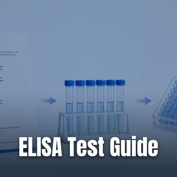
In clinical care, a serum ferritin blood test measures total iron reserves. Phlebotomists take a venous blood sample from the arm, which lab machines analyze with antibody tests like ELISA or immunochemiluminescence [1, 12]. Lab reports list findings using nanograms of protein per milliliter of blood, a scale matching micrograms per liter [13]. This number gives doctors an objective measure of stored iron [4].
Reference intervals for normal ferritin vary between commercial labs and patient groups [1]. WebMD writer Kim Painter notes standard adult ranges between 24 and 336 ng/mL for men and 24 to 307 ng/mL for women [3]. MedicineNet editor Dr. Melissa Conrad Stöppler lists normal adult male ranges from 20 to 250 ng/mL, while values for women aged 18 to 39 span 10 to 120 ng/mL because of menstrual blood losses [4]. Newborn infants show much higher numbers, with Cleveland Clinic reporting normal infant values up to 650 ng/mL during the first six months of life [1].
In September 2026, the American Society of Hematology released updated clinical guidelines to identify iron shortage before anemia takes hold [5, 6]. Pediatric hematologist Dr. Angela Weyand from University of Michigan Medical School explained that old statistical reference curves failed because iron deficiency is remarkably common, affecting roughly 2 billion people worldwide and 14% of United States adults in a 2025 JAMA review. The new guidance sets a deficiency threshold of 30 ng/mL or lower for adults, while recommending 20 ng/mL for young children aged 9 months to 4 years and 50 ng/mL for high-risk pregnant individuals [5, 6].
Why Do Stored Iron Levels Drop?
Depleted iron stores occur when daily iron loss exceeds intake, draining ferritin reserves [6]. The leading cause of iron deficiency in adults is ongoing blood loss from heavy menstrual periods, hidden bowel bleeding, or frequent blood donations [3, 8]. Poor intestinal absorption also causes low ferritin in people with celiac disease or inflammatory bowel disorders. Poor dietary intake and increased iron demands during pregnancy also deplete stores quickly [4, 8].
When ferritin drops, people often feel run down long before blood counts reveal anemia. Dr. Caroline Cromwell, a hematologist at Weill Cornell Medicine and NewYork-Presbyterian, explains that low iron stores sap energy, causing fatigue, dizziness, headaches, restless legs, and shortness of breath [3, 7]. Sela Breen reported in Mindbodygreen that online communities coined the phrase ferritin face to describe appearance changes like dry pale skin, brittle nails, thinning hair, and dark under-eye circles. Some patients also experience pica, developing compulsive cravings to chew ice or eat dirt and clay [3, 7].
Fixing low ferritin means finding why iron was lost rather than taking blind supplements [6]. Care starts with oral iron pills, though some patients suffer from stomach upset. Dr. Karim Haddad of Atlas Health notes that pairing iron with vitamin C aids absorption, while severe cases require intravenous iron infusions [7, 8]. Doctors also order a complete blood count (CBC) to check red cell health [1, 8].
Elevated Ferritin and Systemic Inflammation
A high ferritin score does not always mean an individual has too much iron [6]. Ferritin acts as an acute-phase reactant, meaning cells make extra protein during systemic inflammation, infection, or tissue damage [4, 13]. When white blood cells spot bacterial endotoxins or inflammatory signals, macrophages boost ferritin synthesis to lock iron away from multiplying germs, denying essential nutrients to infectious pathogens [13]. Consequently, patients with rheumatoid arthritis, liver disease, or leukemia can show high ferritin even when usable iron is scarce [4, 5].
Genuine iron overload stems from genetic illnesses such as hereditary hemochromatosis, where the intestines absorb excessive iron from daily meals. In men with hemochromatosis, unmanaged buildup can accumulate a 20-gram iron burden by middle age, far exceeding the 3 to 4 grams found in healthy adult bodies. This surplus iron collects inside vital organs, damaging the liver, heart, joints, and pancreas. As documented by WebMD and MedicineNet, this toxic mineral buildup can trigger liver cirrhosis, heart failure, diabetes mellitus, severe joint pain, unexplained weight loss, and bronze darkening of the skin [3, 4]. Repeated blood transfusions also cause secondary iron overload [3, 5].
To separate inflammatory spikes from real iron overload, physicians run broad testing panels. Clinicians measure total iron-binding capacity (TIBC), transferrin saturation, and C-reactive protein to evaluate inflammation. When ferritin passes 300 ng/mL alongside elevated transferrin saturation, doctors perform genetic screens or liver scans to measure tissue iron [5, 8].
- WEBSITE Cleveland Clinic. (2018, May 1). What is a ferritin test? Cleveland Clinic. [Article Link]
- REPORT SCOPe. (2026, September 12). SCOPe 2.08: Structural classification of proteins — extended (PDB entry 1fha). Structural Classification of Proteins. [Article Link]
- WEBSITE Painter, K. (2023, November 14). What is a ferritin blood test? What do the results mean? WebMD. [Article Link]
- WEBSITE Stöppler, M. C. (2024, May 21). Ferritin blood test (high, low, normal levels): Meaning, chart. MedicineNet. [Article Link]
- WEBSITE Nall, R. (2018, August 13). Ferritin level blood test: Purpose, procedure, and results. Healthline. [Article Link]
- ONLINE NEWS Rizzo, N. (2026, October 5). Everyone is talking about ferritin levels. How much does it really matter for your health? TODAY. [Article Link]
- ONLINE NEWS Breen, S. (2026, November 7). What is ferritin face? We asked an MD for the truth about the viral term. Mindbodygreen. [Article Link]
- WEBSITE Haddad, K. (2026, November 1). What is ferritin? How it works and why you need it. Atlas Health. [Article Link]
- BOOK CHAPTER Haas, K. (2018, May 17). Iron storage: Ferritin. Chemistry LibreTexts. [Article Link]
- ACADEMIC JOURNAL Granier, T., Langlois d’Estaintot, B., Gallois, B., Chevalier, J.-M., Précigoux, G., Santambrogio, P., & Arosio, P. (2003). Structural description of the active sites of mouse L-chain ferritin at 1.2 Å resolution. JBIC Journal of Biological Inorganic Chemistry, 8(1), 105–111. [Article Link]
- ACADEMIC JOURNAL Langlois d’Estaintot, B., Santambrogio, P., Granier, T., Gallois, B., Chevallier, J. M., Précigoux, G., Levi, S., & Arosio, P. (2004). Crystal structure and biochemical properties of the human mitochondrial ferritin and its mutant Ser144Ala. Journal of Molecular Biology, 340(2), 277–293. [Article Link]
- WEBSITE The Blood Project. (2024, January 22). Serum ferritin. The Blood Project. [Article Link]
- WEBSITE Wikipedia. (n.d.). Ferritin. In Wikipedia. [Article Link]
- WEBSITE Wikipedia. (n.d.). Ferritin light chain. In Wikipedia. [Article Link]


