






Understanding what is myasthenia gravis begins with how motor nerves talk to voluntary muscles across the human body. The condition is a chronic autoimmune disorder where faulty antibodies block or destroy communication ports on muscle cell membranes, leaving muscle fibers unable to contract normally. This breakdown produces fluctuating muscle weakness that worsens during physical activity and improves after periods of rest. Worldwide, the disorder affects roughly 20 out of every 100,000 people, most commonly emerging in women under age 40 and in men over age 60 [1]. While symptoms can range from drooping eyelids to severe breathing difficulties, modern targeted medical therapies now help most individuals regain functional strength and maintain active daily lives [2].
What Is Myasthenia Gravis?
The name of the disorder comes from the Greek word mys, meaning muscle, and asthenia, meaning weakness, joined with the Latin word gravis, meaning serious [14]. In clinical medicine, myasthenia gravis represents a type-II hypersensitivity autoimmune response that selectively targets voluntary skeletal muscles [6]. The immune system spares the heart muscle [3].
Global epidemiological surveys report that myasthenia gravis affects 50 to 200 people per million, with between 3 and 30 new cases diagnosed per million individuals each year [14]. In the United States, roughly 60,000 people live with the diagnosis at any given moment [1]. Across the Atlantic, the British charity Myaware supports around 3,000 members and estimates that approximately 20,000 people live with myasthenic conditions in the United Kingdom [13]. Over recent decades, observed frequency rates have climbed steadily because clinicians now possess better diagnostic assays and identify mild cases earlier [4, 14].
Although the disorder can emerge at any point in life, epidemiological records reveal a distinct two-peak distribution based on sex and age. Symptoms arise most often in women younger than 40 and in men older than 60 [1, 4]. True genetic inheritance is rare; only 3 to 5 percent of affected individuals report a relative with myasthenia gravis or another autoimmune condition like systemic lupus erythematosus or autoimmune thyroid disease [4]. In pregnant patients, maternal antibodies can cross the placenta to cause transient neonatal myasthenia in newborns, but those symptoms usually resolve within several weeks as maternal antibodies leave the infant’s bloodstream [2, 3].

Breakdown at the Neuromuscular Junction
To understand how muscle contractions fail, one must examine the anatomy of the motor nerve terminal. As a motor axon nears a muscle fiber, it loses its protective myelin sheath and expands into a terminal bouton holding about 200,000 synaptic vesicles. Each vesicle measures 300 to 500 Angstrom units across [6].
Inside each vesicle sits a single quantum of acetylcholine, which equals about 10,000 neurotransmitter molecules. Synaptic vesicles rest in three distinct storage pools: a primary store of about 1,000 quanta positioned at presynaptic active zones for immediate release, a mobilization store of 10,000 quanta that replaces depleted reserves within seconds, and a tertiary reserve store of over 100,000 quanta situated further up the axon. When an action potential arrives, voltage-gated calcium channels open and let calcium ions trigger vesicle fusion, emptying acetylcholine across the narrow 50 to 75 nanometer synaptic cleft to bind with nicotinic acetylcholine receptors packed along the crests of postsynaptic junctional folds before acetylcholinesterase clears the cleft [6].
In people with myasthenia gravis, IgG1 and IgG3 autoantibodies bind to the main immunogenic region on the alpha subunits of the receptor [6, 14]. This attack activates the complement system, which flattens the junctional folds and speeds up receptor degradation [14]. Other antibodies directly block acetylcholine binding. Because muscle cells cannot recycle damaged receptors, up to 80% of receptor sites can disappear from the membrane. Signals fail to cross threshold. Muscle fibers stop contracting normally as electrical communication breaks down [2, 6].
Early Signs and Ocular MG Patterns
In about two-thirds to 75% of patients, the initial symptoms of myasthenia gravis strike the delicate muscles controlling the eyes [1, 15]. The most common early complaint is drooping of one or both eyelids, known clinically as ptosis, which occurs when the levator palpebrae superioris muscle weakens [14, 15]. Misalignment of extraocular muscles frequently causes double vision as lid drooping advances [15].
Patients experience double vision, or diplopia, because weak extraocular muscles cannot align both eyes correctly on visual targets [15]. These ocular difficulties tend to worsen when reading, driving, or looking at bright lights [14]. Eye muscles prove especially vulnerable to neuromuscular failure because they contract rapidly and rely on unusually high baseline nerve firing rates. Furthermore, extraocular motor units possess fewer acetylcholine receptors per endplate than larger skeletal muscles in the limbs. Because binocular vision demands precise alignment without relying on joint position sensors, even a tiny motor deficit produces obvious double vision. Physical exams often reveal asymmetric lid droop or Cogan’s lid twitch, where the upper lid twitches after looking down for ten to fifteen seconds [15].

When muscle weakness stays confined entirely to the eye muscles, doctors classify the condition as ocular myasthenia. In about 80% to 85% of these individuals, weakness spreads to other muscle groups within two years, developing into generalized myasthenia gravis [14, 15]. However, if symptoms remain strictly limited to the eyes for more than three years, there is a 94% likelihood that the disorder will never spread to the rest of the body [15]. For others, facial weakness makes smiling look like a snarl, and chewing fibrous food becomes exhausting [14].
Why Symptoms Worsen With Activity
The hallmark characteristic of myasthenia gravis is muscle weakness that fluctuates dynamically throughout the day, worsening during physical exertion and recovering after rest [1, 2]. This phenomenon represents true muscle fatigability rather than ordinary mental exhaustion or sleepiness [14]. Healthy muscles resist this fatigue through deep transmitter reserves [2].
During sustained muscle contractions in healthy individuals, the nerve terminal naturally releases slightly fewer acetylcholine quanta with each successive impulse as the primary vesicle store temporarily empties [2, 6]. In a normal muscle endplate, abundant receptors ensure contractions stay strong despite this slight drop in neurotransmitter volume. In myasthenic muscles, however, the loss of functional acetylcholine receptors eliminates the biological safety margin that protects everyday movements [2, 6]. As continuous exertion depletes readily available acetylcholine, the reduced chemical signal cannot stimulate enough remaining receptors to reach the endplate threshold [6]. Consequently, muscle fibers drop out of the contraction, causing visible weakness to deepen as an activity continues [1]. Holding arms overhead or walking up stairs becomes progressively harder with each attempt [3].
Patients typically report feeling their strongest in the morning after a full night of restorative sleep [1, 2]. Resting gives the nerve terminal time to mobilize acetylcholine vesicles from secondary stores back into the active release zones [6]. By late afternoon or evening, after hours of continuous speaking, blinking, and walking, available transmitter pools run low and muscle weakness peaks [1, 14]. Taking scheduled rest breaks during daily routines helps restore transmitter stores and lets individuals recover temporary physical strength [1, 3].


Role of the Thymus Gland
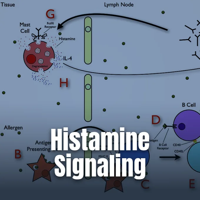
The thymus gland, located in the chest behind the breastbone, plays a central role in guiding immune cell maturation early in life [1, 2]. In adults with myasthenia gravis, this immune organ often exhibits striking abnormalities [2]. Approximately two-thirds of patients show thymic hyperplasia, where the gland contains active germinal centers filled with antibody-producing B cells [1, 4].
In addition to hyperplasia, roughly 10% to 15% of patients harbor a thymoma, a tumor of the thymic epithelial tissue that is usually benign but can occasionally become invasive [1, 2]. For decades, surgeons removed the thymus to improve symptoms, yet definitive proof from randomized trials remained elusive until the international MGTX trial appeared in 2016. Led by Dr. Gil Wolfe, professor of neurology at the University at Buffalo, researchers evaluated 126 patients with generalized nonthymomatous myasthenia gravis who received either extended transsternal thymectomy plus prednisone or prednisone alone. Blinded assessments over three years showed that patients who underwent surgery had lower Quantitative Myasthenia Gravis scores and required substantially lower average doses of corticosteroids to maintain clinical stability [12]. The surgical procedure helped rebalance abnormal immune signaling across the body [2].
Shortly after publication in the New England Journal of Medicine, statisticians discovered an error in how time-weighted prednisone doses were calculated for patients who had successfully tapered off the medication. The journal issued a formal correction notice clarifying the point estimates, but the revised calculation did not weaken the study’s conclusions. “The recalculation didn’t reverse any of the findings; it actually strengthened them, making thymectomy look even better,” Dr. Wolfe said [12]. Today, thymectomy remains an established recommendation for thymoma cases and selected generalized patients [1].
Diagnostic Testing and Antibody Panels
Diagnosing myasthenia gravis requires combining clinical history with specialized laboratory and electrodiagnostic tests [1, 3]. Blood testing for circulating antibodies against the acetylcholine receptor provides the most common confirmation, detecting abnormal proteins in roughly 85% to 90% of generalized cases [2, 7]. However, only about half of ocular patients test positive on this screen [2].
When anti-AChR tests return negative, clinicians test for antibodies against muscle-specific tyrosine kinase (MuSK) or low-density lipoprotein receptor-related protein 4 (LRP4), which identify distinct autoimmune subsets requiring tailored treatment plans [2, 4]. At the bedside, physicians assess muscle fatigability through sustained upward gaze or repetitive motor movements [15]. When eyelid drooping is present, doctors can perform a simple ice pack test by applying a cold compress over the closed eyelid for two to five minutes. Because cooling inhibits acetylcholinesterase enzymes and improves acetylcholine receptor binding, droopy eyelids temporarily lift in response to direct cold. This straightforward non-invasive test offers high diagnostic utility when complex laboratory work remains pending [2].
Electrodiagnostic evaluations verify impaired neuromuscular transmission when blood tests remain inconclusive. Repetitive nerve stimulation delivers small electrical impulses to a motor nerve, producing a characteristic decremental drop in muscle action potentials as neurotransmitter stores deplete [2, 3]. Single-fiber electromyography measures transmission timing across individual muscle fibers, providing the most sensitive electrophysiological test available [15]. Historically, physicians administered intravenous edrophonium in the Tensilon test to observe temporary strength gains, but clinicians no longer use this test because the drug is no longer manufactured in the United States [2].
Preventing Sudden MG Respiratory Crisis
The most dangerous complication of myasthenia gravis is a myasthenic crisis, an acute state of respiratory failure where the diaphragm and chest wall muscles become too weak to move air [1, 3]. Approximately one in five patients experiences at least one crisis episode during their lifetime. Breathing turns shallow and swallowing difficulties can cause choking or aspiration of saliva [1, 14].
When respiratory muscles fail, patients require emergency hospital admission and mechanical ventilation with a breathing machine to sustain oxygen levels until medical therapies take effect [1, 2]. Crises rarely strike without a warning trigger [14]. Systemic infections, especially respiratory viruses and influenza, represent the most frequent cause of sudden symptom deterioration [1, 2]. Fever, emotional stress, pregnancy, and physical trauma can also destabilize previously controlled disease [1, 14]. In an emergency setting, doctors use rapid rescue interventions such as plasmapheresis, which filters pathogenic antibodies directly out of the plasma, or high-dose intravenous immunoglobulin infusions. These emergency treatments lower circulating autoantibody levels within days, providing temporary symptom relief that lasts for several weeks [2, 3].

Many common prescription medications can impair neuromuscular transmission and trigger dangerous flares in myasthenic patients. Macrolide antibiotics like azithromycin, fluoroquinolones like ciprofloxacin, and aminoglycosides like gentamicin are known to worsen weakness, whereas cephalosporins and tetracyclines are safe alternatives. Oncology patients receiving cancer checkpoint inhibitors like pembrolizumab face notable risks; studies show these therapies can induce severe new-onset myasthenia, causing respiratory failure in up to 45% of affected individuals. Cardiovascular drugs also warrant vigilance; statins slightly increase flare risks in patients over 60, and beta blockers raise exacerbation odds roughly 2.7-fold [14].
Modern Medications for Symptom Relief
Standard medical management begins with acetylcholinesterase inhibitors like pyridostigmine, sold under the brand name Mestinon [2, 3]. By slowing the breakdown of acetylcholine in the synaptic cleft, pyridostigmine allows neurotransmitters to linger longer and stimulate surviving receptors [1, 2]. However, because enzyme inhibitors do not stop autoimmune destruction, most patients also require immunosuppressive medications [14].
Clinicians commonly prescribe corticosteroids like prednisone, or steroid-sparing agents such as azathioprine and mycophenolate mofetil to dampen abnormal antibody production [2, 3]. A major advance arrived with targeted neonatal Fc receptor (FcRn) blockers that accelerate the removal of harmful antibodies. In December 2021, the FDA approved Vyvgart (efgartigimod alfa-fcab) for adult generalized patients with anti-AChR antibodies [5, 7]. “There are significant unmet medical needs for people living with myasthenia gravis, as with many other rare diseases,” said Billy Dunn, M.D., director of the Office of Neuroscience in the FDA’s Center for Drug Evaluation and Research [5]. In the Phase 3 ADAPT trial, 68% of treated patients achieved meaningful functional improvement on the MG-ADL scale compared to 30% on placebo [7]. In June 2023, the FDA approved a subcutaneous formulation, Vyvgart Hytrulo, which delivers treatment in a brief injection [8, 9].
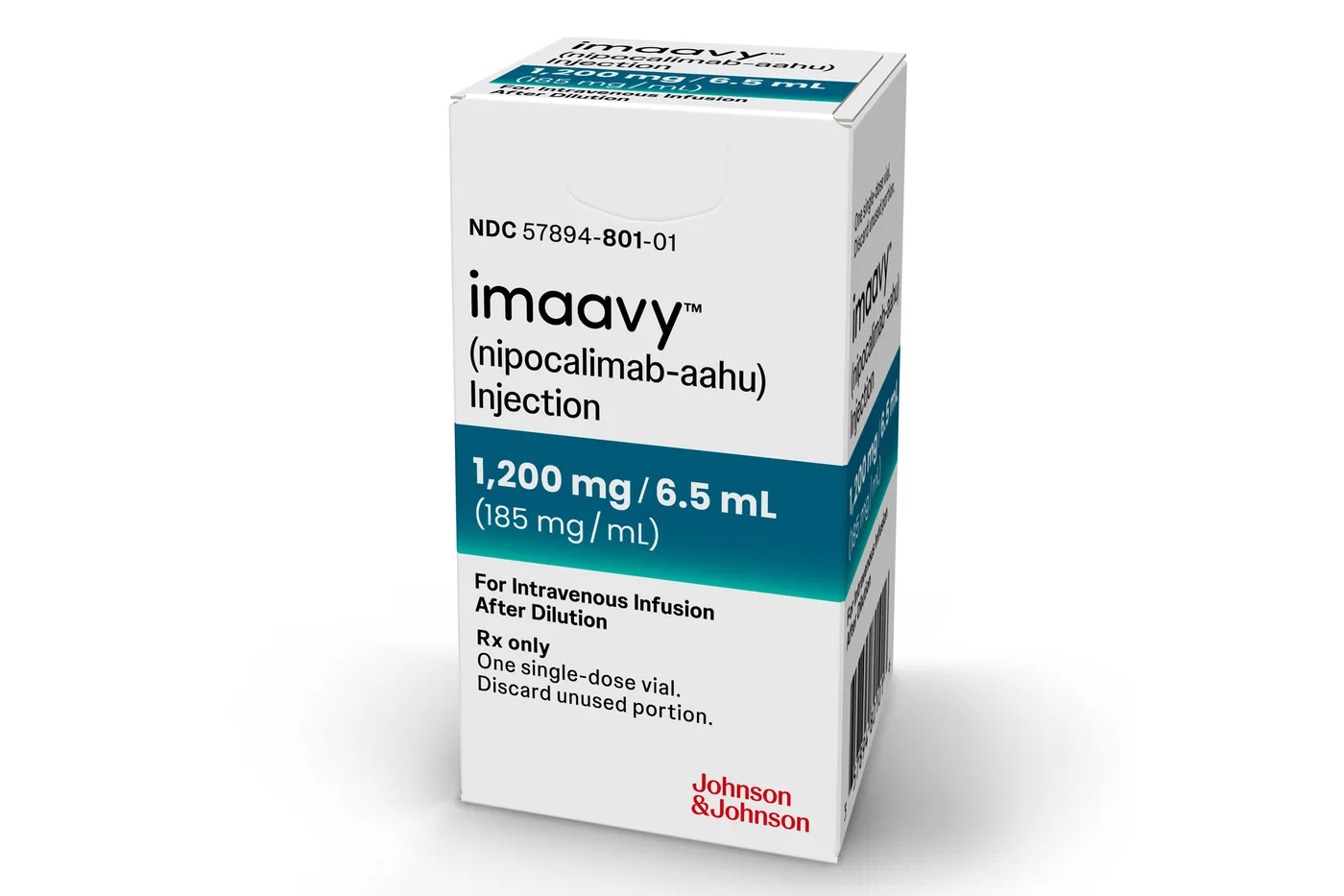
Treatment choices widened further with rozanolixizumab, approved as Rystiggo in June 2023 for adults with AChR or MuSK antibodies. In the Phase 3 MycarinG trial, rozanolixizumab significantly reduced muscle weakness scores compared to placebo [10]. In April 2025, the FDA approved Johnson & Johnson’s Imaavy (nipocalimab-aahu) for adults and pediatric patients aged 12 and older. In clinical trials, nipocalimab cut total serum IgG levels by 69% over 24 weeks. These targeted biologic therapies give doctors precise tools to suppress pathogenic antibodies while helping patients maintain active lives [11].
- WEBSITE Cleveland Clinic. (2023). Myasthenia Gravis: What Is It? [Article Link]
- WEBSITE Shmerling, R. H. (2018). Myasthenia Gravis. Harvard Health Publishing. [Article Link]
- WEBSITE MedlinePlus. (2025). Myasthenia gravis: MedlinePlus Medical Encyclopedia. National Library of Medicine. [Article Link]
- WEBSITE MedlinePlus Genetics. (2016). Myasthenia Gravis. National Library of Medicine. [Article Link]
- PRESS RELEASE U.S. Food and Drug Administration. (2021). FDA Approves New Treatment for Myasthenia Gravis. [Article Link]
- WEBSITE Medscape. (2026). Myasthenia Gravis: Background, Anatomy, Pathophysiology. [Article Link]
- PRESS RELEASE argenx. (2021). argenx Announces U.S. Food and Drug Administration (FDA) Approval of VYVGART in Generalized Myasthenia Gravis. [Article Link]
- PRESS RELEASE Halozyme Therapeutics. (2023). Halozyme Announces argenx Receives FDA Approval for VYVGART Hytrulo With ENHANZE for Subcutaneous Use in Generalized Myasthenia Gravis. PR Newswire. [Article Link]
- PRESS RELEASE argenx. (2023). argenx Announces U.S. Food and Drug Administration Approval of VYVGART Hytrulo Injection for Subcutaneous Use in Generalized Myasthenia Gravis. [Article Link]
- PRESS RELEASE UCB. (2023). UCB Announces U.S. FDA Approval of RYSTIGGO for the Treatment of Adults With Generalized Myasthenia Gravis. PR Newswire. [Article Link]
- PRESS RELEASE Johnson & Johnson. (2025). Johnson & Johnson Receives FDA Approval for IMAAVY, a New FcRn Blocker in Generalized Myasthenia Gravis. [Article Link]
- ONLINE NEWS Stern, V. (2017). Big Corrections Usually Weaken Findings. But a Recent NEJM One Strengthened Them, Author Says. Retraction Watch. [Article Link]
- WEBSITE Myaware. (2023). Support and Information for People Living With Myasthenia. [Article Link]
- WEBSITE Wikipedia contributors. (2026). Myasthenia Gravis. Wikipedia, The Free Encyclopedia. [Article Link]
- WEBSITE Wikipedia contributors. (2026). Ocular Myasthenia. Wikipedia, The Free Encyclopedia. [Article Link]


