





Readers asking what is the cerebellum find an egg-sized organ nestled low in the skull behind the pons and brainstem. It takes up barely 10 percent of total human brain volume, yet it holds more than half of all neurons in the central nervous system [4]. Anatomists note that it packs roughly 3.6 times as many nerve cells as the cerebral neocortex into tightly folded sheets called folia. It does not start muscle actions on its own, functioning instead as an internal clock and calibration device that refines motor balance, posture, and temporal accuracy. In aquatic species like Mormyrid fishes, this hindbrain region expands until it matches or exceeds the cerebrum in overall size [8].
- What Is the Cerebellum in Human Anatomy?
- Three Functional Zones of the Cortex
- Circuit Layers Within the Cortical Fold
- Deep Nuclei Outgoing Pathways
- How Purkinje Cells Guide Motor Precision
- Motor Learning in the Marr-Albus Model
- Clinical Damage and the White Cerebellum Sign
- Case Studies in Total Cerebellum Absence
What Is the Cerebellum in Human Anatomy?
Tucked into the posterior cranial fossa beneath the occipital lobes of the cerebrum, the organ links to the brainstem through three paired stalks known to anatomists as cerebellar peduncles [4]. A tough dura mater sheet called the cerebellar tentorium separates it from the cerebrum above. Under formal IFAA classification, it carries code A14.1.07.001 within the metencephalon [7]. It measures about 6 cm by 5 cm by 10 cm, filling 100 to 150 cubic cm of space [8].
Unfolding the tightly crumpled gray matter of the human cerebellar cortex would yield a continuous flat sheet measuring 1 meter in length and 10 cm across [10]. High-resolution MRI records an expansive surface area of 730 square cm, though broader anatomical estimates range between 500 and 1000 square cm [8]. The organ receives nearly 200 million input fibers from sensory receptors and motor control centers, dwarfing the human optic nerve, which carries only one million fibers [10]. Dr. James Knierim of Johns Hopkins University and McGovern Medical School at UTHealth points out in Neuroscience Online that this massive convergence allows the hindbrain to track body posture in real time [4].
How does such a compact tissue volume handle so much incoming data? The answer rests within its core of branching white matter, once called the arbor vitae or tree of life [8]. Myelinated axon tracts route signals into four deep nuclei embedded within the interior before exiting through three pairs of peduncles that steer all nerve traffic forward without internal loops [10]. Dr. James Knierim explains that the superior, middle, and inferior cerebellar peduncles keep inputs and outputs flowing in an organized stream [4].
Three Functional Zones of the Cortex
Two transverse fissures split the cortical surface into three anatomical lobes. The primary fissure divides the anterior lobe from the posterior lobe, while the posterolateral fissure marks off the small flocculonodular lobe resting beneath the main body [4]. Down the center runs a narrow ridge known as the vermis, from the Latin word for worm, which separates the two lateral halves [8]. Ten smaller lobules marked by Roman numerals subdivide these zones into discrete processing units that neurobiologists use to map sensory inputs across the entire human body [4], [8].
The oldest evolutionary sector is the vestibulocerebellum, matching the flocculonodular lobe. Also called the archicerebellum, it takes balance inputs from semicircular canals in the inner ear and projects to brainstem vestibular nuclei. The vestibulo-ocular reflex shifts the eyes in the opposite direction when the head turns, keeping visual images steady on the retina during fast body movement or running [4].
Along the center lies the spinocerebellum, which takes in continuous sensory streams from the spinal cord, muscle spindles, and joint receptors. By pooling these inputs, it modulates descending motor pathways to steady posture and smooth limb motion during ongoing strides [4]. Further outward, the broad lateral halves form the cerebrocerebellum, the newest phylogenetic division that links directly to the cerebral cortex [8]. Neurologist Jeremy D. Schmahmann of Harvard and researcher Mario Manto at FNRS-Belgium document that this lateral zone helps plan voluntary motor sequences while guiding cognitive language syntax and rapid verbal choices [1], [8].
Circuit Layers Within the Cortical Fold
A uniform circuit pattern repeats through every fold of the cortex. Three distinct cellular layers make up this continuous sheet, moving from the innermost granular layer through the Purkinje cell boundary to the outermost molecular zone. Inside the granular layer sit 50 billion granule cells, a massive count representing more than half of all neurons in the entire human brain packed tightly together [4].
Above the granule layer sits the Purkinje cell layer, a sheet only one cell thick that holds the bodies of specialized neurons first identified by Czech anatomist Jan Evangelista Purkyně in 1837 [8]. Each cell displays a sprawling apical dendritic tree that reaches up into the molecular layer like an open fan. Looked at from the front, the dendritic branches spread wide; looked at from the side, they stay flat. Purkinje cells align in parallel rows like leaves stacked inside a book, allowing millions of passing parallel fibers to cross each dendritic tree at right angles [4], [8].
The top molecular layer holds Purkinje dendrites and thousands of parallel fibers, which represent the bifurcated axons of granule cells rising from below. Two kinds of inhibitory interneurons, stellate cells and basket cells, populate this outer zone and release GABA to sculpt the exact temporal window during which Purkinje cells fire [8].
Deep Nuclei Outgoing Pathways
All signals leaving the cortex must pass through the deep cerebellar nuclei embedded in the central white matter. These four paired structures act as the sole output gates for the entire hindbrain organ, meaning a focal lesion here matches the loss of the whole cortex. From medial to lateral, anatomists identify the fastigial nucleus, the interposed nuclei, and the large dentate nucleus [4]. Dr. Duane E. Haines said in The Cerebellum that these internal nuclei link the hindbrain directly to motor tracts running down the spinal cord and up to the thalamus [1].
The fastigial nucleus rests closest to the midline, receiving inhibitory input from the vermis and projecting to vestibular nuclei and reticular centers to balance posture. Next to it sit the interposed nuclei, composed of the emboliform nucleus and the globose nucleus, which send corrective signals to the contralateral red nucleus [4], [10].
The largest output center is the dentate nucleus, which receives fibers from the lateral hemispheres and projects onward to the red nucleus and the ventrolateral thalamus. Unlike the cerebrum, which governs the opposite half of the body, this hindbrain system directs ipsilateral muscle control. Sensory inputs enter from the left side of the body, and corrective motor signals return to the left side. Output fibers heading to the contralateral red nucleus cross the midline, but the descending rubrospinal tract crosses right back below the brainstem, producing a double-crossed circuit that keeps bodily movement stable [4].

How Purkinje Cells Guide Motor Precision
Two separate input channels bring information into the cortex. Mossy fibers arrive from the pons, spinal cord, and vestibular system. They synapse onto thousands of tiny granule cells, which in turn send their axons upward into the molecular layer to distribute incoming signals across parallel fibers [4]. Each Purkinje cell receives thousands of weak inputs from parallel fibers, gathering broad sensory context across the nervous system [8].
In contrast to mossy fibers, climbing fibers originate only from the inferior olivary nucleus in the medulla. A single climbing fiber climbs up the proximal dendrites of one Purkinje cell, creating one of the strongest excitatory connections in the mammalian nervous system [4]. When that climbing fiber fires, it triggers a massive electrical event called a complex spike across the entire dendritic tree. That burst signals an unexpected event or motor error, informing the cell that an ongoing movement failed to reach its intended goal [8]. Researcher Marco Molinari said in Essentials of Cerebellum and Cerebellar Disorders that this error message drives motor adaptation [2].
Purkinje cells provide the sole output from the cortex, and their signals are purely inhibitory [4]. They release the neurotransmitter GABA directly onto deep nuclei to suppress their baseline firing rates [8]. By sculpting the timing and intensity of this inhibition, Purkinje cells carve out clean bursts of excitation in output centers to ensure that muscles contract with fluid precision without limb shaking [4], [10].
Motor Learning in the Marr-Albus Model
Beyond smoothing routine steps, the hindbrain drives motor learning and physical skill acquisition. When a person learns to hit a baseball or play violin, early attempts are clumsy and slow [4]. In the late 1960s and early 1970s, computational theorists David Marr and James Albus constructed a mathematical model to explain how cerebellar synapses adapt through trial and error. They proposed that climbing fibers deliver a teaching signal that alters synaptic strength, changing how Purkinje cells respond to incoming parallel fiber messages [8].
When a motor act misses its target, the inferior olive detects the mismatch between intention and outcome. It sends an instructional error signal along climbing fibers to active Purkinje cells [8]. Synchronous firing weakens active parallel fiber synapses, initiating a lasting physiological process called long-term depression that helps subsequent motor commands follow corrected neural pathways with greater accuracy [4], [8].
Predictive calibration extends beyond limbs into cognitive tasks [4]. The lateral cerebrocerebellum activates during language tests and abstract reasoning. Brain scans show the lateral halves light up when a person must pick a verb that fits a noun, like matching sit with chair [8]. In Springer Cham’s volume Essentials of Cerebellum and Cerebellar Disorders, editors Donna L. Gruol, Noriyuki Koibuchi, and Ying Shen state that cognitive circuits use the same forward internal models seen in motor timing, applying one uniform calculation method to physical action and mental thought [2].
Clinical Damage and the White Cerebellum Sign
Injuries to this hindbrain center cause motor problems without causing paralysis. Patients often suffer from ataxia, an inability to coordinate voluntary muscle contractions that leaves steps clumsy and wide while disrupting balance and fine finger control. Damage to midline tissue disrupts posture and gait, while lateral damage impairs dexterity, causing dysmetria where patients overshoot physical targets and struggle with rapid alternating movements [4].
In emergency neuroimaging, a finding called the white cerebellum sign provides clear diagnostic clues during childhood brain trauma. Also known as the dense cerebellum sign or reversal sign, it appears on computed tomography scans of infants and young children suffering from severe anoxic brain injury following drowning or cardiac arrest. The cerebellum itself does not gain physical density. Instead, diffuse brain edema wipes out the normal border between gray matter and white matter across the cerebral hemispheres. Because cerebral water erases normal tissue contrast, the cerebellum, brainstem, and thalami stand out as starkly white after drowning, bacterial meningitis, encephalitis, birth asphyxia, or status epilepticus [9].
Blast injuries from explosive artillery also harm cerebellar cells. Researchers Elaine Peskind, David Cook, and James Meabon at VA Puget Sound Health Care System and University of Washington studied 33 combat veterans deployed to Iraq and Afghanistan. Veterans with more blast exposures showed less cerebellar glucose metabolism. In blast-exposed mice, the team found blood-brain barrier leaks and lost Purkinje cells. Molecular biologist David Cook said that blast trauma shares genetic risk mechanisms with Alzheimer’s disease through the APOE gene and its E4 allele, while clinical psychiatrist Elaine Peskind said that cerebellar dysfunction contributes to veteran irritability, memory loss, and combat stress [5].
Case Studies in Total Cerebellum Absence
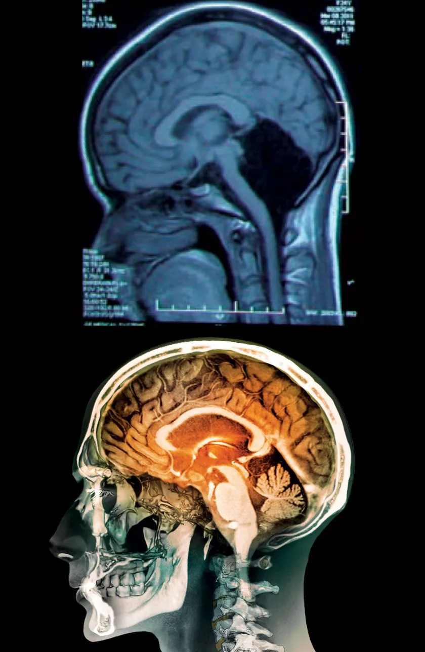
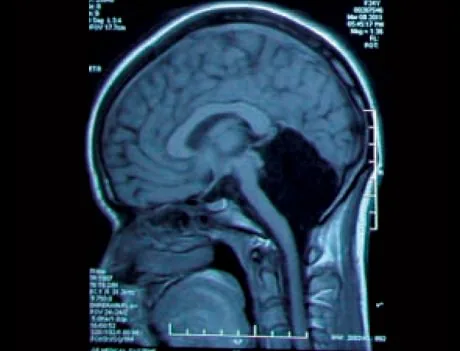
The brain’s ability to adapt to structural loss appears in rare cases of cerebellar agenesis, where an individual is born without a cerebellum. In 2014, medical staff at the Chinese PLA General Hospital of Jinan Military Area Command in Shandong Province treated a 24-year-old woman suffering from dizziness and nausea. When doctors carried out a CAT scan, they discovered that her entire cerebellum was missing, with the empty posterior cranial cavity filled completely with cerebrospinal fluid. Her mother reported that she had not walked until age seven and her speech was intelligible only by age six [6].
The woman showed only mild motor deficits and slightly slurred speech, joining an elite group of just nine people known in medical literature to have reached adulthood without a cerebellum. Most people with congenital agenesis die young during infancy [6]. Dr. Mario Manto at the Free University of Brussels in Belgium said that such rare cases reveal how the cerebral cortex can adapt to take over missing cerebellar work [1], [6].
Today, medical specialists track more than 300 distinct cerebellar disorders, ranging from inherited ataxias to vascular strokes. In Section 2 of the four-volume Handbook of the Cerebellum and Cerebellar Disorders published by Springer Dordrecht, editors Mario Manto, Jeremy D. Schmahmann, Ferdinando Rossi, Donna L. Gruol, and Noriyuki Koibuchi gather 106 review chapters detailing anatomical connections. Reviewer Joseph I. Sirven said that this work summarizes all known pathologies of the hindbrain [1]. Archived open-access journals like Cerebellum & Ataxias from BioMed Central and Springer Nature continue to catalog clinical trials [3]. Unraveling cerebellar timing helps scientists understand how the brain moves the body and shapes human thought [1], [4].
- BOOK Manto, M., Schmahmann, J. D., Rossi, F., Gruol, D. L., & Koibuchi, N. (Eds.). (2013). Handbook of the Cerebellum and Cerebellar Disorders. Springer Dordrecht. [Article Link]
- BOOK Gruol, D. L., Koibuchi, N., Manto, M., Molinari, M., Schmahmann, J. D., & Shen, Y. (Eds.). (2016). Essentials of Cerebellum and Cerebellar Disorders: A Primer For Graduate Students. Springer Cham. [Article Link]
- ACADEMIC JOURNAL Cerebellum & Ataxias. (2014-2021). BioMed Central and Springer Nature. [Article Link]
- WEBSITE Knierim, J. (2020). Cerebellum (Section 3, Chapter 5). Neuroscience Online: An Electronic Textbook for the Neurosciences. Department of Neurobiology and Anatomy, McGovern Medical School at UTHealth. [Article Link]
- ONLINE NEWS Yuhas, D. (2016). Veterans of Iraq, Afghanistan Show Brain Changes Related to Explosion Exposure. Scientific American. [Article Link]
- ONLINE NEWS Thomson, H. (2014). Woman of 24 Found to Have No Cerebellum in Her Brain. New Scientist. [Article Link]
- WEBSITE Federative International Programme on Anatomical Terminologies. (2013). Cerebellum (TA98 A14.1.07.001). IFAA. [Article Link]
- WEBSITE Wikipedia contributors. (2026). Cerebellum. Wikipedia, The Free Encyclopedia. [Article Link]
- WEBSITE Wikipedia contributors. (2026). White cerebellum sign. Wikipedia, The Free Encyclopedia. [Article Link]
- WEBSITE Wikipedia contributors. (2026). Anatomy of the cerebellum. Wikipedia, The Free Encyclopedia. [Article Link]


