





When a healthy blood vessel tears, tiny blood cells called platelets stick to the injured wall while blood proteins join forces to build a stable fibrin seal. In hemophilia blood clotting, this vital defense stalls because the body makes too little working clotting factor [2]. The disorder stems from altered genes that cut levels of factor VIII or factor IX, leaving blood unable to solidify quickly during bleeding episodes. Without enough active factor, clots form at a crawl or break apart too soon, putting patients at risk of prolonged bleeding after tooth extractions, deep cuts, or minor surgery [1].
- How Hemophilia Blood Clotting Breaks Down
- Mechanisms in the Tenase Clotting Pathway
- Inheritance Patterns in Classic Hemophilia
- Why Do Joint Bleeds Cause Synovial Damage?
- Blood Tests at Specialized Treatment Centers
- Preventive Care and Factor Concentrates
- Can Gene Therapy Cure Severe Haemophilia?
- New Subcutaneous Medicines and Daily Living
How Hemophilia Blood Clotting Breaks Down
Clinical doctors at every federally funded Hemophilia Treatment Center, or HTC, define hemophilia as an inherited condition marked by poor clotting and internal bleeding. When vascular walls break, plasma proteins bind in step-by-step order to plug the gap, but missing just one factor slows the entire repair chain. Inherited hemophilia affects about 1 in every 5,000 male births worldwide, touching families across all backgrounds. CDC surveillance published by J. M. Soucie, C. H. Miller, and B. Dupervil in the journal Haemophilia, pages 487-93, showed that about 33,000 males live with the disorder in the United States [2].
Symptoms span a wide clinical range. Mild cases bleed only after dental surgery or heavy trauma, while severe cases suffer repeated bleeds into deep joints without warning [11].
Historical records trace this bleeding tendency back to early medical writings. An inherited bleeding disorder in infant boys appeared in Jewish Talmudic texts during the second century. Modern study began in 1803 when American physician John Otto reported on affected kindreds, followed by clinical reviews by Nasse in 1820 and laboratory clotting tests by Wright in 1893. In 1937, researchers Patek and Taylor isolated a plasma fraction called antihemophilic factor, which scientists later proved was factor VIII [10]. In 1952, doctors recognized factor IX deficiency in a young patient named Stephen Christmas, giving hemophilia B its long-standing name of Christmas disease [15].
Mechanisms in the Tenase Clotting Pathway
Normal clotting works through a chain reaction called the coagulation cascade, relying on intrinsic and extrinsic pathways that merge to produce a firm fibrin mesh. When a vessel breaks, tissue factor binds circulating factor VII to turn on factor X, while contact with damaged tissue turns on factor XII, which triggers factor XI, leading activated factor XI to switch on factor IX in a calcium-dependent step. Activated factor IX then pairs with activated factor VIII on platelet surfaces to build the tenase enzyme complex. This tenase unit rapidly activates factor X, which combines with factor Va to transform prothrombin into thrombin [10].
Thrombin acts as the master enzyme of the cascade. It cuts soluble fibrinogen into tough fibrin strands that weave over the initial platelet plug [10].
Factor VIII travels in human blood joined to von Willebrand factor, a large shielding protein that keeps it from breaking down too quickly in the bloodstream. In normal plasma, factor VIII lasts for 12 hours, but without von Willebrand factor, its lifespan shrinks to just 2 hours. Cells in the liver make factor VIII, adding sulfates to tyrosine units before releasing it into circulation [10]. While earlier guides examined the causes, signs, and modern treatments for hemophilia, clinical management also depends on tracking how specific clotting factor deficits alter the molecular cascade.
Inheritance Patterns in Classic Hemophilia
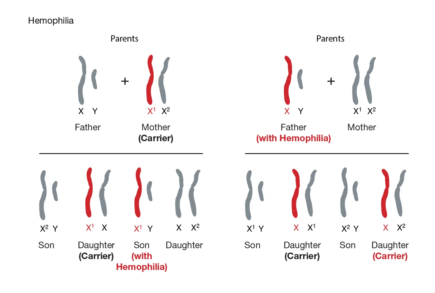
The genetics of classic hemophilia, or hemophilia A, follow an X-linked pattern because the F8 and F9 genes sit on the X chromosome [2]. Women carry two X chromosomes, while men carry one maternal X chromosome and one paternal Y chromosome. Since the Y chromosome holds no clotting factor genes, a boy who gets an altered X chromosome from his mother will have the disease [3]. Men with hemophilia pass their altered X chromosome to all daughters, who become carriers, but never to their sons [11].
Female carriers usually make enough factor from their second healthy X chromosome to prevent severe bleeding problems [3]. Yet through a natural cell process called Lyonization, each female cell shuts down one X chromosome at random during early growth in the womb. If this shut-down process turns skewed, silencing the healthy chromosome in most liver cells, a carrier will have low factor levels and suffer heavy menstrual bleeding or easy bruising [4]. During pregnancy, maternal screening helps obstetric teams distinguish inherited coagulopathies from other gestational complications, including maternal health risks linked to preeclampsia.

A family history does not appear in every newborn. In about one-third of all newborns diagnosed with the condition, the mutation arises out of nowhere as a spontaneous genetic change [2]. In severe hemophilia A, about 45 to 50 percent of boys share an intron 22 inversion in the F8 gene on chromosome Xq28 [13]. Hemophilia B, caused by changes in the F9 gene on chromosome Xq27, affects about 1 in 20,000 newborn boys worldwide [3]. In 1990, Oxford researchers George Brownlee and Merlin Crossley showed that specific gene faults cause hemophilia B Leyden, a rare type where childhood bleeding eases after puberty [15].
Why Do Joint Bleeds Cause Synovial Damage?
Bleeding into joint cavities, known to doctors as hemarthrosis, is the most common and painful trouble seen in severe bleeding conditions [14]. The knees, ankles, and elbows bear continuous physical loads during childhood play, causing tiny blood vessels in the joint lining to stretch and burst [11].
When pooled blood sits inside a joint space, red blood cells break down and shed iron directly into the synovial lining cells, triggering inflammation that makes the tissue swell and grow abnormal extra blood vessels. This swollen joint tissue, known as hemophilic synovitis, bleeds again after the slightest knock [14]. A joint that suffers four bleeds within six months is called a target joint [10]. Repeated bleeding wears away smooth joint cartilage and damages the bone underneath [14].
Without treatment, damaged cartilage leads to chronic stiffness, bone cysts, and permanent joint crippling called hemophilic arthropathy. In the journal Haemophilia, orthopedic surgeon E. C. Rodriguez-Merchan and his team stressed that early preventive treatment is the best way to safeguard joint cartilage from breakdown [14]. In Australia, where Haemophilia Foundation Australia supports more than 2,800 diagnosed people, early home care keeps children active in sports and school [11].


Blood Tests at Specialized Treatment Centers
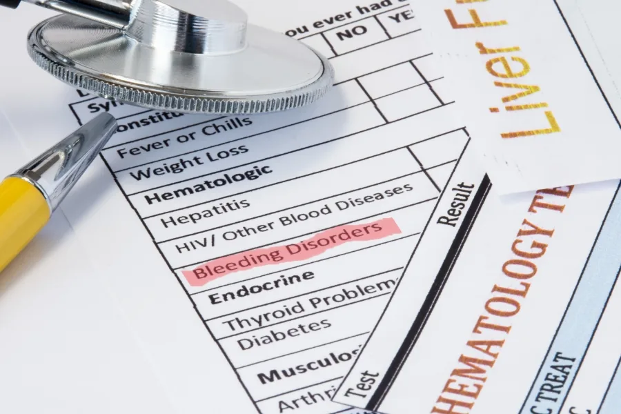
A doctor suspects a bleeding issue when a baby boy bleeds heavily after routine circumcision or develops deep bruises when learning to crawl. Hospital labs run a complete blood count along with prothrombin time and activated partial thromboplastin time tests [2]. In hemophilia A and B, prothrombin time and platelet counts look normal, but partial thromboplastin time is slow because intrinsic clotting is blocked [13]. Once a slow partial thromboplastin time shows up, special factor assay tests measure exact protein activity in blood plasma [2].
Doctors group disease severity by the amount of active factor circulating in a person’s blood [10]. Normal factor levels in healthy people range from 50 to 150 percent [11].
People with mild hemophilia have 5 to 40 percent factor activity, bleeding mainly after tooth pulls, deep cuts, or major operations [11]. Those with moderate disease hold between 1 and 5 percent factor levels, while severe disease means having less than 1 percent of normal clotting factor [10]. In chapter 134 of Hematology: Basic Principles and Practice, experts explain that severe patients face regular spontaneous bleeds into muscles and joints [7]. Writing in BMJ, Dr. K. Fijnvandraat and fellow Dutch hematologists urged teams to give factor right away during head trauma before waiting for scan images [1].
Preventive Care and Factor Concentrates
The standard treatment for bleeding disorders is factor replacement therapy, which infuses the missing protein directly into a vein [2]. Patients use either recombinant factor grown in sterile lab cell cultures or plasma-derived factor gathered from human blood donors and treated with heat or chemicals to kill viruses. Before 1985, donated blood pools exposed many hemophilia patients to hepatitis and HIV, but modern safety steps and lab-made proteins have removed those viral dangers [10]. At home, trained parents and patients inject factor concentrates into veins to treat acute bleeding or prevent it entirely [11].
Treatment plans fall into on-demand therapy or routine prophylaxis. On-demand treatment halts bleeding after an injury, while prophylaxis provides regular weekly infusions to keep factor levels above 1 percent [11].
A major hurdle in factor therapy is the rise of neutralizing antibodies known as inhibitors. About 15 to 20 percent of boys with hemophilia make immune inhibitors that treat replacement factor like a foreign threat and destroy it [2]. When high inhibitor levels block standard factor infusions, hematologists turn to bypassing agents like recombinant activated factor VII [10]. For mild hemophilia A, doctors often prescribe desmopressin, an injected or nasal drug that prompts blood vessels to release stored factor VIII [7].
Can Gene Therapy Cure Severe Haemophilia?
Recent clinical trials using harmless adeno-associated viruses have brought medicine closer to long-term gene fixes for bleeding disorders. In 2017, Professor John Pasi led a clinical trial at Barts Health NHS Trust and Queen Mary University of London for severe factor VIII deficiency. Thirteen British men received a single intravenous drip carrying healthy F8 gene instructions into their liver cells. Eleven of the thirteen men reached near-normal factor VIII levels, and all thirteen stopped their weekly factor injections [6].
Trial patient Jake Omer, a 29-year-old father from Billericay in Essex, had injected factor three times weekly since early childhood. Following his single infusion, his factor levels climbed and his joints stayed healthy without regular needle sticks [6].
“The gene therapy has changed my life,” Omer said, adding that he could walk two miles daily without joint pain. Professor Jo Martin, President of The Royal College of Pathologists, praised the trial team at Royal London Hospital [6]. In factor IX deficiency, the B-AMAZE trial published in the New England Journal of Medicine by Professor Pratima Chowdary of Royal Free Hospital and Professor Amit Nathwani of UCL Medical Sciences tested an adeno-associated virus drug named FLT180a [7]. Patient Elliott Mason saw his factor IX jump from 1 percent to normal levels after a one-hour drip. Clive Smith, chairman of The Haemophilia Society, welcomed the data while urging caution [8].
New Subcutaneous Medicines and Daily Living
Newer non-factor drugs protect against bleeds without relying on replacement factor infusions or intravenous access [5]. In 2018, the FDA approved Hemlibra, known generically as emicizumab, an engineered antibody that mimics factor VIII by bridging factor IXa and factor X. In the HAVEN 3 clinical trial with 89 patients, weekly Hemlibra injections cut the yearly bleed rate by 96 percent compared to on-demand care [17]. Patients take Hemlibra through a small needle under the skin once a week or once every four weeks, making care simpler for children and families.
Another innovative drug class uses small-interfering RNA to silence antithrombin, a natural body protein that stops clots from forming. Lowering antithrombin allows more thrombin to form, rebalancing clotting without needing replacement factors [5].
In March 2025, the FDA approved Qfitlia, known as fitusiran, for people 12 and older with hemophilia A or B, with or without inhibitors. In the ATLAS clinical program, fitusiran cut yearly bleeds by 71 percent in patients without inhibitors and 73 percent in those with inhibitors. Injections are given under the skin as few as six times a year, with antithrombin tracked using the Siemens Healthcare Diagnostics INNOVANCE blood assay. “Today’s approval of Qfitlia is significant for patients with hemophilia because it can be administered less frequently than other existing options,” said Dr. Tanya Wroblewski, deputy director at the FDA Center for Drug Evaluation and Research. At comprehensive treatment centers, teams led by doctors like Dr. Guy Young at Children’s Hospital Los Angeles help families combine these breakthrough medicines with physical therapy and preventive dental care [5].
- ACADEMIC JOURNAL Fijnvandraat, K., Cnossen, M. H., Leebeek, F. W., & Peters, M. (2012). Diagnosis and management of haemophilia. BMJ, 344, e2707. [Article Link]
- WEBSITE Centers for Disease Control and Prevention. (2025). About Hemophilia. [Article Link]
- WEBSITE National Library of Medicine. (2022). Hemophilia: MedlinePlus Genetics. [Article Link]
- WEBSITE Centers for Disease Control and Prevention. (2024). Information on Hemophilia for Women. [Article Link]
- PRESS RELEASE U.S. Food and Drug Administration. (2025). FDA Approves Novel Treatment for Hemophilia A or B, with or without Factor Inhibitors. [Article Link]
- ONLINE NEWS Barts Health NHS Trust. (2017). Groundbreaking gene therapy trial set to cure haemophilia A. [Article Link]
- ACADEMIC JOURNAL Carcao, M., Gomez, K., Matino, D., & Pierce, G. F. (2023). Hemophilia A and B. In R. Hoffman et al. (Eds.), Hematology: Basic Principles and Practice (8th ed., chap 134). Elsevier. [Article Link]
- ONLINE NEWS Gallagher, J. (2022). Transformational therapy cures haemophilia B. BBC News. [Article Link]
- ONLINE NEWS Gallagher, J. (2017). Haemophilia A trial results ‘mind-blowing’. BBC News. [Article Link]
- WEBSITE Medscape. (2026). Hemophilia A (Factor VIII Deficiency): Background, Pathophysiology, Etiology. [Article Link]
- WEBSITE Department of Health & Human Services. (1999). Haemophilia. Better Health Channel. [Article Link]
- PRESS RELEASE Sanofi. (2025). Qfitlia approved as the first therapy in the US to treat hemophilia A or B with or without inhibitors. [Article Link]
- WEBSITE Wikipedia. (2026). Haemophilia. [Article Link]
- ACADEMIC JOURNAL Rodriguez-Merchan, E. C., Jiménez-Yuste, V., Aznar, J. A., Hedner, U. K., Knobe, K. E., Lee, C. A., Ljung, R. C. R., Querol, F., Santagostino, E., Valentino, L. A., & Caffarini, A. (2011). Joint protection in haemophilia. Haemophilia, 17(s2), 1-23. [Article Link]
- WEBSITE Wikipedia. (2026). Haemophilia B. [Article Link]
- WEBSITE National Human Genome Research Institute. (2026). Hemophilia. [Article Link]
- PRESS RELEASE U.S. Food and Drug Administration. (2019). FDA approves emicizumab-kxwh for hemophilia A with or without factor VIII inhibitors. [Article Link]


