




Understanding what is tau protein begins with its basic role as a microtubule-associated factor that preserves track stability inside axons and regulates cargo flow throughout central nervous system neurons. Discovered in 1975 as a heat-stable factor needed for tubulin assembly, tau lacks a rigid three-dimensional shape in its normal soluble state. In healthy brain cells, it binds to tubulin to support the cytoskeleton while staying dynamic enough to let motor proteins pass [6]. But when chemical alterations disrupt this balance, it detaches from tubules and gathers into paired helical filaments known as PHFs, forming solid tangles that impair synapses and drive neurodegenerative illnesses [7].
What Is Tau Protein?
Tau stands for tubulin associated unit. It defines a family of soluble proteins that bind to tubulin in human cells, with the highest abundance occurring in the cerebral cortex and smaller amounts residing in glial cells like astrocytes and oligodendrocytes [6].
Cellular biologists distinguish tau from other cytoskeletal elements by its high solubility and resistance to heat denaturation. In standard laboratory tests, the molecule stays soluble even after boiling, a feature that helped researchers isolate it from mammalian brains in 1975. In healthy neurons, tau stays mostly within the axonal compartment. Dendrites maintain only tiny amounts that help organize postsynaptic scaffold complexes. The molecule carries a net positive charge on its microtubule-binding domain, which enables electrostatically driven attachments to the negatively charged outer surface of tubulin protofilaments [6].
When tau functions normally, it keeps axonal tracks stable. But it does not lock them into an immobile state. Microtubules need to disassemble and regrow during growth cone elongation and synaptic plasticity, meaning tau detachment must occur in a controlled rhythm. Because tau proteins possess little secondary structure in solution, they adapt flexibly to interacting partners and undergo rapid shape shifts. If cells lose the ability to regulate these attachments, cargo movement stalls along the axon. Nerve terminals then face energetic failure [6]. Research shows that abnormal chemical changes cause permanent tau detachment, turning a protective scaffold into paired helical filaments (PHFs) that disrupt brain function [7].
Gene Structure and Splicing Isoforms
The MAPT gene encodes tau. Located on chromosome 17q21, this genomic region contains 16 exons, with 11 exons actively contributing to transcription in the adult human brain while alternative splicing across exons 2, 3, and 10 yields six distinct tau protein isoforms [6].
At the N-terminal end, alternative splicing inserts zero, one, or two segments of 29 amino acids, creating inserts of 29 or 58 amino acids designated as 0N, 1N, or 2N variants. Near the C-terminal end, alternative splicing of exon 10 determines whether the molecule carries three or four tandem repeat domains [7]. This produces 3R or 4R variants. Each repeat domain consists of 31 or 32 amino acids that interface with tubulin subunits. The longest isoform in the central nervous system possesses two N-terminal inserts and four binding repeats, forming the 2N4R variant that spans 441 amino acids. In contrast, the shortest adult variant lacks N-terminal inserts and carries three repeats, forming the 0N3R variant with 352 amino acids. These isoforms range between 352 and 441 amino acids, providing distinct binding affinities that regulate cytoskeletal stability [6].
Human populations carry two major genomic configurations of the MAPT locus, designated as the H1 and H2 haplogroups, which differ by a large inversion polymorphism. The H2 lineage appears almost exclusively in people of European ancestry, whereas the H1 haplotype occurs worldwide. Genetic studies indicate that the H1 variant, particularly the H1H1 homozygous genotype, correlates with an increased risk for developing Alzheimer’s disease and progressive supranuclear palsy [7]. In cortical neurons derived from induced pluripotent stem cells, the H1H1 haplotype yields higher alpha-synuclein expression. This finding provides a mechanistic clue for why tau genetics influence multiple brain diseases [6].
Microtubule Stabilization
The main physiological job of tau is to promote tubulin assembly and preserve the structural integrity of axonal microtubules. Isoforms with four binding domains stabilize microtubules far more effectively than isoforms with three repeats, because the additional repeat provides an extra binding anchor for tubulin. Healthy brains balance 3R and 4R isoforms [7]. Under normal physiological conditions, the adult brain expresses an equal ratio of 3R and 4R isoforms, but disruptions that alter this proportion weaken cytoskeletal architecture and impair axonal transport systems [3]. By attaching along the outer surface of tubulin protofilaments, tau bridges adjacent tubulin dimers and suppresses depolymerization to keep long axons intact while preserving cellular flexibility [6].
Tau does not act alone in maintaining neuronal architecture, as other microtubule-associated proteins can compensate when tau levels fall. In laboratory experiments, young tau knockout mice grow without visible anatomical defects in brain structure, suggesting that related proteins step in to preserve baseline microtubule functions. But as these knockout mice age, they begin to develop muscle weakness, hyperactivity, and deficits in fear conditioning [6].
Aged knockout mice also display altered sleep-wake behavior, marked by longer periods of wakefulness and decreased non-rapid eye movement sleep time. Beyond its mechanical duties along the cytoskeleton, tau participates in regulating messenger RNA translation and synaptic signaling. In human, mouse, and Drosophila models, tau acts as a negative regulator of protein synthesis by binding to ribosomes, where it interacts directly with ribosomal protein rpS6. It also facilitates habituation during non-associative learning while regulating long-term memory [6].
Phosphorylation and Kinase Regulation
Human tau contains 79 potential serine and threonine phosphorylation sites along its longest amino acid sequence, with roughly 30 of these sites carrying phospho groups in healthy adult brain tissue. Kinases add phospho groups to alter protein charge, while phosphatases remove them to restore microtubule binding. Specifically, phosphatases like PP2A dephosphorylate Ser396 to maintain axonal stability [6].
Enzymes control this phosphate cycle. Protein kinase N, PAR-1 kinase, and glycogen synthase kinase-3beta catalyze transfer of phospho groups from adenosine triphosphate to specific amino acid sidechains on tau [8]. Specifically, PAR-1 kinase phosphorylates serine 262 and serine 356, a modification that primes the molecule for subsequent tagging by CDK5 and GSK-3. Phosphatases like PP2A and PP2B remove phospho groups from residues like serine 396. Fetal brain tissue exhibits higher baseline phosphorylation than adult tissue, showing notable developmental plasticity before mature phosphatases become fully active in the central nervous system [6].
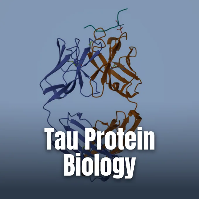
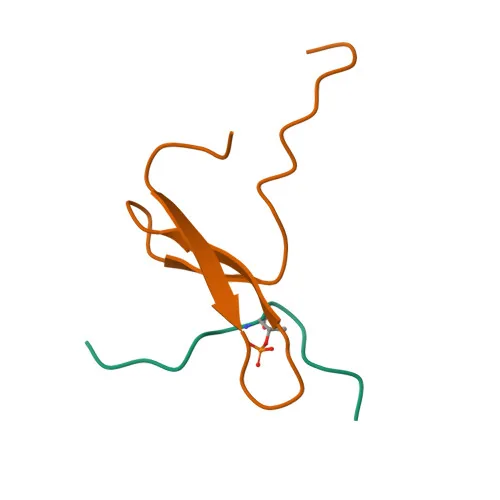
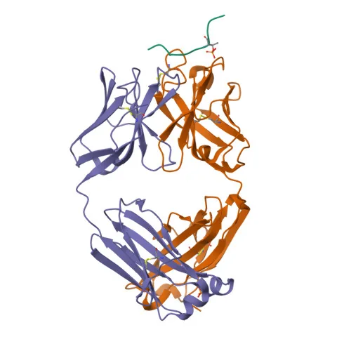
Structural studies show how regulatory enzymes recognize phosphorylated tau sequences. At Institut Pasteur de Lille, Wintjens, Wieruszeski, Lippens, and Landrieu published findings in the Journal of Biological Chemistry (J Biol Chem), indexed on PubMed, using nuclear magnetic resonance spectroscopy to examine the Pin1 WW domain bound to a human tau phosphopeptide containing threonine 231. Their measurements identified a dissociation equilibrium constant of 230 micromolar, showing how Pin1 binds the peptide in a trans conformation across the pThr-Pro bond [4]. At Pfizer Global Research & Development in Cambridge, Shih, Finlay, and their team analyzed a chicken antibody fragment, pT231/pS235_1, which binds phosphorylated tau with a dissociation constant of 0.35 nM. Their crystal structure, resolved at 1.9 Å resolution, revealed a bowl-like CDR-H2 domain that fits the phospho-threonine-231 residue [5].

How Zinc Drives Liquid-Liquid Phase Separation
Zinc binds tau without heparin co-factors. Andrei Yu Roman, François Devred, Deborah Byrne, Romain La Rocca, and Philipp O. Tsvetkov examined tau behavior in vitro in the presence of zinc ions to track temperature-dependent structural shifts. Using turbidimetry, they observed that zinc induces a reversible oligomerization of tau into non-amyloid assemblies that disassemble promptly when scientists add a zinc chelator or lower the temperature. Their findings showed that zinc can guide tau down an alternative folding pathway distinct from classical paired helical filament formation [1].
Building on this work, Dahbia Yatoui and fellow researchers mapped the thermodynamics of zinc coordination on the protein. Using isothermal titration calorimetry and dynamic light scattering, they confirmed that tau carries three distinct zinc-binding sites. A high-affinity zinc site resides within the repeat region spanning R2 and R3, while lower-affinity sites exist at the N-terminal and C-terminal regions [2].
Their experiments showed that tau aggregation specifically requires coordinating zinc at the high-affinity site across the R2R3 region [2]. But when zinc binds to any two of the three sites, it drives liquid-liquid phase separation (LLPS). This process allows soluble tau molecules to condense into concentrated liquid droplets under physiological conditions. Over time, these droplets become gel-like and can form solid clumps. By coordinating up to three zinc ions, tau shifts between soluble forms, condensed liquid droplets, and solid seeds [6].
How Tau Protein Spreads Between Neurons
Pathological tau does not stay inside a single sick neuron. It travels through connected brain networks [6, 7]. When diseased neurons break down, they release monomeric and aggregated tau, including distinct human tau strains, into the surrounding interstitial fluid and cerebrospinal fluid. This extracellular release depends on neuronal activity, allowing active circuits to export abnormal proteins into surrounding brain tissue [6].
Monomers enter cells through LRP1. Normal physiological tau monomers enter recipient cells primarily by binding to low-density lipoprotein receptor-related protein 1. In sharp contrast, large pathological fibrils enter recipient neurons through macropinocytosis, a distinct uptake mechanism that requires interaction with heparan sulfate proteoglycans on the cell surface. Once inside, internal tau fibrils act as seeds. They template native tau misfolding. Tracing this spreading pathway in disease models, Asai and colleagues showed that tau pathology moves from the entorhinal cortex to the hippocampal formation during early stages. Microglia also participate in moving tau between brain regions, though the precise mechanism of microglial transport remains under active study [6].
This transmission pattern resembles prion cascades. Extracellular tau aggregates spread between interconnected brain circuits through mechanisms resembling prion-like transmission in mice, corrupting normal monomers into solid fibrillar assemblies that disrupt synaptic function and block nerve impulses [6, 7]. Localized misfolding gradually expands into broad network failure across the cerebral cortex [7].
Pathological Aggregation in Tauopathies
Tau diseases form a broad family known collectively as tauopathies. Clinicians divide these conditions into primary and secondary categories. In primary tau diseases, tau aggregation represents the main disease event, whereas secondary tau diseases involve tau accumulation driven by another upstream pathology such as amyloid deposition [7].
Alzheimer’s disease serves as the classic secondary tauopathy, where amyloid-beta peptide deposition initiates an upstream cascade that triggers downstream hyperphosphorylation and neurofibrillary tangle formation [7]. This transformation turns soluble proteins into paired-helical-filament (PHF) tau and paired helical filaments (PHFs). During Alzheimer’s progression, at least 19 amino acids on tau become abnormally phosphorylated. Pre-tangle phosphorylation occurs at serine 199, serine 202, and serine 409, while intra-tangle phosphorylation targets serine 396 and threonine 231 [6]. In frontotemporal lobar degeneration, spherical inclusions called Pick bodies appear inside swollen neurons. Pick bodies contain hyperphosphorylated 3R tau isoforms, displaying characteristic gel electrophoresis bands at 60, 64, and 69 kilodaltons [7].
Other primary tauopathies predominantly involve the accumulation of four-repeat isoforms, placing them in the class of 4R tauopathies. At Ludwig-Maximilians-Universität München, Thomas W. Rösler, Günter U. Höglinger, and colleagues classified these disorders by their clinical and neuropathological features [3]. Progressive supranuclear palsy is a rare 4R disorder affecting 5 to 6.4 per 100,000 people, presenting with supranuclear gaze palsy, postural instability, and tufted astrocytes. Corticobasal degeneration also belongs to the 4R group, characterized by cortical ballooned cells, basal ganglia degeneration, and a prominent tau doublet at 64 and 68 kilodaltons. In addition, argyrophilic grain disease represents a late-onset 4R tauopathy detected in 4 to 13 percent of dementia autopsies [7]. The specific isoform composition of these paired helical filaments determines which brain networks degenerate [3].
Diagnostic Biomarkers for Tau Proteins
Accurate detection of abnormal tau is valuable for identifying neurodegenerative illnesses early in their clinical course. Medical centers use biofluid analyses of cerebrospinal fluid to evaluate total tau and phosphorylated tau concentrations in patients with cognitive decline. In patients with Alzheimer’s disease, cerebrospinal fluid typically exhibits elevated tau protein levels together with decreased amyloid-beta levels, creating a reliable diagnostic signature that distinguishes them from healthy controls [7]. Recent advances also examine tau protein levels in peripheral blood, which could make clinical screening more accessible and less invasive than lumbar punctures. Tracking these biofluid markers in vivo allows clinicians to follow disease progression over time and assess whether candidate therapies alter circulating protein levels [3].
PET imaging maps tau deposits. Radiotracers that bind specifically to paired helical filaments allow clinicians to measure the spatial burden of tangles across cortical and subcortical regions directly inside the living brain. But PET scanning remains expensive and involves minor radiation exposure, which restricts how often clinics can repeat scans during routine patient monitoring [7].
Drug trials target tau kinases. Experimental approaches explore inhibiting tau kinases like GSK-3 and CDK5, or stimulating phosphatases like PP2A to prevent hyperphosphorylation while other studies test modulating autophagy to clear toxic clumps [8]. By understanding how tau shifts from a stabilizing microtubule partner into a pathological seed, scientists continue working toward targeted treatments that preserve neuronal connectivity and brain function [3].
- ACADEMIC JOURNAL Roman, A. Y., Devred, F., Byrne, D., La Rocca, R., Ninkina, N. N., Peyrot, V., & Tsvetkov, P. O. (2019). Zinc Induces Temperature-Dependent Reversible Self-Assembly of Tau. Journal of Molecular Biology, 431(4), 687-695. [Article Link]
- ACADEMIC JOURNAL Yatoui, D., Tsvetkov, P. O., La Rocca, R., Baksheeva, V. E., Allegro, D., Breuzard, G., Ferracci, G., Byrne, D., & Devred, F. (2022). Binding of two zinc ions promotes liquid-liquid phase separation of Tau. International Journal of Biological Macromolecules, 223, 1223-1229. [Article Link]
- ACADEMIC JOURNAL Rösler, T. W., Tayaranian Marvian, A., Brendel, M., Nykänen, N., Höllerhage, M., Schwarz, S. C., Hopfner, F., Koeglsperger, T., Respondek, G., Schweyer, K., Levin, J., Villemagne, V. L., Barthel, H., Sabri, O., Müller, U., Meissner, W. G., Kovacs, G. G., & Höglinger, G. U. (2019). Four-repeat tauopathies. Progress in Neurobiology, 180, 101644. [Article Link]
- REPORT Wintjens, R., Wieruszeski, J., Drobecq, H., Lippens, G., & Landrieu, I. (2001). Solution structure of Pin1 WW domain complexed with human tau phosphothreonine peptide. Worldwide Protein Data Bank. [Article Link]
- ACADEMIC JOURNAL Shih, H. H., Tu, C., Cao, W., Klein, A., Ramsey, R., Fennell, B. J., Lambert, M., Ní Shúilleabháin, D., Autin, B., Kouranova, E., Laxmanan, S., Braithwaite, S., Wu, L., Ait-Zahra, M., Milici, A. J., Dumin, J. A., LaVallie, E. R., Arai, M., Corcoran, C.,. Finlay, W. J. (2012). An Ultra-specific Avian Antibody to Phosphorylated Tau Protein Reveals a Unique Mechanism for Phosphoepitope Recognition. Journal of Biological Chemistry, 287(53), 44425-44434. [Article Link]
- WEBSITE Wikipedia contributors. (2026). Tau protein. Wikipedia, The Free Encyclopedia. [Article Link]
- WEBSITE Wikipedia contributors. (2026). Tauopathy. Wikipedia, The Free Encyclopedia. [Article Link]
- WEBSITE Wikipedia contributors. (2026). Tau-protein kinase. Wikipedia, The Free Encyclopedia. [Article Link]


