





The central question of what is neuroplasticity leads directly to a core finding of modern brain science: the nervous system does not lock into a fixed form once youth ends [1]. Instead, living neural networks alter their synaptic links and functional pathways in direct response to daily learning, sensory shifts, and head trauma throughout adulthood [4]. Early anatomists saw the adult human brain as an unyielding organ with permanent circuits that could never repair themselves. Decades of laboratory tests and patient studies confirmed that neural maps reshape their layout over a person’s entire life [1].
- What Is Neuroplasticity?
- Anatomical Evidence in Brain Tissue
- Synaptic Strengthening at Dendritic Spines
- Why Neuroplasticity Matters for Lifelong Learning
- Computerized Cognitive Training Tools
- Therapeutic Interventions in Clinical Practice
- Rethinking Neural Plasticity in Cell Models
- Maladaptive Rewiring in Addiction
What Is Neuroplasticity?
The broader idea of plasticity entered modern science in 1890 when William James published his classic text The Principles of Psychology with Henry Holt and Company in New York. James wrote that organic matter was “a structure weak enough to yield to an influence, but strong enough not to yield all at once” [1]. Polish neurobiologist Jerzy Konorski coined the exact term neural plasticity in a 1948 Cambridge University Press volume on conditioned reflexes [2]. Experimental evidence dates back even earlier to 1793, when Italian anatomist Michele Vincenzo Malacarne trained selected birds and dogs for several years before examining their cranial structures. Dissections proved that cerebellums in trained animals were distinctly larger than those of untrained littermates, providing early proof that mental training reshapes brain tissue [1].
Spanish anatomist Santiago Ramón y Cajal developed the neuron doctrine, showing that individual nerve cells form the basic building blocks of brain tissue. Cajal argued that nonpathological shifts occur in adult brains, even though peers like Walther Spielmeyer and Max Bielschowsky insisted that mature brain tissue cannot form new cells. Later studies by researchers JT Wall and J Xu showed that neural shifts occur across all levels of sensory pathways rather than merely inside the cerebral cortex. In 1943, Warren McCulloch and Walter Pitts drew on work by Nicolas Rashevsky to propose artificial neural learning rules where new synapses form when cells fire simultaneously [1].
Christopher Shaw and Jill McEachern reported that researchers divide these adaptations into two forms: anatomical plasticity and functional plasticity [1]. Anatomical changes alter the shape and size of dendrites, whereas functional changes let healthy circuits take over jobs from damaged areas [7]. Both processes work hand in hand to help people adapt to new life conditions.
Anatomical Evidence in Brain Tissue
Clear physical proof of adult brain flexibility arrived in 1964 when Marian Diamond and her team at the University of California, Berkeley, showed that enriched cages made rodent cerebral cortices thicker. Soon after, Nobel laureates David Hubel and Torsten Wiesel found that sewing one kitten eyelid shut made the visual cortex rewire its inputs toward the open eye. Neuroscientist Michael Merzenich showed that this flexibility lasts into adulthood by mapping hand regions in monkeys alongside Clinton Woosley. Merzenich, who won the 2016 Kavli Prize in Neuroscience, asserted that “If the brain map could normalize its structure in response to abnormal input, the prevailing view that we are born with a hardwired system had to be wrong. The brain had to be plastic” [1].
Sensory tools proved this adaptability in human patients. In the 1960s, Paul Bach-y-Rita made a chair with vibrating nubs that turned camera views into skin taps, letting blind users discern spatial outlines. Stroke doctor Shepherd Ivory Franz showed that undamaged areas take over motor duties from dead tissue [1].
Daily spatial learning alters human brain anatomy, as shown by Eleanor Maguire in tests on London taxi drivers. Maguire found that drivers who memorized the city street grid had more grey matter in the posterior hippocampus than non-drivers. Navigational demands correlated directly with years spent driving cabs across the metropolitan street network. The famous investigation proved that intense memory work reshapes localized brain centers in healthy adult humans [1].

Synaptic Strengthening at Dendritic Spines
Microscopic rewiring follows cell rules stated in 1949 by Canadian psychologist Donald Hebb in his book The Organization of Behavior [1]. Hebbian learning holds that when pre- and postsynaptic cells fire together, the link between them grows stronger. Long-term potentiation, known as LTP, increases signal transmission efficiency between firing cells, while long-term depression, or LTD, systematically weakens neglected synapses to eliminate obsolete connections [3].
During long-term potentiation, glutamate opens post-synaptic N-methyl-D-aspartate receptors, letting calcium flow into the dendritic spine to trigger intracellular signaling. The resulting calcium influx pulls extra AMPA receptors into the synaptic wall, making the post-synaptic neuron far more sensitive to subsequent glutamate releases from the neighboring axon. With repeated use, thin spines turn into stable mushroom shapes that last for months or years [3]. Psychologist Karl Lashley saw pathway shifts in rhesus monkeys in 1923, and modern spine tracking proves that structural changes at synapses store lasting cognitive memories [1].
Biochemical reviews by Gulyaeva in the journal Biochemistry Moscow show that plasticity uses signaling cascades, protein phosphorylation, and tissue remodeling. In particular, brain-derived neurotrophic factor, or BDNF, helps build proteins at active synaptic spots to strengthen communication links. Studies by Rozov and Balaban detailed how GABAergic synapses and atypical protein kinases regulate long-term memory formation within hippocampal circuits [2]. Local astrocytes and microglia clean up transmitters to keep these cellular changes orderly.
Why Neuroplasticity Matters for Lifelong Learning
Understanding why neuroplasticity matters explains how adults pick up fresh work skills, master hard concepts, and heal after head injuries. In a lifespan review for Brain Sciences, Marzola, Melzer, Pavesi, Gil‐Mohapel, and Brocardo wrote that neuroplasticity helps older brains balance out cell loss by rewiring surviving circuits. While infant brains make vast numbers of temporary synapses, adult networks keep the ability to reshape pathways when faced with real mental tasks [4]. Spanish doctor Justo Gonzalo observed dynamic adaptability in brain injury cases in the 1940s, noting how sensory networks recruit unassigned cortex to restore sight and touch in patients during Stratton reversing glass experiments [1].
Mental effort builds defense against normal age decline. Studies show that learning another language builds cognitive reserve by forcing people to switch words and focus attention across complex tasks. Continuous lexical retrieval strengthens white matter tracts and helps seniors stay mentally independent throughout their lives [5].
Researchers Park and Bischof observed that older minds build neural scaffolding to maintain performance when older circuits wear down. Brain scans show that older adults call on both frontal brain lobes during memory tests where young people use just one. Bilateral prefrontal recruitment proves that the aging brain adapts by recruiting supplementary neural pathways to maintain cognitive performance rather than simply surrendering to anatomical deterioration [5]. Scaffolding helps seniors handle everyday demands with confidence.



Computerized Cognitive Training Tools
Digital training tools offer structured ways to spark brain plasticity at home. In a research paper for the International Journal of Engineering Pedagogy spanning pages 50 to 62, Marios Pappas and Athanasios Drigas examined a decade of digital tools, concluding that phone apps and video games produce real gains in attention and memory. These computer tools help both healthy adults and patients with memory loss by providing structured, repetitive cognitive tasks that deliver immediate performance feedback across multiple testing sessions [6].
A major puzzle in brain science is whether game practice transfers to everyday life tasks. Park and Bischof reported that while task gains last over time, transfer to other skills is small unless users tackle fresh, demanding tasks [5]. Learning difficult real-world skills—such as playing a musical instrument or programming complex software—triggers wider brain rewiring because it challenges sensory, motor, and cognitive faculties simultaneously. Rich and demanding environments push the brain to link separate cortex areas to solve real challenges.
Brisk aerobic exercise acts as a chemical boost for brain remodeling. Workouts trigger the release of BDNF, spur new cell birth in the hippocampus, and bring more blood to the head. Regular physical workouts paired with tricky mental hobbies give people a practical path to keep their minds sharp across their entire lives [4].
Therapeutic Interventions in Clinical Practice
Clinicians work to translate laboratory discoveries into routine hospital treatments. An NIH Blueprint for Neuroscience Research workshop led by Cramer and twenty-four leading scientists mapped out clinical therapies for stroke, paediatric disorders, mental health conditions, and traumatic brain injury. The expert panel concluded that combining physical rehabilitation with non-invasive brain stimulation or targeted pharmacological agents helps patients regain lost motor abilities by promoting adaptive cortical reorganization [7].
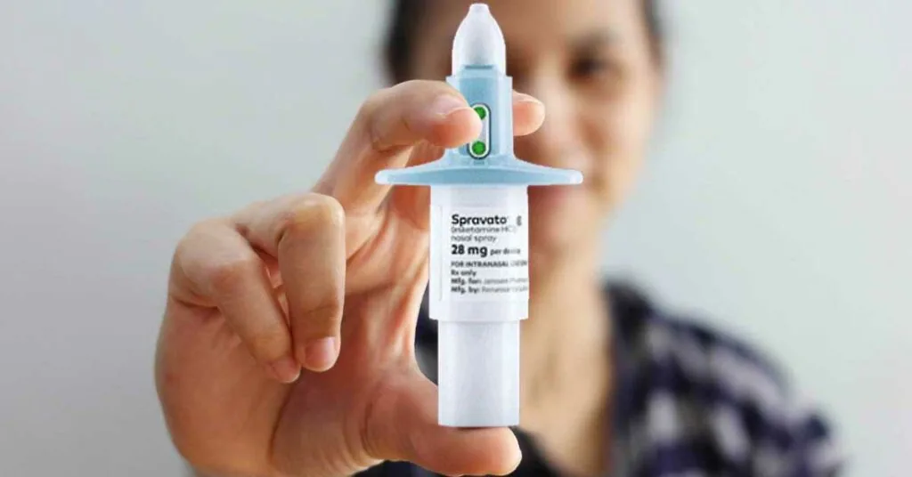
Fast-acting drugs target synaptic receptors to lift deep depressive moods. At the Neuroplasticity MD clinic in Fort Lee and Montclair, New Jersey, medical director Dr. Yuli Fradkin, who trained at Beth Israel, Tufts, and Rutgers, uses intravenous ketamine to relieve major depression, helping over 70% of patients. “This is a game changer,” said Dr. John Krystal of Yale School of Medicine, who pioneered ketamine research. Unlike older pills that alter serotonin over several weeks, ketamine blocks NMDA receptors to prompt an immediate rush of glutamate, building new synapses within hours [9].
Psychedelic drugs also spur rapid structural changes. In a comprehensive review for Neuropsychopharmacology, Calder and Hasler showed that classic psychedelics like psilocybin and LSD promote rapid dendritogenesis and synaptogenesis in the prefrontal cortex and hippocampus. Therapeutic doses turn on plasticity genes like BDNF, helping people break free from trapped thought loops during clinical therapy sessions [8].
Rethinking Neural Plasticity in Cell Models
Most computer models treat the brain as a net of passive synapses whose weights change with traffic. In an unreviewed arXiv preprint from Cornell University, researcher Gabriele Scheler challenged this view, proposing a fundamental paradigm shift from synapse-centric architectures toward a neuron-centric biological model [10]. The theoretical work notes that synapse-centric accounts date back to early computational theories from Peter Putnam and Robert W. Fuller [1]. Scheler argued that viewing each cell as an active, self-programming computer explains biological plasticity better than treating it as a simple wire link [10].
Scheler’s model divides signal transfer from memory storage across three cell zones. These distinct compartments include outer surface receptors on dendritic spines, protein signaling paths in the sub-membrane cytoplasm, and genetic information housed inside the cell nucleus. Under this view, a neuron sorts and filters incoming pulses through inner chemistry before altering its outer membrane. Intracellular gating prevents random electrical noise from wiping out stored memories, protecting network stability while still allowing the cell to record meaningful behavioral experiences [10].
These cellular principles guide experimental computing projects, including innovative initiatives combining electronic hardware with lab-grown brain tissue to construct hybrid biological processors. Clarifying whether memory sits in outer synaptic weights or deep inner chemistry helps engineers build smarter chips. Researchers De Pitta and Volterra demonstrated that glial networks further coordinate signal processing beyond simple binary switches [2].
Maladaptive Rewiring in Addiction
Plasticity does not always bring healthy results; under bad inputs, it drives harmful habits. In chronic drug addiction diagnosed under models like the Diagnostic and Statistical Manual of Mental Disorders, repeated drug use hijacks reward paths in the ventral tegmental area and the nucleus accumbens. While dopamine surges in the ventral tegmental area settle down after seven to ten days, long-term structural changes take root inside the nucleus accumbens where they persist for months. Heavy substance abuse weakens synapses within the nucleus accumbens, and these stubborn cellular changes explain why intense drug cravings flare up repeatedly during prolonged withdrawal periods [3].
Brain immune cells shape how well synapses adapt under stress. Singhal and Baune showed in Frontiers in Cellular Neuroscience that chronic psychological stress, aging, and bodily infections turn microglia into an overactive, proinflammatory state. Overactive microglia release inflammatory signals that damage synapses and trim healthy links, deepening depression [11].
In progressive conditions like Alzheimer’s disease, toxic protein clumps and mitochondrial damage wear away synaptic networks. Scientists are testing ways of drugging previously unreachable proteins in neurodegenerative illnesses to eliminate toxic molecular aggregates before permanent cellular damage takes place. Targeted drug therapies paired with non-invasive mental drills that stimulate remaining brain plasticity offer patients a dependable clinical way to hold onto memory and daily skills [4].
- WEBSITE Wikipedia contributors. (2026). Neuroplasticity. Wikipedia, The Free Encyclopedia. [Article Link]
- ACADEMIC JOURNAL Gulyaeva, N. V. (2017). Molecular mechanisms of neuroplasticity: An expanding universe. Biochemistry (Moscow), 82(3), 237–242. [Article Link]
- WEBSITE Wikipedia contributors. (2026). Addiction-related structural neuroplasticity. Wikipedia, The Free Encyclopedia. [Article Link]
- ACADEMIC JOURNAL Marzola, P. E. R., Melzer, T. M., Pavesi, E., Gil‐Mohapel, J., & Brocardo, P. S. (2023). Exploring the Role of Neuroplasticity in Development, Aging, and Neurodegeneration. Brain Sciences, 13(12), 1610. [Article Link]
- ACADEMIC JOURNAL Park, D. C., & Bischof, G. N. (2013). The aging mind: neuroplasticity in response to cognitive training. Dialogues in Clinical Neuroscience, 15(1), 109–119. [Article Link]
- ACADEMIC JOURNAL Pappas, M. A., & Drigas, A. S. (2019). Computerized Training for Neuroplasticity and Cognitive Improvement. International Journal of Engineering Pedagogy (iJEP), 9(4), 50–62. [Article Link]
- ACADEMIC JOURNAL Cramer, S. C., Sur, M., Dobkin, B. H., O’Brien, C. P., Sanger, T. D., Trojanowski, J. Q., Rumsey, J. M., Hicks, R. R., Cameron, J. L., Chen, D., Chen, W., Cohen, L. G., deCharms, C., Duffy, C. J., Eden, G. F., Fetz, E. E., Filart, R., Freund, M., Grant, S. J.,. Mayberg, H. S. (2011). Harnessing neuroplasticity for clinical applications. Brain, 134(6), 1591–1609. [Article Link]
- ACADEMIC JOURNAL Calder, A. E., & Hasler, G. (2022). Towards an understanding of psychedelic-induced neuroplasticity. Neuropsychopharmacology, 48(1), 104–112. [Article Link]
- WEBSITE Neuroplasticity MD. (2026). Ketamine Treatment New Jersey. [Article Link]
- PREPRINT Scheler, G. (2022). Sketch of a novel approach to a neural model. arXiv. [Article Link]
- ACADEMIC JOURNAL Singhal, G., & Baune, B. T. (2017). Microglia: An Interface between the Loss of Neuroplasticity and Depression. Frontiers in Cellular Neuroscience, 11, 270. [Article Link]
Cover image: http://www.ccnlab.net / Wikimedia Commons, CC BY-SA 4.0


