






Does eating wheat bread cause lasting harm in certain people? Celiac disease autoimmunity is an immune reaction triggered by eating gluten proteins found in wheat, rye, and barley. In people carrying specific immune genes, gluten peptides cross the gut lining and incite an abnormal immune attack that steadily flattens tiny gut villi. This tissue harm shrinks the surface area of the small bowel, which leads to poor nutrient uptake, chronic diarrhea, and systemic illness across the body. In the journal The Lancet, volume 391, pages 70–81, researchers Lebwohl, Sanders, and Green reported that the condition affects roughly 1% of people worldwide, but most cases remain undiagnosed [1].
Forms of Coeliac Disease
Medical consensus defines the disorder as an immune enteropathy driven by dietary gluten in genetically prone hosts. It differs from wheat allergy. In 2012, an international consensus panel led by Jonas Ludvigsson published the Oslo definitions in the journal Gut, volume 62, pages 43–52, establishing standardized global terminology to classify distinct clinical presentations across gastroenterology practice [9].
Classical forms feature plain signs of poor nutrient absorption, including foul-smelling diarrhea, pale stools, belly swelling, and rapid weight loss in early life [8]. By contrast, non-classical disease brings problems outside the digestive tract. Patients often face chronic fatigue, low iron levels, mouth sores, or aching joints without obvious belly pain. In subclinical cases, people carry bowel lesions but report no noticeable distress. Many adults live for years with low energy, thinking poor health is normal until a gluten-free diet restores their baseline strength [7]. In 2003, Lo, Lebwohl, and Green published findings in Digestive Diseases and Sciences, volume 48, pages 395–8, showing that adult presentations often diverge from typical childhood malnutrition and require broader clinical suspicion among primary care doctors [1].
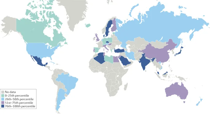
Screening studies indicate that celiac disease autoimmunity occurs across the world, affecting about one in 133 people in North America according to large cohort investigations conducted by Alessio Fasano at Columbia University [5]. In the journal Clinical Gastroenterology and Hepatology, volume 16, pages 823–36, Singh, Arora, and Catassi showed that global prevalence reached approximately 1.4% on blood tests and 0.7% on biopsy records [14]. Public awareness remains incomplete across many healthcare regions [4].
Discovery of the Grain Trigger
Descriptions of bowel wasting reach back to antiquity, when the Greek physician Aretaeus of Cappadocia first recorded the illness in the first century A.D.. He called it koiliakos, after the Greek word for abdomen, to depict people suffering from chronic diarrhea and bodily decline. Centuries later, British doctor Samuel Gee gave a clinical lecture in London in 1887, later published in St Bart Hosp Rep in 1888, describing a chronic digestive illness in children aged one to five. Gee noted that diet might cause the wasting. Yet he wrongly advised feeding sick children thin slices of toasted bread, which kept them unwell [5].
The true dietary trigger came to light during World War II through clinical work by Dutch pediatrician Willem-Karel Dicke in the Netherlands. When wartime bread shortages cut grain supplies, child mortality from the disease plunged from greater than 35% to virtually zero. Dicke published these findings after the war [5].
In 1953, Dicke joined biochemists Weijers and van de Kamer to publish classic papers in Acta Paediatrica, volume 42, pages 34–42, showing that wheat protein caused fat malabsorption. Earlier in 1932, Haas had written in JAMA, volume 99, pages 448–52, about dietary treatment for childhood diarrhea. Later in 1989, Booth reviewed this history in the BMJ, volume 297, pages 1646–9, tracing how post-war biochemists isolated gluten as the toxic plant protein [1]. Their work transformed a fatal wasting illness into a condition managed through food choices [5].
Biological Mechanisms of Gut Damage
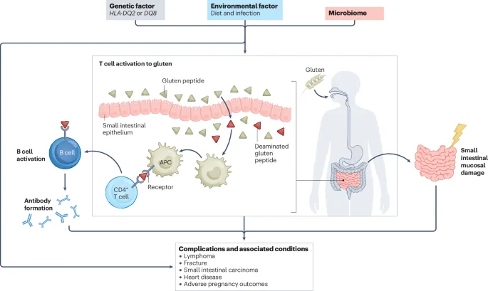
Dietary gluten consists of plant storage proteins found in wheat, rye, and barley [7]. In botanical taxonomy, these crops belong to the tribe Triticeae, whose grain proteins share similar peptide chains. Because these proteins contain high amounts of proline and glutamine, human digestive enzymes cannot break them down completely into single units [8].
Beneath the gut lining, an enzyme called tissue transglutaminase removes an amino group from glutamine, turning it into glutamic acid [8]. This chemical modification creates a strong binding affinity between the altered gluten peptides and human leukocyte antigen class II molecules on antigen-presenting cells, which then display HLA-DQ2 or HLA-DQ8 molecules to CD4 helper T cells in the bowel mucosa [5]. Activated helper T cells release pro-inflammatory cytokines, including interferon-gamma, which stimulates killer intraepithelial white cells to attack healthy intestinal epithelial cells and destroy the mucosal barrier. These combined cellular attacks steadily erode the microscopic architecture of the intestinal wall, leaving the mucosal surface severely blunted [1].
Continuous immune cell attack flattens the microscopic villi into smooth tissue, a state known as villous atrophy. Microscopic villi normally provide a large surface area for absorbing carbohydrates, fats, minerals, and vitamins into bloodstream vessels. As the villi flatten into scar tissue, the small bowel loses its capacity to process ingested meals, which triggers osmotic diarrhea, belly pain, and systemic shortages of key minerals [3].
Genetic and Environmental Factors
Inherited genes play an important role in the onset of celiac disease autoimmunity, with over 90% of patients carrying the HLA-DQ2 allele and most others carrying HLA-DQ8 [8]. Even so, these genes alone do not cause the disease. Roughly 30% to 40% of healthy people carry HLA-DQ2 or HLA-DQ8, yet only about 3% of carriers ever develop gut damage [13].
Because genes alone do not explain why disease emerges, scientists actively investigate environmental triggers that disrupt mucosal tolerance in early life [8]. Factors under study include gut bacteria, childhood viral infections such as reovirus, and infant feeding patterns [7]. Pediatric studies show that the timing of gluten introduction does not prevent the illness in children carrying genetic risks, whereas transient viral infections or microbial shifts may induce localized gut stress that breaks oral tolerance to dietary peptides in genetically primed hosts. Early life exposures shape long-term immunity in complex ways [8].
In the journal Theoretical and Applied Genetics, volume 121, pages 1527–39, Dutch researcher van den Broeck and co-workers studied 36 modern European wheat varieties and 50 old landraces grown a century ago. They discovered that modern wheat breeding boosted the expression of the Glia-α9 epitope, a gluten fragment that triggers strong T-cell reactions in most patients. While modern wheat retained similar total protein levels, this shift in breeding may have raised immune reactions across modern food supplies [12].


Symptoms Across Life Stages
Symptoms vary widely by age, diet, and how long the gut has been inflamed [2]. In toddlers aged nine to twenty-four months, symptoms usually appear shortly after cereal foods enter the diet. Sick infants suffer from chronic diarrhea with pale, greasy stools, swollen bellies, repeated vomiting, and poor appetite. Over time, poor nutrient uptake leads to muscle wasting, thin limbs, low energy, and stunted height [14]. In 1932, Haas documented these early digestive wasting patterns in JAMA, volume 99, pages 448–52, noting how severe nutritional deficits altered childhood growth trajectories and caused physical decline [1]. Children falter without medical care.
In school-age children and teenagers, the disease often presents without typical stomach trouble. Pediatric doctors often find celiac cases during tests for unexplained short stature, delayed puberty, mouth ulcers, or low iron that oral pills fail to fix. Permanent teeth often display yellow enamel discoloration or horizontal grooving caused by early nutritional loss [2].
In adults, symptoms are often subtle and lead to mistaken labels such as irritable bowel syndrome [8]. Grown patients frequently report brain fog, ongoing tiredness, bloating, migraines, and aching joints rather than watery diarrhea. Many undiagnosed adults consider themselves well simply because they have grown used to feeling run down. Once they cut gluten from their meals, their energy returns and prior complaints fade away [7].

Comorbidities of Coeliac Disease
Long-term bowel harm leads to problems that touch many body systems [3]. Poor uptake of vitamin D and calcium triggers secondary hyperparathyroidism, which thins bone mineral mass and raises the odds of early osteoporosis [1]. Low iron and poor folate absorption cause stubborn anemia [7].
In Nature Reviews Gastroenterology & Hepatology, Swedish researcher Jonas Ludvigsson and colleagues reviewed how the disease connects to other autoimmune disorders. People with the condition face higher risks of type 1 diabetes and autoimmune thyroid problems like Hashimoto thyroiditis [3]. The skin illness dermatitis herpetiformis causes very itchy blisters on elbows and knees due to IgA deposits in skin tissue. Neurological complications include gluten ataxia, nerve pain in limbs, and higher rates of seizures [1]. Extraintestinal manifestations of celiac disease autoimmunity often involve multiple organ systems outside the digestive tract and require careful long-term clinical monitoring [3].
Active bowel inflammation also harms reproductive health in both women and men. Untreated women experience higher rates of delayed periods, early menopause, trouble conceiving, and miscarriages during pregnancy. Registry data analyzed by European doctors show that healing the gut lining through diet helps reduce these reproductive risks. Complete mucosal recovery protects maternal health [3].
Diagnostic Tests for Coeliac Disease
Testing must take place while the patient continues to eat gluten-rich foods, because cutting gluten before tests clears antibodies and lets gut tissue heal, causing false-negative readings [10]. Doctors begin with a simple blood draw to test for IgA antibodies against tissue transglutaminase, known as anti-tTG IgA [8].
In adults, positive blood tests require upper endoscopy with biopsies of the small intestine to confirm mucosal injury [10]. Doctors take one or two samples from the duodenal bulb and four from the lower duodenum to catch patchy harm. Pathologists read the samples under the microscope using the Marsh classification, looking for intraepithelial white cells, enlarged crypts, and villus flattening. Genetic testing for HLA-DQ2 and HLA-DQ8 carries a high negative predictive value, effectively ruling out the condition when absent. In individuals with confirmed IgA deficiency, clinicians deploy IgG-based assays such as deamidated gliadin peptide antibodies to secure an accurate diagnosis and prevent missed cases [8].

In pediatric medicine, guidelines from ESPGHAN allow a no-biopsy route when anti-tTG IgA levels exceed ten times the normal cutoff and a second blood test confirms endomysial antibodies [4]. Despite refined testing tools, diagnostic delays remain long across healthcare systems. In the journal BMC Gastroenterology, volume 11, a Swedish study by Norström and colleagues showed that the mean delay to diagnosis from first symptoms was 9.7 years [11].
Dietary Treatment and Management
At present, the only safe treatment is a lifelong gluten-free diet [10]. Cutting wheat, rye, and barley stops the immune attack, allowing gut villi to regrow and restore normal nutrient absorption [3]. Patients eat plain meat, poultry, fish, eggs, beans, fruit, vegetables, and safe grains such as rice, corn, millet, sorghum, and buckwheat [7].
Following a gluten-free meal plan brings daily practical hurdles, including higher food bills, fewer dining options, and risks of cross-contact in shared home kitchens [4]. Support groups such as Coeliac UK at Westminster provide food guides, advice helplines, and dining accreditation schemes to help families manage their choices safely [6]. Regular checkups with clinical dietitians and annual blood antibody tests help track gut healing. Adhering to safe food habits lets children resume normal growth and helps adults regain daily vigor [7]. Safe nutrition restores gut health.
In about 10% to 20% of patients, symptoms linger despite diet efforts, a state called non-responsive celiac disease [10]. While stray gluten intake causes most issues, a few patients develop refractory celiac disease with persistent villous atrophy [1]. To help them, researchers Sollid and Khosla published research in the Journal of Internal Medicine, volume 269, pages 604–13, studying non-dietary pills such as oral digestive enzymes to break down stray gluten peptides before they harm the bowel. Understanding celiac disease autoimmunity drives ongoing research into protective drugs for patients facing accidental gluten exposure across everyday dining environments [13].
- BOOK CHAPTER Varma, S., & Krishnareddy, S. (2022). Uncomplicated Celiac Disease. Refractory Celiac Disease, 5-19. [Article Link]
- BOOK CHAPTER De Villasante, G. C. (2022). Classical and Non-classical Forms of CD in Paediatrics. Advances in Celiac Disease, 37-52. [Article Link]
- ACADEMIC JOURNAL Ludvigsson, J. F., Yao, J., Lebwohl, B., Green, P. H. R., Yuan, S., & Leffler, D. A. (2025). Coeliac disease: complications and comorbidities. Nature Reviews Gastroenterology & Hepatology. [Article Link]
- ACADEMIC JOURNAL Makharia, G. K., Singh, P., Catassi, C., Sanders, D. S., Leffler, D., Ali, R. A. R., & Bai, J. C. (2022). The global burden of coeliac disease: opportunities and challenges. Nature Reviews Gastroenterology & Hepatology. [Article Link]
- ONLINE NEWS Fasano, A. (2009). Celiac Disease Insights: Clues to Solving Autoimmunity. Scientific American. [Article Link]
- WEBSITE Coeliac UK. (2026). No life limited by coeliac disease. [Article Link]
- WEBSITE Wikipedia contributors. (2026). Coeliac disease. Wikipedia, The Free Encyclopedia. [Article Link]
- ACADEMIC JOURNAL Caio, G., Volta, U., Sapone, A., Leffler, D. A., De Giorgio, R., Catassi, C., & Fasano, A. (2019). Celiac disease: a comprehensive current review. BMC Medicine, 17(1). [Article Link]
- ACADEMIC JOURNAL Ludvigsson, J. F., Leffler, D. A., Bai, J. C., Biagi, F., Fasano, A., Green, P. H., Hadjivassiliou, M., Kaukinen, K., Kelly, C. P., Leonard, J. N., Lundin, K. E. A., Murray, J. A., Sanders, D. S., Walker, M. M., Zingone, F., & Ciacci, C. (2012). The Oslo definitions for coeliac disease and related terms. Gut, 62(1), 43-52. [Article Link]
- ACADEMIC JOURNAL Al-Toma, A. A., Volta, U., Auricchio, R., Castillejo, G., Sanders, D. S., Cellier, C., Mulder, C. J. J., & Lundin, K. E. A. (2019). European Society for the Study of Coeliac Disease (ESsCD) guideline for coeliac disease and other gluten‐related disorders. United European Gastroenterology Journal, 7(5), 583-613. [Article Link]
- ACADEMIC JOURNAL Norström, F., Lindholm, L., Sandström, O., Nordyke, K., & Ivarsson, A. (2011). Delay to celiac disease diagnosis and its implications for health-related quality of life. BMC Gastroenterology, 11(1). [Article Link]
- ACADEMIC JOURNAL van den Broeck, H. C., de Jong, H. C., Salentijn, E. M. J., Dekking, L., Bosch, D., Hamer, R. J., Gilissen, L. J. W. J., van der Meer, I. M., & Smulders, M. J. M. (2010). Presence of celiac disease epitopes in modern and old hexaploid wheat varieties: wheat breeding may have contributed to increased prevalence of celiac disease. Theoretical and Applied Genetics, 121(8), 1527-1539. [Article Link]
- ACADEMIC JOURNAL Sollid, L. M., & Khosla, C. (2011). Novel therapies for coeliac disease. Journal of Internal Medicine, 269(6), 604-613. [Article Link]
- ACADEMIC JOURNAL Brown, J. R. G., & Singh, P. (2018). Coeliac disease. Paediatrics and International Child Health, 39(1), 23-31. [Article Link]


