




Celiac disease is a chronic systemic autoimmune disorder triggered by eating dietary gluten, a composite storage protein found naturally in wheat, barley, and rye grains [4]. When susceptible people eat foods made from these grains, their immune system damages the lining of the small intestine [1]. Tiny, fingerlike structures called villi flatten out and lose their ability to take in nutrients from meals [2]. This intestinal damage leaves the body short of iron, folate, and calcium, creating health problems that reach far beyond the gut [3]. The disorder affects roughly 1% of people worldwide, yet most people with the condition do not know they have it [6].
- Gluten Peptides in the Upper Bowel
- Immune Attacks on Small Intestinal Villi
- Genetic Roots and the HLA Complex
- How Celiac Disease Symptoms Present
- Extraintestinal Manifestations and Deficiencies
- The Path to Clinical Diagnosis
- Why Undiagnosed Cases Escaped Detection
- Managing Celiac Disease on a Gluten-Free Diet
Gluten Peptides in the Upper Bowel
Dietary gluten consists of storage proteins found in wheat, barley, and rye grains [4]. In wheat, the alcohol-soluble part of gluten is called gliadin [3]. Human stomach acid and pancreatic enzymes easily break down most dietary proteins into simple amino acids. Gluten resists digestion. Because human gastric juice and pancreatic proteases cannot split the dense peptide bonds surrounding proline residues, long protein fragments travel intact straight into the upper small intestine. Its protein chains carry unusually high concentrations of proline and glutamine, two amino acids that resist breakdown by normal digestive proteases in the human stomach and pancreas [2].
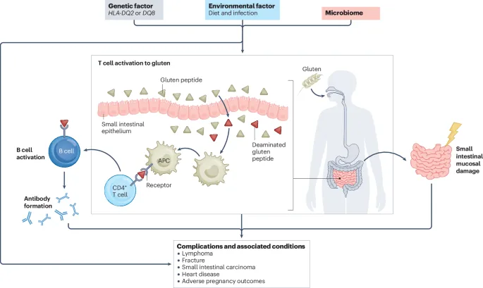
Long gluten fragments travel intact through the small bowel [2]. One common fragment contains 33 amino acids and passes through the stomach without breaking down [3]. In healthy people, these intact fragments stay inside the gut cavity and leave the body without causing trouble [2]. In susceptible individuals, intact fragments slip across the epithelial cell layer into the lamina propria below [3]. That breach sets off local immune reactions [7].
Once inside mucosal tissue, the fragments meet an enzyme called tissue transglutaminase, or TG2. By modifying neutral glutamine residues into negatively charged glutamic acid through deamidation, this enzyme enables the altered gliadin fragments to bind with exceptionally high affinity to class II human leukocyte antigen molecules on antigen-presenting cells [3, 7]. Binding is otherwise weak. Helper T cells stay quiet without it [2]. Once this tight bond forms, however, tissue transglutaminase initiates an inflammatory chain reaction that steadily damages the surrounding intestinal mucosa [3].

Immune Attacks on Small Intestinal Villi
Antigen-presenting cells in the lamina propria display the modified gliadin fragments to helper CD4+ T lymphocytes. Helper T cells recognize these presented peptide complexes as dangerous foreign invaders and promptly release potent inflammatory signals, particularly the proinflammatory cytokine interferon gamma. This chemical signal recruits more immune cells into the mucosal layer [3]. The gut becomes inflamed as immune defenses treat wheat proteins like an active bacterial infection [2].
Epithelial cells under stress produce interleukin 15, a signaling protein that stimulates intraepithelial lymphocytes. These lymphocytes take up positions between epithelial cells and display natural killer cell receptors like NK-G2D. They begin attacking and destroying healthy gut lining cells [3]. Cell loss quickly outpaces the gut’s normal repair rate [2]. As surface enterocytes perish and shed into the lumen, the epithelial barrier breaks down, allowing even more intact gluten fragments to enter the underlying tissue and sustain the inflammatory response [7].
Villi absorb our meals [1]. When persistent intraepithelial lymphocyte attacks strip away these microscopic fingerlike projections, a destructive process termed villous atrophy occurs, while the adjacent intestinal crypts deepen and expand in a futile attempt by mucosal stem cells to replace dying enterocytes [3]. Duodenal biopsies show flattened mucosa [1]. Because this surface loss cripples the intestine’s capacity to absorb fats, vitamins, and minerals, essential nutrients pass straight through the digestive tract unabsorbed [2].
Genetic Roots and the HLA Complex
Genetic predisposition is required for the condition to develop [7]. Almost every patient carries genes for one of two specific human leukocyte antigen variants: HLA-DQ2 or HLA-DQ8. About 90 to 95 percent of patients carry HLA-DQ2, while the rest carry HLA-DQ8. These molecules have positively charged pockets that bind negatively charged gliadin peptides [3]. No genes, no disease [1].
Carrying HLA-DQ2 or HLA-DQ8 does not mean a person will get sick [4]. Roughly 30 to 40 percent of healthy people carry these same genes and eat wheat without any symptoms. Only about 2 to 3 percent of people with the risk genes go on to develop intestinal damage [2]. This means HLA testing works best to rule the condition out. When genetic testing shows that someone lacks both HLA-DQ2 and HLA-DQ8, doctors can comfortably conclude that celiac disease is not the cause of their symptoms [1].

Twin studies show that identical twins have a concordance rate of roughly 75 percent, while non-identical twins show concordance rates near 10 to 30 percent [3]. Non-HLA genes also influence disease susceptibility by altering how immune cells respond to stress [7]. Genes are not alone [4]. Childhood viral infections, changes in the gut microbiome, or the timing of infant feeding may trigger the disease later in life [7]. Dr. Alessio Fasano said that the disorder requires a triad of factors: the environmental trigger of gluten, predisposing HLA genes, and a leaky intestinal barrier that allows foreign peptides to reach immune sentinels [2].
How Celiac Disease Symptoms Present
Symptoms of celiac disease differ widely from person to person [1]. In infants and young children, classical signs typically emerge several months after parents introduce solid cereals or baby foods containing wheat, barley, or rye into their daily diet [3]. Common pediatric signs include chronic diarrhea, poor appetite, and pale, greasy stools that smell unusually foul [1, 4]. Infants may also suffer from vomiting, stomach swelling, and cranky behavior [1].
Because damaged villi cannot absorb food, affected children fall behind on weight gain and normal height. Paediatricians frequently observe delayed puberty, muscle wasting, and permanent enamel defects in teeth that develop during active disease [1, 8]. When malabsorption persists, children can develop severe malnutrition despite eating regular meals. Early diagnosis allows children to regain lost weight and catch up on normal growth patterns within months of changing their diet [4].
In adults, symptoms look quite different from the classical childhood presentation [4]. Instead of chronic diarrhea, adults often report vague symptoms like abdominal cramping, gas, indigestion, or persistent constipation [1]. Many adult patients experience silent or subclinical celiac disease, meaning they have flattened villi and positive blood tests but no noticeable digestive complaints. Undiagnosed individuals often get used to feeling fatigued, assuming low energy is just part of normal daily life. Only after removing gluten from their meals do they realize how much chronic inflammation had been dragging them down [4]. Gemma Castillejo de Villasante reported that modern serological antibody testing has uncovered a substantial population of children and adults who present with subtle extraintestinal complaints rather than obvious diarrhea [8].



Extraintestinal Manifestations and Deficiencies
Nutrient deficiencies can cause problems in almost any organ system. Because the upper small intestine is where the body absorbs iron, flattened villi often cause iron deficiency anemia [4]. Patients feel exhausted, look pale, and suffer from breathlessness [1]. Pills cannot fix it. Standard oral iron supplements often fail to restore red blood cell counts because the damaged duodenal lining cannot absorb the mineral into circulation [4].
Impaired absorption of vitamin D and calcium robs the bones of essential minerals. Researchers Sanskriti Varma and Suneeta Krishnareddy documented that malabsorption commonly causes severe micronutrient deficits, leading to secondary hyperparathyroidism, progressive osteopenia, and premature bone fractures. Some patients develop an intensely itchy, blistering rash called dermatitis herpetiformis on their elbows, knees, and buttocks [1, 7]. This skin condition is the cutaneous form of gluten sensitivity, caused by IgA antibody deposits in the skin. A skin biopsy can confirm dermatitis herpetiformis and point directly to gluten-driven bowel disease [7].
The disease can also affect the nervous system and internal organs. Patients may suffer from tingling or numbness in their hands and feet, headaches, depression, or gluten ataxia, a coordination disorder [4]. Jonas F. Ludvigsson and colleagues observed that individuals living with biopsy-verified enteropathy face elevated long-term risks of systemic complications, including pneumonia, severe sepsis, and chronic liver abnormalities [5]. Many patients develop mild liver enzyme elevations, known as celiac hepatitis, which resolve after the gut lining heals on a gluten-free diet [4]. People diagnosed with celiac enteropathy show a well-documented genetic overlap with other autoimmune conditions, facing higher lifetime risks of developing type 1 diabetes, autoimmune thyroid disease, and Addison disease [1, 5].

The Path to Clinical Diagnosis
Diagnosing the condition requires specific blood tests followed by an examination of the gut lining. The primary screening tool is a blood test for anti-tissue transglutaminase antibodies, or tTGA, using the IgA class [1, 7]. Doctors also use anti-endomysial antibody tests to check uncertain results [1]. These antibody tests accurately identify active intestinal inflammation in most patients [7].
IgA deficiency complicates testing. Because roughly 3 to 5 percent of celiac patients lack sufficient immunoglobulin A, standard IgA-based antibody tests often produce false-negative results, forcing diagnostic laboratories to measure total serum IgA levels and deploy specialized IgG antibody panels whenever overall levels fall short [3]. Dietary habits matter immensely. Patients must keep eating normal daily amounts of bread or pasta prior to blood draws, because eliminating gluten clears circulating antibodies from the bloodstream and masks active disease [1, 4].
If blood tests suggest the disease, gastroenterologists perform an upper endoscopy to take small tissue samples from the duodenum [1]. A pathologist checks the tissue under a microscope to confirm villous blunting and crypt deepening [3]. If a patient already stopped eating gluten, doctors must perform a gluten challenge, having the patient eat gluten daily for several weeks so the immune reaction reappears before the biopsy [4]. While pediatric guidelines in Europe allow some children with very high antibody levels to skip the endoscopy, duodenal biopsy remains the standard confirmation test for adults [7]. A follow-up test after starting treatment confirms healing [1].
Why Undiagnosed Cases Escaped Detection
Medical records show that celiac disease was recognized long before modern immunology explained it. In the first century A.D., Greek physician Aretaeus of Cappadocia described an abdominal illness he named koiliakos. Centuries later, in an 1887 lecture, British physician Samuel Gee gave the first modern description of the chronic indigestion in children. Gee was mistaken about treatment. He suspected that dietary errors caused the illness, but inadvertently prescribed thin slices of toasted bread [2].
The breakthrough connecting cereal grains to gut damage arrived during World War II. Dutch paediatrician Willem-Karel Dicke noticed that wartime bread shortages in the Netherlands dropped celiac mortality rates among children from greater than 35 percent to essentially zero. When wheat became available again after the war ended, mortality climbed right back to former levels. Dicke’s clinical observations helped researchers isolate wheat gluten as the primary culprit behind the intestinal damage [2].
For decades, medical teaching assumed celiac disease was rare outside Europe, estimating its frequency at fewer than 1 in 3,000 or 1 in 10,000 people [2, 3]. In 2003, Alessio Fasano and colleagues published a North American study of over 13,000 people showing that 1 in 133 apparently healthy individuals actually had the disease. Cases had been missed because subtle symptoms often lack obvious digestive distress [2]. In another cohort study tracking healthy young adults from 1948 to 1954 in the United States, Rubio-Tapia and colleagues found that undiagnosed celiac disease raised 45-year all-cause mortality nearly fourfold, with a hazard ratio of 3.9 compared to healthy peers [3]. Today, researchers recognize that roughly 1% of the global population is affected [6].
Managing Celiac Disease on a Gluten-Free Diet
Wheat must go. Following a strict gluten-free diet offers the only known medical therapy for the disorder [1, 7]. Patients must remove all foods, drinks, and medications containing wheat, rye, and barley [1]. While pure, uncontaminated oats are safely tolerated by most patients, cross-contact during commercial harvesting, transport, and grain milling remains a serious concern that requires careful label inspection [3, 4]. When gluten is removed, the gut lining begins to heal, and symptoms gradually fade away [1].
In children, mucosal healing often happens within 3 to 6 months after starting the diet. Adult recovery takes longer, typically requiring 2 to 3 years for villi to regain their full height. Sticking to the diet can be difficult because wheat is a common ingredient in many processed foods [1]. Gluten-free groceries are also expensive, and dining out carries risks of accidental exposure [6]. Patient advocacy groups like the National Celiac Association and Coeliac UK offer dietary resources, help families navigate menus, and campaign for safer gluten-free food options in markets [9].
Up to 20 percent of patients experience persistent symptoms despite trying to follow the diet [6]. Most of these non-responsive cases trace back to accidental gluten ingestion or other conditions like lactose intolerance [1, 4]. However, about 5 percent of patients have refractory celiac disease, where intestinal inflammation continues despite at least 12 months on a confirmed gluten-free diet [3, 4]. Type 1 refractory disease has normal intraepithelial lymphocytes, while Type 2 features abnormal lymphocytes and carries an increased risk of intestinal lymphoma [3]. Because diet alone is difficult to maintain and does not resolve every case, scientists are actively studying non-dietary therapies, such as oral digestive enzymes and tight junction modulators, to provide extra protection against gluten exposure [6].
- WEBSITE MedlinePlus. (2026). Celiac disease – sprue. MedlinePlus Medical Encyclopedia. [Article Link]
- ONLINE NEWS Fasano, A. (2009). Celiac disease insights: Clues to solving autoimmunity. Scientific American. [Article Link]
- WEBSITE Medscape. (2025). Celiac disease (sprue): Background, pathophysiology, etiology. Medscape Reference. [Article Link]
- WEBSITE Wikipedia contributors. (2026). Coeliac disease. Wikipedia, The Free Encyclopedia. [Article Link]
- ACADEMIC JOURNAL Ludvigsson, J. F., Yao, J., Lebwohl, B., Green, P. H. R., Yuan, S., & Leffler, D. A. (2025). Coeliac disease: complications and comorbidities. Nature Reviews Gastroenterology & Hepatology, 22, 1-15. [Article Link]
- ACADEMIC JOURNAL Makharia, G. K., Singh, P., Catassi, C., Sanders, D. S., Leffler, D., Ali, R. A. R., & Bai, J. C. (2022). The global burden of coeliac disease: opportunities and challenges. Nature Reviews Gastroenterology & Hepatology, 19, 313-328. [Article Link]
- BOOK CHAPTER Varma, S., & Krishnareddy, S. (2022). Uncomplicated celiac disease. In Refractory Celiac Disease (pp. 5-19). Springer, Cham. [Article Link]
- BOOK CHAPTER de Villasante, G. C. (2022). Classical and non-classical forms of CD in paediatrics. In Advances in Celiac Disease (pp. 35-50). Springer, Cham. [Article Link]
- WEBSITE Coeliac UK. (2026). Coeliac UK: No life limited by coeliac disease. Coeliac UK. [Article Link]


